Review
. 2023 Apr;24(4):213-232.
doi: 10.1038/s41583-023-00675-z. Epub 2023 Feb 15.

Developmental mechanisms underlying the evolution of human cortical circuits

Affiliations
Review

Developmental mechanisms underlying the evolution of human cortical circuits

Pierre Vanderhaeghen et al. Nat Rev Neurosci. 2023 Apr.

Abstract

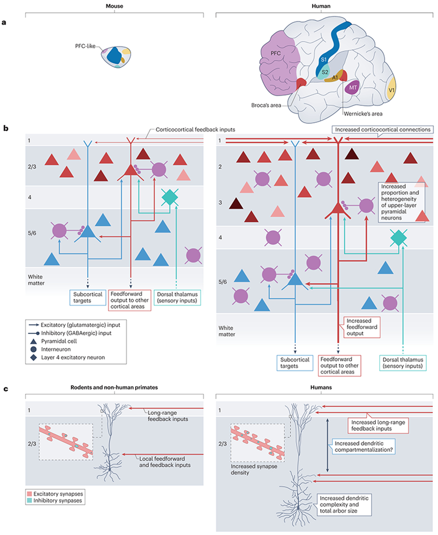
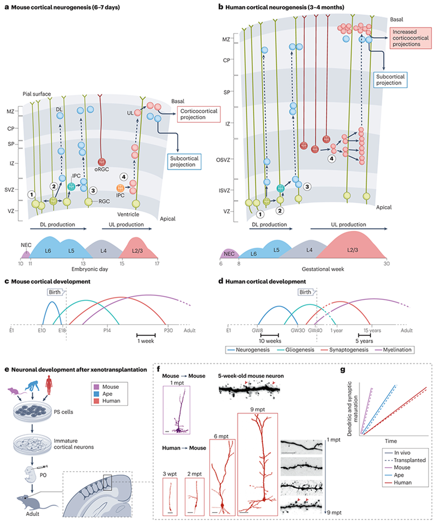
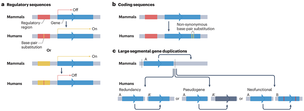
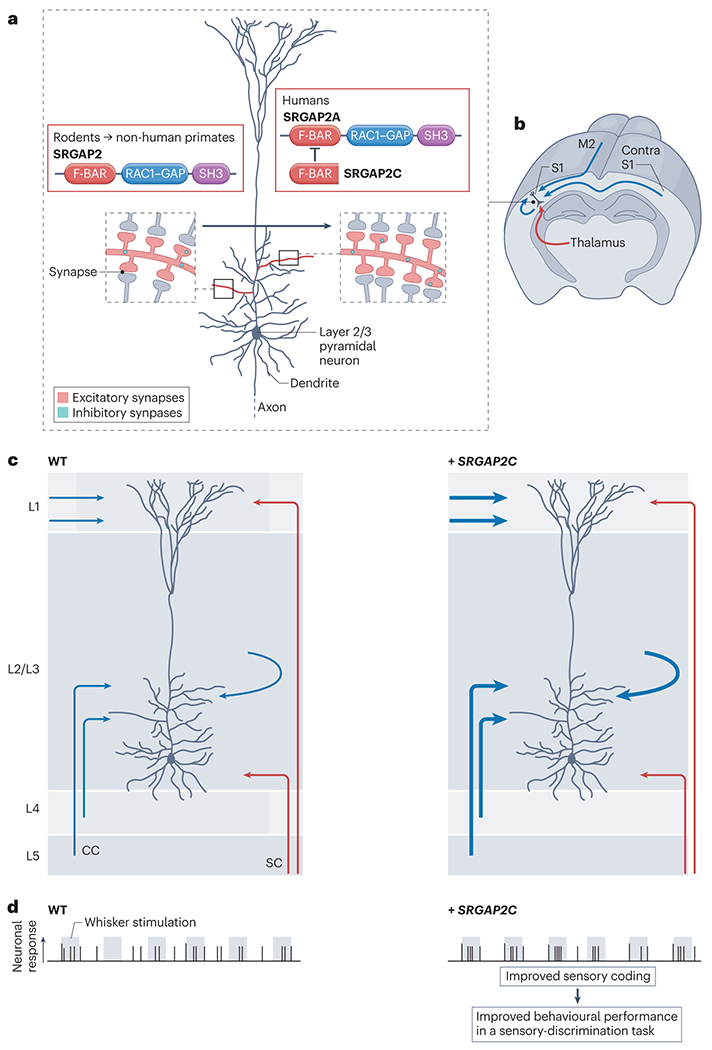
The brain of modern humans has evolved remarkable computational abilities that enable higher cognitive functions. These capacities are tightly linked to an increase in the size and connectivity of the cerebral cortex, which is thought to have resulted from evolutionary changes in the mechanisms of cortical development. Convergent progress in evolutionary genomics, developmental biology and neuroscience has recently enabled the identification of genomic changes that act as human-specific modifiers of cortical development. These modifiers influence most aspects of corticogenesis, from the timing and complexity of cortical neurogenesis to synaptogenesis and the assembly of cortical circuits. Mutations of human-specific genetic modifiers of corticogenesis have started to be linked to neurodevelopmental disorders, providing evidence for their physiological relevance and suggesting potential relationships between the evolution of the human brain and its sensitivity to specific diseases.


Conflict of interest statement

Competing interests

The authors declare no competing interests.

Figures

Fig. 1|
Fig. 1|. Cortical circuit evolution.
a, Schematic illustration of the expansion of cortical area size that has taken place in the human brain, when compared to the mouse (brains depicted approximately to scale). The expanded size of the human neocortex is accompanied by an increase in the number of cortical areas driven by the emergence of new cortical areas (such as Wernicke’s area and Broca’s areas). In addition there has been an increase in the size of certain cortical areas, including a pronounced expansion of the prefrontal cortex (PFC) compared to its putative homologous regions (PFC-like regions) in the mouse. b, In mammals, cortical neurons are organized in 6 layers that are generated in an inside-first outside-last manner: early born neurons generate deep layer (DL, layers 5/6) pyramidal neurons (PNs) (blue), then thalamo-recipient layer 4 neurons (green) and finally upper layer (UL, layers 2/3) PNs (shades of red). DL neurons project mostly to sub-cortical targets such as the dorsal thalamus (from layer 6) and the striatum, spinal cord and other sub-cortical targets (from layer 5). UL layer PNs project mostly locally to layer 5 and to other cortical areas (via feedforward cortico-cortical projections). Layer 2/3 UL neurons receive inputs from long-range feedback projections from other cortical areas in layer 1. The human cortex is characterized by an increased number of feedforward and feedback cortico-cortical connections (indicated by thicker lines in the right panel) . c, Layer 2/3 UL pyramidal neurons in the human cortex are larger, more complex (increased branching) and have a a longer apical dendrite than those present in mice and other mammals (including other non-human primates). It has been proposed that the longer apical dendrite of UL in human cortex leads to increased dendritic compartmentalization because the the apical tuft is located further away from the soma, although the evidence for this remains controversial (indicated by the ?)–,. Human UL PNs also receive more excitatory synapses and inhibitory synapses compared to other mammals,.
Fig. 2|
Fig. 2|. Species-specific features of human cortical development.
a, Mouse cortical neurogenesis lasts approximately a week,. Radial glial neural progenitors (also known as radial glial cells (RGC) in the ventricular zone (VZ) divide symmetrically to expand their pool (step 1) or divide asymmetrically to generate neurons (step 2). Following the migration of the neurons along the radial glia scaffold, this generates first the deep layer (DL) neurons destined to reside in layers 5/6 and to project sub-cortically. In later steps of neurogenesis, mainly through the generation of intermediate progenitor cells (IPC) in the subventricular zone (SVZ), additional DL neurons (step 3) and the upper layer (UL) neurons destined to reside in layers 2/3 (step 4) and to form cortico-cortical projections are produced. A specialized type of radial glial progenitors called outer radial glia (oRG), which lose their apical attachment at the ventricular surface but keep their basal endfeet at the pial surface, are found in mouse cortex but are extremely rare. b, In human cortex, neurogenesis lasts for approximately 4 months, with a more prolonged period of neuronal generation . oRGC are found in increased numbers in non-human primates, and in particular in the human cortex, contributing to the increased generation of layer 2/3 UL neurons in these species. cd, Comparison of the timeframe of the sequential events that characterize mouse and human corticogenesis . In the human cortex all of the developmental events shown — including neurogenesis, gliogenesis (formation of astrocytes), synaptogenesis and the myelination of axons by oligodendrocytes — are highly neotenic. In human cortex, synaptogenesis (which includes synapse formation and pruning) is not complete until apprixmately 15 years after birth. e-f, In xenotransplantation experiments, cortical neurons derived from pluripotent stem cells (PSCs) of various species are transplanted into the neonatal cortex of immunodeficient mice, followed by their analysis in the months following transplantation (e). These studies have revealed the intrinsically slow and neotenic features of human induced pluripotent stem (iPS)-cell or embryonic stem (ES)-cell derived pyramidal neurons, compared to those derived from ape or mouse stem cells. When mouse or ape PSC-derived cortical pyramidal neurons are transplanted into the mouse neonatal cortex, they develop mature morphological features in about one month thus following the timeline of mouse cortical neurons . However, when human PSC-derived cortical pyramidal neurons are xenotransplanted into mouse cortex, their differentiation takes place over more than 6-9 months (f) mimicking the protracted maturation of cortical neurons in the developing human cortex ,. The schematic chart in panel g, illustrates the timeline of dendrite and synapse maturation observed in vivo for the indicated species compared to xenotransplantated cortical neurons from the corresponding species into mouse cortex. Results from these xenotransplantation experiments indicate that transplanted neurons from each species differentiate at a similar pace to their in vivo equivalents, suggesting that the mechanisms controlling the species-specific timing of development are largely intrinsic to the neurons. CP: cortical plate; E : embryonic day ; GW : gestational week; IZ: intermediate zone; MZ: marginal zone ; ISVZ/OSVZ ; inner and outer subventricular zone. The mouse transplanted neuron image in panel f is adapted with permission from . The human transplanted neuron images in panel f are adapted, with permission, from . The mouse and human dendritic spine images in panel f are adapted, with permission from .
Fig. 3|
Fig. 3|. Genetic modifiers of human brain evolution.
a, Human-specific base-pair substitutions are often found in regulatory regions (enhancers and promoters), where they can alter spatio-temporal patterns of gene expression. b, Another class of human-specific genetic modifiers are non-synonymous base-pair substitutions in exons that result in changes in amino-acid composition in the corresponding protein coding region specifically in the human genome. c, Human-specific gene duplications can lead to the production of new gene paralogs. These duplications can lead to a new (A’) nearly identical gene copy that increases gene dosage (redundancy), cause the copied gene to lose its function, becoming a pseudogene (because of a loss in regulatory sequences or transcription start site), or produce a gene that has acquired a new function through truncation or fusion with other coding sequences (neo-functionalization). As illustrated in Table 1, a number of genetic modifiers in each of these classes have been shown experimentally to result in alterations in cellular functions in the developing or adult brain.
Fig. 4|
Fig. 4|. Example of a human-specific modifier of cortical development and function
a-b, SRGAP2 (known as SRGAP2A in humans) is a postsynaptic protein that contains three functional domains. SRGAP2 is located at both excitatory and inhibitory synapses in mammalian cortical pyramidal neurons, where it promotes the maturation of the synapses while limiting their density. A human-specific truncated paralog of this protein, SRGAP2C, binds to and inhibits all known functions of SRGAP2A, leading to neotenic synaptic development and increased synapse density (as shown on the right side of the figure) when expressed in mouse pyramidal neurons,. c, The introduction of SRGAP2C into mouse layer 2/3 pyramidal neurons drives an increase in the number of excitatory synapses as a result of a specific increase in cortico-cortical (CC) synaptic connections from both feedforward and feedback projections (shown in blue, with increased connections indicated by a thicker line) but not from subcortical inputs (SC, shown in red). d, The changes in circuit architecture induced in mice transgenically expressing SRGAP2C in all cortical pyramidal neurons lead to increased reliability of sensory coding, illustrated here as the fraction of action potentials that are induced during sensory stimulations (shown in grey) in layer 2/3 pyramidal neurons. Mice expressing SRGAP2C also show improved learning, compared to wild-type (WT) littermates, in a whisker-based sensory discrimination task.

References

    1. Wood B & E KB. Hominin taxic diversity: Fact or fantasy? Am J Phys Anthropol 159, S37–78 (2016). 10.1002/aipa.22902 - DOI - PubMed
    1. Changeux JP, Goulas A & Hilgetag CC A Connectomic Hypothesis for the Hominization of the Brain. Cereb Cortex 31, 2425–2449 (2021). 10.1093/cercor/bhaa365 - DOI - PMC - PubMed
    1. Defelipe J The evolution of the brain, the human nature of cortical circuits, and intellectual creativity. Front Neuroanat 5, 29 (2011). 10.3389/fnana.2011.00029 - DOI - PMC - PubMed
    1. Lichtman JW & Denk W The big and the small: challenges of imaging the brain’s circuits.Science 334, 618–623 (2011). - PubMed
    1. Geschwind DH & Rakic P Cortical evolution: judge the brain by its cover. Neuron 80, 633–647 (2013). - PMC - PubMed

Publication types