Review
. 2023 May;18(5):1584-1620.
doi: 10.1038/s41596-023-00802-1. Epub 2023 Feb 15.

Generating human blastoids modeling blastocyst-stage embryos and implantation

Affiliations
Review

Generating human blastoids modeling blastocyst-stage embryos and implantation

Heidar Heidari Khoei et al. Nat Protoc. 2023 May.

Abstract

Human early development sets the stage for embryonic and adult life but remains difficult to investigate. A solution came from the ability of stem cells to organize into structures resembling preimplantation embryos-blastocysts-that we termed blastoids. This embryo model is available in unlimited numbers and could thus support scientific and medical advances. However, its predictive power depends on how faithfully it recapitulates the blastocyst. Here, we describe how we formed human blastoids that (1) efficiently achieve the morphology of the blastocyst and (2) form lineages according to the pace and sequence of blastocyst development, (3) ultimately forming cells that transcriptionally reflect the blastocyst (preimplantation stage). We employ three different commercially available 96- and 24-well microwell plates with results similar to our custom-made ones, and show that blastoids form in clinical in vitro fertilization medium and can be cryopreserved for shipping. Finally, we explain how blastoids replicate the directional process of implantation into endometrial organoids, specifically when these are hormonally stimulated. It takes 4 d for human blastoids to form and 10 d to prepare the endometrial implantation assay, and we have cultured blastoids up to 6 d (time-equivalent of day 13). On the basis of our experience, we anticipate that a person with ~1 year of human pluripotent stem cell culture experience and of organoid culture should be able to perform the protocol. Altogether, blastoids offer an opportunity to establish scientific and biomedical discovery programs for early pregnancy, and an ethical alternative to the use of embryos.


Conflict of interest statement

Competing interests

The Institute for Molecular Biotechnology, Austrian Academy of Sciences has filed patent application EP21151455.9 describing the protocols for human blastoid formation and for the blastoid–endometrium interaction assay. H.K., A.J., H.H.K. and N.R. are the inventors on this patent. All other authors declare no competing interests.

Figures

Extended Data Fig. 1
Extended Data Fig. 1. Triple inhibition of Hippo, ERK and TGFβ pathways leads to efficient and robust formation of human blastoids.
Time course bright-field images of PXGL hPSCs aggregates and blastoid formation within AggreWell (top) and microwell arrays (bottom) in PALLY medium. Scale bars, 400 μm.
Extended Data Fig. 2
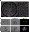
Extended Data Fig. 2. Human blastoids formation in IVF medium and vitrification.
a, Bright-field image of human blastoids formed after 48 h stimulation with PALLY medium followed by the use of IVF medium (G2, Vitrolife) for the last 2 d. Scale bars, 400 μm. b, Bright-field image of control (top) and vitrified-thawed human blastoid (bottom) and after 2 d extended culture on Matrigel-coated plate. Scale bars, 100 μm. c, Confocal immunofluorescence image of OCT4 (yellow) and aPKC (gray) in control (top) and vitrified-thawed human blastoid (bottom) cultured on Matrigel-coated plate for 2 d, counterstained with phalloidin marking F-actin (cyan). Arrows point to the pro-amniotic-like cavity. Scale bar, 100 μm.,
Fig. 1
Fig. 1. A schematic of the time window of human peri-implantation development modelled by blastoids.
Five days after fertilization, the developing embryo is known as a blastocyst. Around this time, blastocyst ‘hatches’ into the uterine cavity and is ready to implant into the uterine wall. Implantation happens around 6-7 days after fertilization, but can only occur if the endometrial wall is prepared by the correct levels of hormones. Blastocyst usually implants with its inner cell mass facing the endometrium, known as the polar side. Human blastoids model the developmental window of days 5–7. This assessment is based on benchmarked morphological and transcriptomic data. The morphological evolution of blastoids reflect the clinical stages B3 to B6. Although blastoids might be co-opting some biological processes (e.g., Hippo inhibition for cell specification) occurring at earlier stages (e.g., morula to blastocyst transition), the projection of transcriptomic signatures of blastoid cells (24, 65, 96 hours) onto large reference maps including multiple stages of human embryonic development currently supports that blastoid cells resemble the cells of day 5 to day 7 embryos.
Fig. 2
Fig. 2. Schematic overview of the protocol. The workflow includes; a, initial PXGL hPSCs seeding and passaging (Steps 1-25), b,
procedure for the formation of human blastoid from hPSCs cultured in PXGL medium (Steps 25-65) and c, Seeding and passaging endometrial organoids (Steps 66-103), OFEL formation (Steps 104-125) and Implantation assay (Steps 126-138) a, Human embryonic stem cells (ESCs) or induced PSCs (iPSCs) cultured in PXGL medium are seeded into feeder-layers for maintenance and passaged several times before human blastoid formation (Steps 1-25). b, hPSCs are dissociated into single cells (Steps 35-47) and plated in non-adherent hydrogel microwells or AggreWell plate to form small aggregates of the cells (steps 48-53). Upon exposure to lysophosphatidic acid (LPA), A83-01 and PD0325901 in a chemically defined medium containing leukaemia inhibitory factor (LIF) and Y-27632 for 2 days, some aggregates form small cavities (steps 54-61). With continuing culture for 2 days in a medium containing LPA and Y-27632, human blastoidss are formed efficiently and consistently (Steps 62-65). N2B27, serum-free medium; PALLY, PD0325901 + A83-01 + LPA + hLIF + Y-27632; LY, LPA + Y-27632. c, Procedure for the formation of open faced endometrial layer (OFEL) from human endometrial organoids. Human endometrial organoids are seeded and passaged before OFEL formation (Steps 66-103) and then are dissociated into single cells and small colonies and plated in Matrigel-coated wells (Steps 104-120). After 1-2 days, the cells become confluent and grow as a monolayer. Once confluency is reached, E2 supplemented medium is added to the cell for 2 days (Steps 121-122) and then, OFELs are stimulated with E2, P4, cAMP, and XAV-939 for 4 days to make them ready for implantation assay (Steps 123-125). In the next step, human blastoids that display blastocyst morphology with compact ICM are selected and transferred onto OFELs (Steps 126-135). Attachment and development of blastoids will be monitored in the next days (Step 136). OFEL, open face endometrial layer. EPC, E2+P4+cAMP.
Fig. 3
Fig. 3. Four basic criteria to validate the formation of blastoids.
Human blastoids should comply to 4 basic criteria (see also Comparison with other methods in the text). (i) Blastoids should form efficiently both in terms of morphology and of specification of the analogs of the 3 lineages, EPI (yellow), PrE (magenta), TE (cyan). A high efficiency suggests that the initial cell state is both intrinsically capable and adequately stimulated. (ii) Blastoids should generate analogs of the 3 lineages according to the developmental sequence (TE/EPI first, pTE/PrE second) and pace (< 4 days) of blastocyst development. By matching the pace and sequence of development, lineages and cells similar to those of the blastocyst form. (iii) Blastoids should form blastocyst stage cellular analogues, but not of post-implantation stages as defined by transcriptome comparison through scRNAseq. To do so, a reference map of human embryos at different stages is essential as using a map restricted to the targeted cells prevents revealing the presence of off-target cells. A reference map is available here: https://petropoulos-lanner-labs.clintec.ki.se/app/shinyblastoids and is used in this figure. Forming cells similar to those of the blastocyst ensures stage-specific mechanisms and functions. (iv) Blastoids should be capable of recapitulating functional features of blastocyst implantation and development. EPI (yellow), PrE (magenta), TE (cyan). Directional attachment of blastoids specifically to primed endometrial layers is a functional evidence for axis formation and for uterus implantation capacity.
Fig. 4
Fig. 4. Morphological characterization of PXGL hPSCs.
a, Representative images of PXGL hPSC colonies, cultured in an optimal hPSC medium (PXGL) and suboptimal culture condition. In the suboptimal culture condition, hPSCs will spontaneously differentiate as seen by loss of colony border integrity, loss of dome-shaped morphology and exhibition of a flat morphology. Scale bars, 100 μm. b, Flow cytometry analysis plot of hPSCs cultured in optimal and suboptimal culture conditions and stained with specific antibodies (SUSD2 and CD75).
Fig. 5
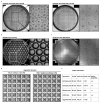
Fig. 5. Triple inhibition of Hippo, ERK and TGFβ pathways leads to efficient and robust formation of human blastoids composed of blastocyst-like cells.
Bright-field image of human blastoids formed innon-adherent hydrogel microwell plates (a), Elplasia plate (b) and Gri3D plate (c). Scale bars, 400 μm.d, Bright-field image of human blastoids formed in non-adherent AggreWell plates after 96 h either with LPA (PALLY medium, left) or without LPA (PALY medium, right). Each well of an AggreWell plate is 400 μm in diameter. Scale bar, 400 μm. e, Information for each plate used for blastoid formation. Hydrogel microwell plates and Gri3D plates have the best optical properties (+++).
Fig. 6
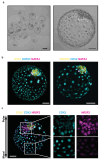
Fig. 6. Human blastoids resemble the morphology of the blastocyst, comprise analogues of the three founding lineages and form an axis.
a, Bright-field images of representative human blastoids harvested from wells (left) and representative image of a typical human blastoid (right), Scale bars, 200 μm (left), 50 μm (right) b, Confocal immunofluorescence image (MIP) of the epiblast (EPI) markers NANOG and OCT4 (yellow), the TE markers CDX2 and GATA3, and the PrE markers GATA4 (magenta) in human blastoids. c, Confocal immunofluorescence image (MIP)of CDX2 (cyan), Polar TE marker NR2F2 (magenta) and NANOG (yellow) in a representative human blastoid. Scale bars, 50 μm. MIP, maximum intensity projection.
Fig. 7
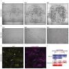
Fig. 7. Representative images of endometrial organoids and monolayers.
a, Bright-field images of time course organoid growth at days 2, 4 and 7 in Matrigel. Scale bars, 500 μm. b, Representative images of OFELs after 2, 4 and 7 days post seeding. Scale bars, 200 μm. c, Confocal immunofluorescence image for the tight junction molecule ZO-1 (Yellow), the adherence junction molecule E-Cadherin (Magenta) in representative stimulated OFELs. Scale bars, 100 μm. d, Heatmap of key cell cycle and secretory epithelial genes differentially expressed between stimulated and non-stimulated OFELs in bulk transcriptome. The endometrial cells were stimulated with hormones in the organoid culture and then were seeded to form OFELs.
Fig. 8
Fig. 8. Human blastoids recapitulate aspects of implantation.
a, Representative phase-contrast images of blastoids after deposition onto non-stimulated (left) or stimulated (right) OFELs. Human blastoid attached to endometrial cells from the polar region (also see Video 1). b, Immunofluorescence stainings for MUC1 (Magenta), a glycoprotein that highly expresses at the epithelial surface of endometrium in the receptive phase, with an attached GFP+ blastoid. Dashed lines indicate the area that trophoblast cells repelled endometrial cells. Scale bars, 100 μm

References

    1. Stamatiadis P, et al. TEAD4 regulates trophectoderm differentiation upstream of CDX2 in a GATA3-independent manner in the human preimplantation embryo. Human Reproduction. 2022 - PubMed
    1. Gerri C, et al. Initiation of a conserved trophectoderm program in human, cow and mouse embryos. Nature. 2020;587:443–447. doi: 10.1038/s41586-020-2759-x. - DOI - PMC - PubMed
    1. Ma H, et al. In vitro culture of cynomolgus monkey embryos beyond early gastrulation. Science. 2019;366 - PubMed
    1. O’Leary T, et al. Tracking the progression of the human inner cell mass during embryonic stem cell derivation. Nat Biotechnol. 2012;30:278–282. - PubMed
    1. Sunde A, et al. Time to take human embryo culture seriously. Hum Reprod. 2016;31:2174–2182. - PubMed

Publication types

LinkOut - more resources