. 2023 Feb;614(7949):732-741.
doi: 10.1038/s41586-023-05711-7. Epub 2023 Feb 15.

A NPAS4-NuA4 complex couples synaptic activity to DNA repair

Affiliations

A NPAS4-NuA4 complex couples synaptic activity to DNA repair

Elizabeth A Pollina et al. Nature. 2023 Feb.

Abstract

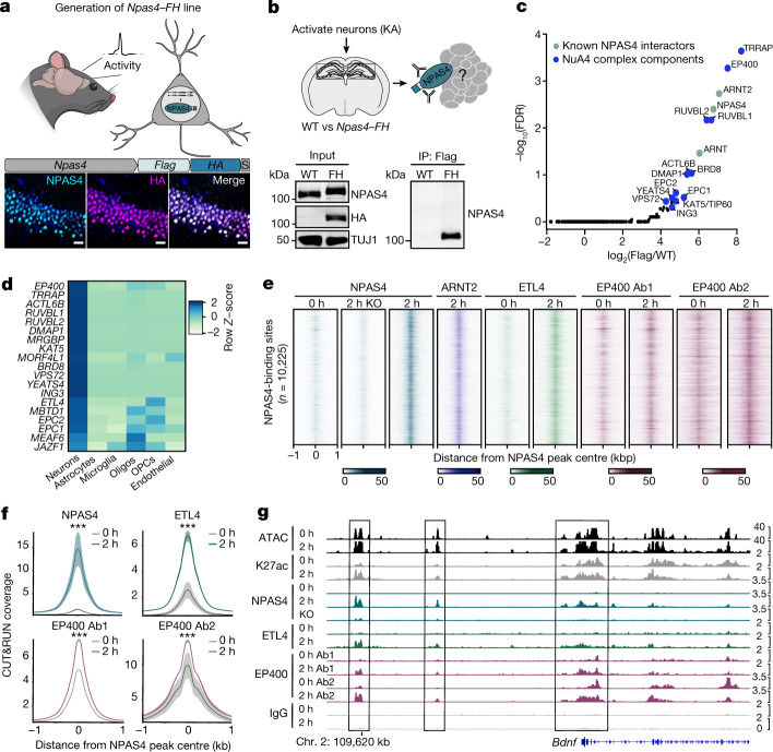
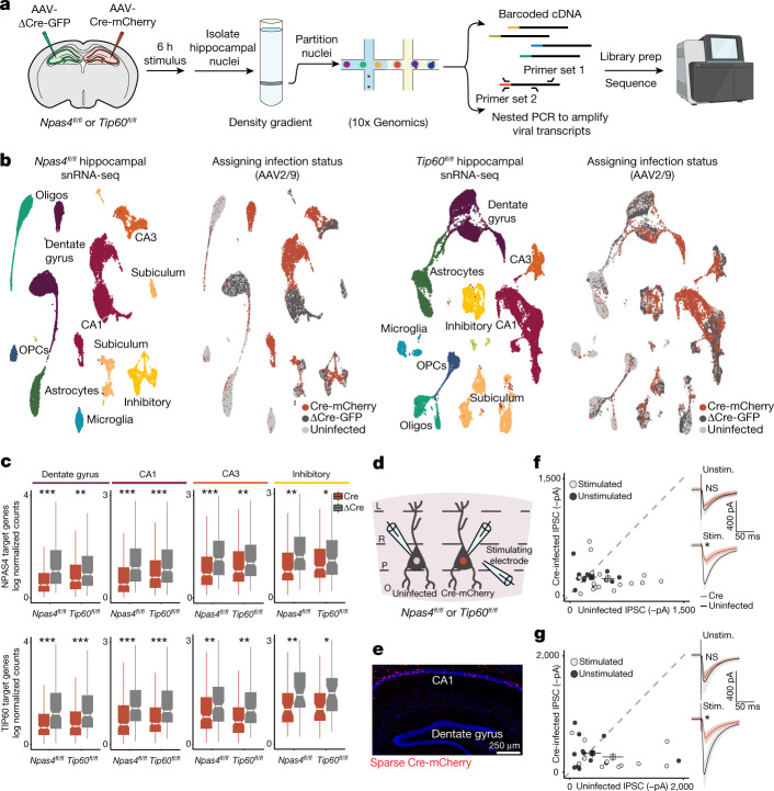
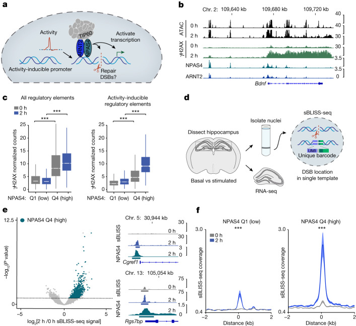
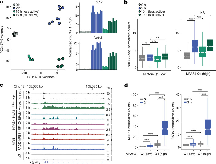
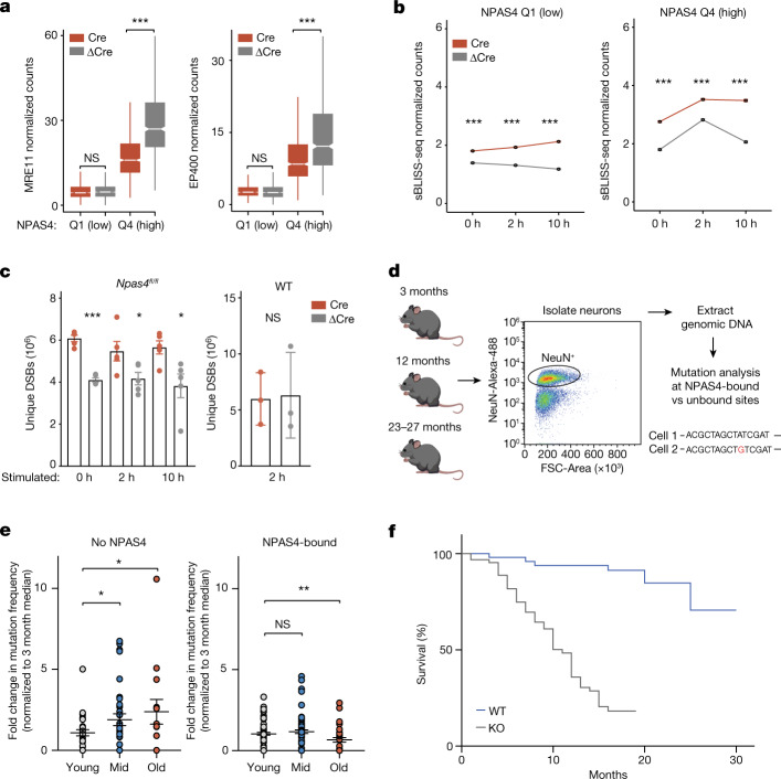
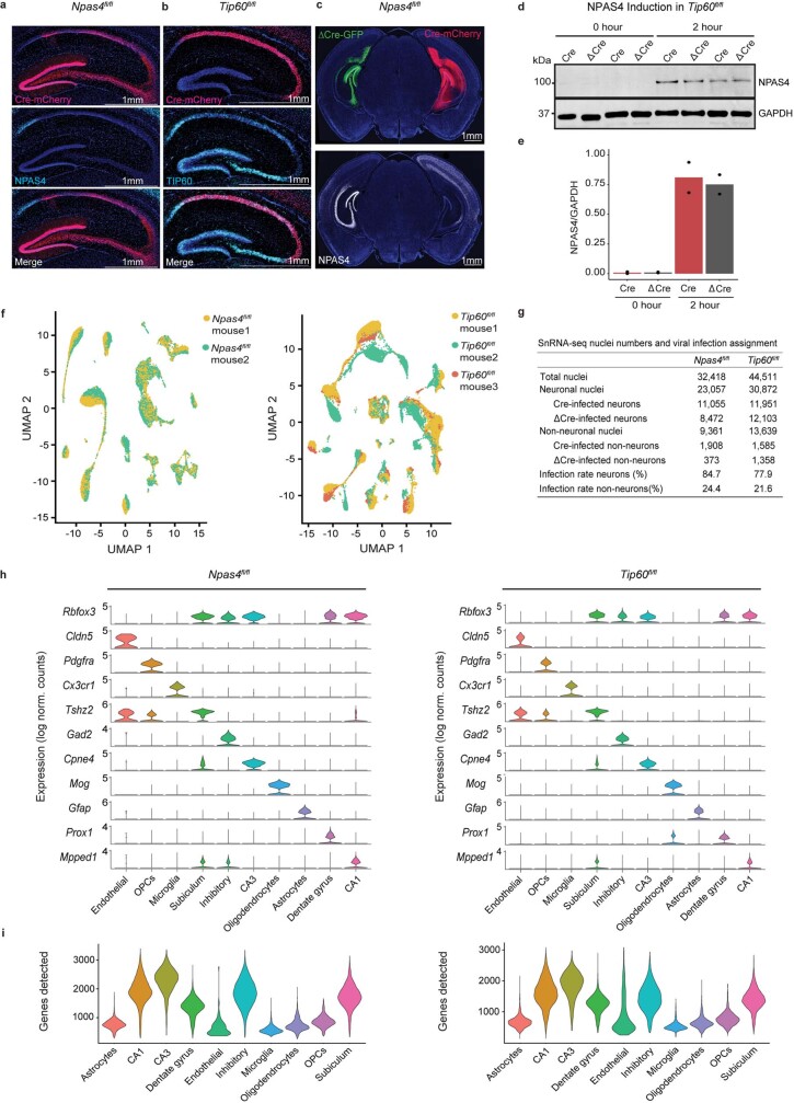
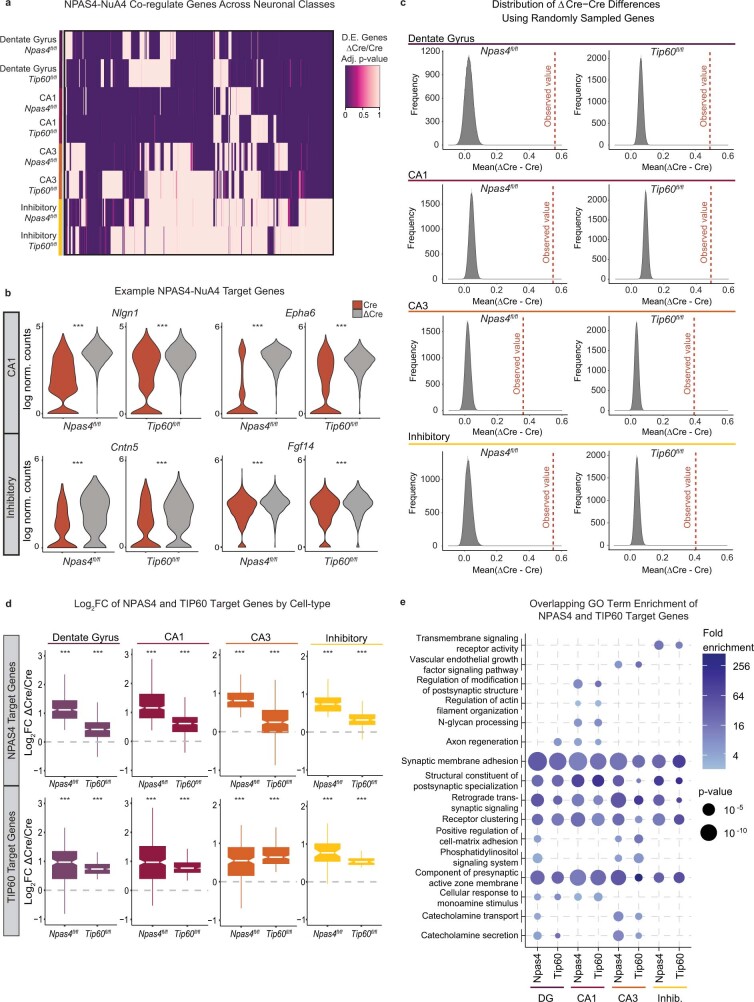
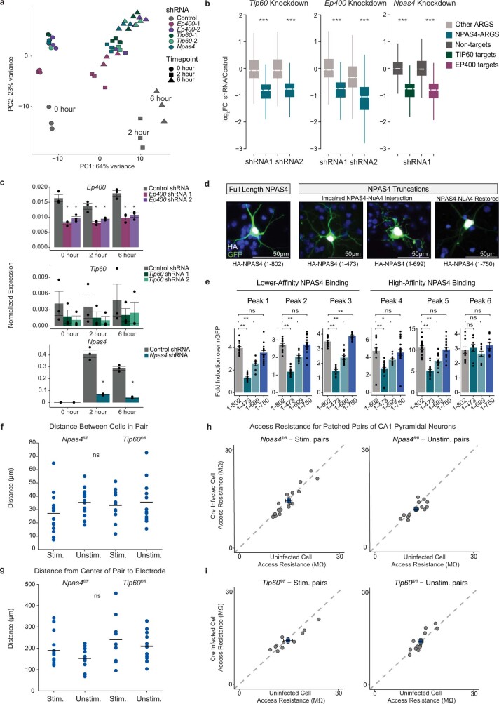
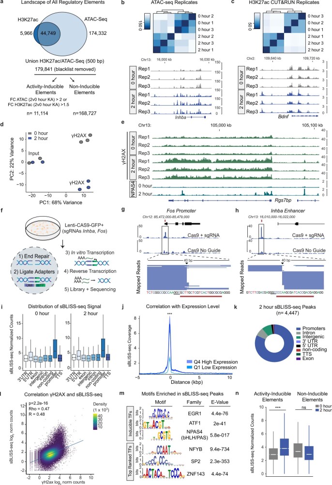
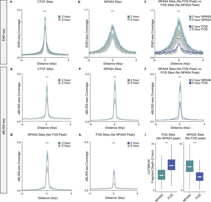
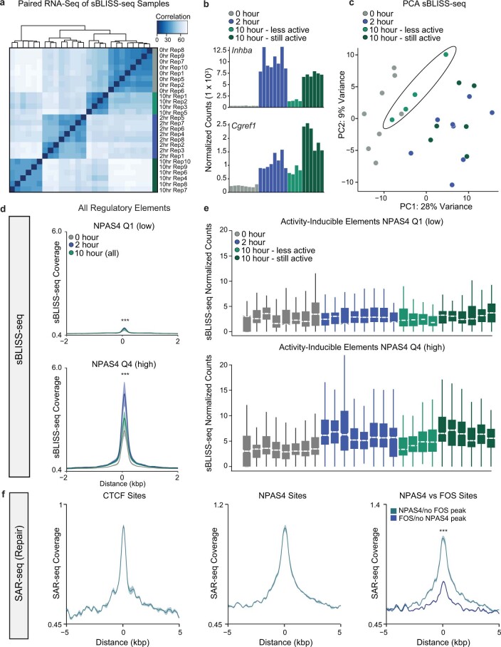
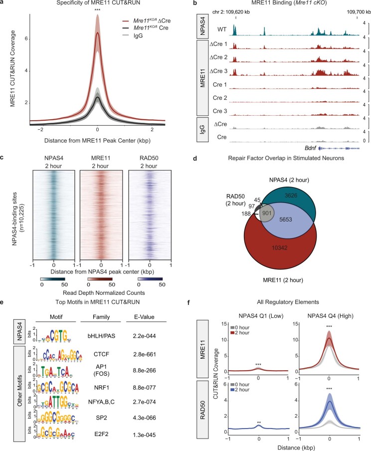
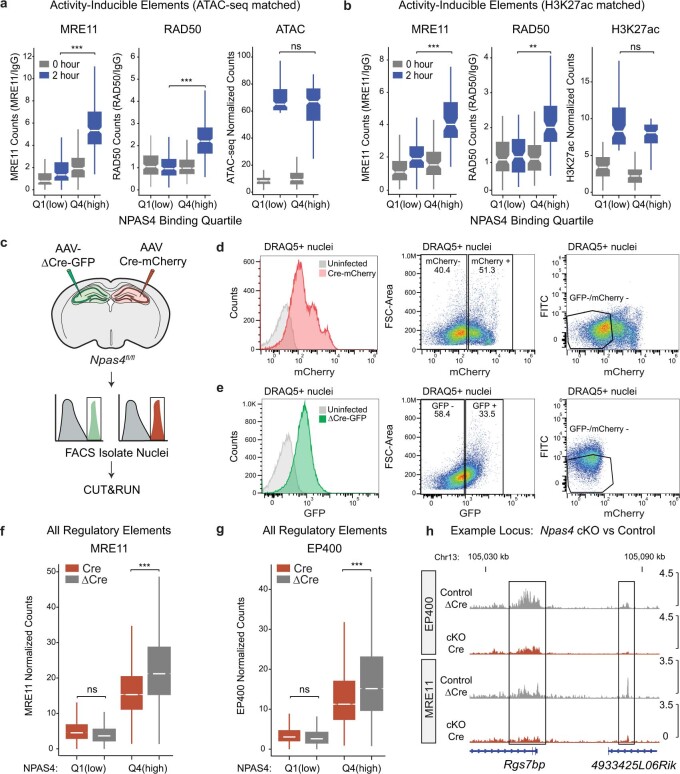
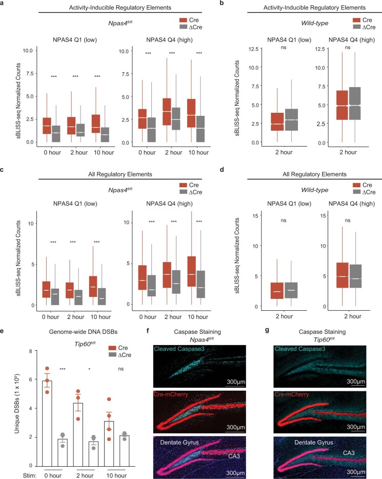
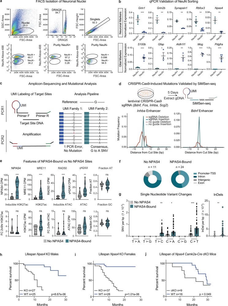
Neuronal activity is crucial for adaptive circuit remodelling but poses an inherent risk to the stability of the genome across the long lifespan of postmitotic neurons1-5. Whether neurons have acquired specialized genome protection mechanisms that enable them to withstand decades of potentially damaging stimuli during periods of heightened activity is unknown. Here we identify an activity-dependent DNA repair mechanism in which a new form of the NuA4-TIP60 chromatin modifier assembles in activated neurons around the inducible, neuronal-specific transcription factor NPAS4. We purify this complex from the brain and demonstrate its functions in eliciting activity-dependent changes to neuronal transcriptomes and circuitry. By characterizing the landscape of activity-induced DNA double-strand breaks in the brain, we show that NPAS4-NuA4 binds to recurrently damaged regulatory elements and recruits additional DNA repair machinery to stimulate their repair. Gene regulatory elements bound by NPAS4-NuA4 are partially protected against age-dependent accumulation of somatic mutations. Impaired NPAS4-NuA4 signalling leads to a cascade of cellular defects, including dysregulated activity-dependent transcriptional responses, loss of control over neuronal inhibition and genome instability, which all culminate to reduce organismal lifespan. In addition, mutations in several components of the NuA4 complex are reported to lead to neurodevelopmental and autism spectrum disorders. Together, these findings identify a neuronal-specific complex that couples neuronal activity directly to genome preservation, the disruption of which may contribute to developmental disorders, neurodegeneration and ageing.


Conflict of interest statement

The authors declare no competing interests.

Figures

Fig. 1
Fig. 1. Neuronal activity assembles the NPAS4–NuA4 complex on chromatin.
a, Top, schematic of generation of the Npas4FH mouse model. Bottom, representative immunohistochemistry (performed in triplicate) for NPAS4 and HA in hippocampus samples from Npas4–FH mice following 2 h of KA stimulation. Scale bars, 25 µm. b, NPAS4–FH and associated protein complexes isolated by anti-Flag immunoprecipitation (IP). Representative western blot (performed in triplicate) with anti-NPAS4 antibody confirms immunoprecipitation only in Npas4–FH mice. For gel source data, see Supplementary Fig. 1. TUJ1 processing control was run on a separate gel. c, NPAS4-interacting proteins. –log10(false discovery rate (FDR)) compared with log2-transformed fold change in peptides obtained by anti-Flag immunoprecipitation in stimulated Npas4–FH tissue compared with wild-type tissue (n = 3 pools of 8–10 mice), followed by mass spectrometry. Green points indicate known NPAS4 interactors. Blue points indicate NuA4 components. d, Expression levels of NuA4 subunits across cell types in human primary motor cortex, displayed as row Z-score. snRNA-seq data published by the Allen Brain Institute. Oligos, oligodendrocytes; OPCs, oligodendrocyte precursor cells. e, CUT&RUN signals for NPAS4–NuA4 components in either unstimulated (0 h) or stimulated (2 h) hippocampal tissue. Each NPAS4-binding site is represented as a horizontal line centred at the peak summit and extended ±1 kb. The colour intensity represents the read depth (normalized to 10 million) as indicated by the scale bar for each factor. Data plotted show the aggregate signal from all replicates per factor. Ab1, antibody 1 (Bethyl A300-541-A); Ab2, antibody 2 (Abcam Ab5201). For eg, n = 3–5 mice pooled per replicate. Replicate numbers provided in Supplementary Table 2. f, Aggregate CUT&RUN coverage (fragment depth per bp per peak) at NPAS4-binding sites. Signals displayed as mean ± s.e.m. ***P < 2.2 × 10−16. P values were calculated from the average signal extracted in a 2 kb window centred on NPAS4 peaks using unpaired, two-tailed Wilcoxon rank-sum tests. g, Integrative Genomics Viewer (IGV) tracks of NPAS4–NuA4 CUT&RUN signal at the activity-inducible gene Bdnf. Data plotted show the aggregate signal from all replicates per factor. The y axis displays normalized coverage scaled for each factor at the chosen locus. Source Data
Fig. 2
Fig. 2. NPAS4–NuA4 regulates activity-dependent transcription and recruitment of somatic inhibition.
a, Scheme of the experimental design. Nuclei were isolated from hippocampi of Npas4fl/fl and Tip60fl/fl mice expressing either Cre-mCherry or ΔCre-GFP, followed by snRNA-seq. An additional PCR step to amplify viral transcripts within the cDNA library was used to assign infection status to each nucleus. b, Left, uniform manifold approximation and projection (UMAP) visualizations of Npas4fl/fl and Tip60fl/fl snRNA-seq datasets. Right, UMAP visualizations of infection status. Npas4fl/fl: 32,418 nuclei from 2 mice, 12,963 Cre-infected, 8,845 ΔCre-infected. Tip60fl/fl: 44,511 nuclei from 3 mice, 13,536 Cre-infected, 13,461 ΔCre-infected. c, Boxplots showing the average expression (Seurat log(e) normalized counts) of NPAS4 or TIP60 target genes (Methods) comparing Cre-infected and ΔCre-infected nuclei within each neuronal celltype. Boxplots show the median (line), inter-quartile range (IQR; box) and 1.5× IQR (whiskers), and notches indicate the median ± 1.58× IQR/sqrt(n). ***P < 5 × 10−10, **P < 5 × 10−4, *P < 0.01. P values were calculated using unpaired, two-tailed Wilcoxon rank-sum tests. Exact P values and cell numbers per cluster provided in the source data. d, Schematic of the recording configuration, showing the stimulating electrode in the centre of the stratum pyramidale to measure IPSCs onto neighbouring Cre-infected and uninfected CA1 pyramidal neurons in either Npas4fl/fl or Tip60fl/fl acute hippocampal slices. L, lacunosum; O, oriens; P, pyramidale; R, radiatum. e, Representative image (performed in triplicate) showing sparse Cre-mCherry expression in the CA1. f,g, Scatterplots of IPSC amplitudes recorded from pairs of neighbouring uninfected and Cre-infected cells in unstimulated (Unstim.; saline) or stimulated (Stim.; low-dose KA) Npas4fl/fl (f) or Tip60fl/fl(g) mice. Points represent an uninfected–Cre-infected pair. Squares represent the average ± s.e.m. Right insets, traces of uninfected or Cre-infected cells. Npas4fl/fl: stimulated: n = 16 pairs from 4 mice; unstimulated: n = 12 pairs from 5 mice. Tip60fl/fl: stimulated: n = 11 pairs from 2 mice; unstimulated: n = 12 pairs from 2 mice. *P = 3.73 × 10−3 for Npas4fl/fl; *P = 0.0209 for Tip60fl/fl. Data are mean ± s.e.m. P values were calculated using unpaired, two-tailed t-tests. The sequencer image in a is from BioRender (https://biorender.com). Source Data
Fig. 3
Fig. 3. NPAS4–NuA4-bound sites undergo recurrent DNA breaks in vivo.
a, A model for the dual function of NPAS4–NuA4 in stimulating transcription and DNA repair in active neurons. b, IGV tracks displaying aggregate signals for ATAC-seq (n = 3), NPAS4 CUT&RUN (n = 5), ARNT2 CUT&RUN (n = 2) and γH2AX ChIP–seq (n = 3) at the activity-inducible gene Bdnf. n = 3–5 mice pooled per replicate. c, Boxplots of average γH2AX ChIP–seq normalized counts (n = 3 pools of 3–5 mice) at all regulatory elements (left) and activity-inducible regulatory elements (right), subset by quartiles of NPAS4 CUT&RUN signal (Methods). All regulatory elements: Q1 = 44,864 sites, Q4 = 7,378 sites. Activity-inducible elements: Q1 = 1,017 sites, Q4 = 764 sites. Boxplots show the median (line), IQR (box) and 1.5× IQR (whiskers), and notches indicate the median ± 1.58× IQR/sqrt(n). ***P < 2.2 × 10−16. P values were calculated using unpaired, two-tailed Wilcoxon rank-sum tests. d, Schematic of sBLISS-seq to map DSBs in brain nuclei. A tissue aliquot from each sBLISS-seq sample was kept for paired RNA-seq analysis. e, Volcano plot depicting the DeSeq2 log2-transformed fold change versus –log10(Benjamini–Hochberg adjusted P value) of sBLISS-seq signals between 0 and 2 h KA stimulation. Dotted line indicates adjusted P < 0.1. Elements with log2-transformed fold change > 0, adjusted P < 0.1 and that are within Q4 of NPAS4 IgG-normalized CUT&RUN signals are indicated in blue. Right inset, IGV tracks of aggregate sBLISS-seq (n = 8 individual mice) and NPAS4 CUT&RUN (n = 5 pools of 3–5 mice) at Cgref1 and Rgs7bp promoters. Coloured bars represent statistically defined peaks. f, Aggregate plots showing sBLISS-seq coverage (fragment depth per bp per peak) at activity-inducible regulatory elements, subset by quartiles of NPAS4 binding. Signals are mean ± s.e.m. (n = 8 individual mice). ***P < 2.2 × 10−16. P values were calculated using the average signal extracted in a 500 bp window around the element centre using unpaired, two-tailed Wilcoxon rank-sum tests.
Fig. 4
Fig. 4. NPAS4–NuA4-bound sites undergo DNA repair as inducible transcription subsides.
a, PCA of paired RNA-seq prepared from the same tissue used to generate the sBLISS-seq time course in hippocampus. Samples clustered according to the stimulation state, with 10 h stimulation samples separating into groups with either dampening or sustained transcriptional induction. DeSeq2-normalized counts of inducible genes Bdnf and Nptx2 shown for each sample (0 h, n = 8; 2 h, n = 8; 10 h, n = 10). One mouse per replicate. b, Boxplots of average sBLISS-seq-normalized counts in 0 h (n = 8), 2 h (n = 8), 10 h less active (n = 4), and 10 h still active (n = 6) samples at activity-inducible regulatory elements, subset by quartiles of NPAS4 binding. One mouse per replicate. Boxplots show the median (line), IQR (box) and 1.5× IQR (whiskers), and notches indicate the median ± 1.58× IQR/sqrt(n). Q1 = 1,017 sites, Q4 = 764 sites. Q1, *P = 0.03435, **P = 2.049 × 10–8, ***P = 3.057 × 10–13; Q4, ***P < 2.2 × 10–16, not significant (NS) = 0.1742. P values were calculated using unpaired, two-tailed Wilcoxon rank-sum tests. c, IGV tracks of sBLISS-seq, γH2AX ChIP–seq, NPAS4, EP400, MRE11 and RAD50 CUT&RUN signals. Data plotted show the aggregate signals from all replicates per factor. The y axis displays normalized coverage scaled for each factor at the chosen locus. CUT&RUN and γH2AX ChIP–seq, n = 3–5 mice pooled per replicate. sBLISS-seq, n = 1 mouse per replicate. Replicate numbers provided in Supplementary Table 2. d, Boxplots of average MRE11 and RAD50 normalized counts at activity-inducible regulatory elements, subset by quartiles of NPAS4 CUT&RUN signals. Q1 = 1,017 sites, Q4 = 764 sites. Boxplots show the median (line), IQR (box) and 1.5× IQR (whiskers), and notches indicate the median ± 1.58× IQR/sqrt(n). n = 3–5 mice pooled per replicate. Replicate numbers provided in Supplementary Table 2. ***P < 2.2 × 10−16. P values were calculated using unpaired, two-tailed Wilcoxon rank-sum tests. Source Data
Fig. 5
Fig. 5. NPAS4–NuA4 disruption impairs genome stability and reduces lifespan.
a, Boxplots of average MRE11 or EP400 CUT&RUN normalized counts in Cre-infected or ΔCre-infected hippocampi of Npas4fl/fl mice at activity-inducible sites, subset by quartiles of NPAS4 binding. Q1 = 1,017 sites, Q4 = 764 sites. Boxplots show the median (line), IQR (box) and 1.5× IQR (whiskers), and notches indicate the median ± 1.58× IQR/sqrt(n). n = 2–3 mice pooled per replicate. Replicate numbers provided in Supplementary Table 2. MRE11 Q1, P = 0.2453; Q4, ***P < 2.2 × 10−16. EP400 Q1, P = 0.9962; Q4, ***P < 2.2 × 10−16. P values were calculated using unpaired, one-tailed Wilcoxon rank-sum tests (ΔCre > Cre). b, Line plot depicting average ± s.e.m. of sBLISS-seq normalized counts in Cre-infected or ΔCre-infected hippocampi of Npas4fl/fl mice, subset by quartiles of NPAS4 binding. Q1 = 1,017 sites, Q4 = 764 sites. Data from n = 5 each for Cre-infected and ΔCre-infected mice. ***P < 2.2 × 10–16. P values were calculated using unpaired, one-tailed Wilcoxon rank-sum tests (Cre > ΔCre). See Extended Data Fig. 12a for boxplot distribution of data. c, Genome-wide breaks in hippocampal nuclei isolated from Npas4fl/fl (n = 5) or wild-type (n = 3) mice infected with Cre or ΔCre virus. Data are mean ± s.e.m. Npas4fl/fl: 0 h, P = 5.24 × 10–6; 2 h, P = 0.044; 10 h, P = 0.022. Wild-type: 2 h, P = 0.906. P values were calculated using two-tailed, unpaired t-tests. d, Collection of hippocampal neuronal nuclei across lifespan. e,Normalized mutation frequency at NPAS4-bound and unbound sites in wild-type mice. Mutation frequency (base changes and insertions and deletions) with age was calculated per site and normalized to the median frequency for that site in young animals. Points represent the normalized rate for one site sampled from one mouse. Data are mean ± s.e.m. Data are from n = 4 (young), 4 (middle aged) and 3 (old) mice. No NPAS4 sites: mid–young, *P = 0.03; old–young, *P = 0.016. NPAS4-bound sites: mid–young, NS = 0.18; old–young, **P = 0.0095. P values calculated using one-tailed, unpaired t-tests. f, Survival of Npas4 wild-type (n = 53; 28 females, 25 males) and Npas4 knockout (n = 64; 37 females, 27 males) mice, showing abbreviated lifespan of Npas4 knockout mice. P = 4.09 × 10–13 by a two-tailed Mantel–Cox test, P = 3.63 × 10–11 by a two-tailed Gehan–Breslow–Wilcoxon test. Source Data
Extended Data Fig. 1
Extended Data Fig. 1. NPAS4 forms a complex with NuA4 across brain regions.
a. Lysate from the hippocampus of KA-stimulated mice, fractionated by molecular weight (MW) using gel filtration and non-denaturing size exclusion chromatography. Western blotting for NPAS4 in the different MW fractions confirms that NPAS4 resides in a high MW complex peaking ~1 MDa in size. Representative image from 3 experiments. See gel source data (Supplementary Fig. 1). b. Sequence validation of Flag HA epitope tag appended to the C-terminus of Npas4 or Arnt2 in the P0 or F1 generation in Npas4-FH or Arnt2-FH mice. c. Left: Immunohistochemistry of NPAS4 and HA antibody staining in hippocampus of Npas4-FH mice, 6 h enriched environment. Scale bars: 100 μm. Right: Validation of Arnt2-FH knockin mouse line by immunohistochemistry in CA1. Scale bar 25 μm. Representative images from 3 experiments. d. Validation by Western blot that NPAS4 and NPAS4-FH have the same induction kinetics and specificity for membrane depolarization-induced calcium signaling. To stimulate cultured cortical neurons, 55 mM KCl was applied for the indicated time points. Representative image from one experiment. See gel source data (Supplementary Fig. 1). e. Confirmation by Western blot of NPAS4-FH expression and Flag immunoprecipitation (IP) in cultured cortical neurons from either wild-type or Npas4-FH mice. Representative image from one experiment. See gel source data (Supplementary Fig. 1). f. Confirmation via anti-Flag immunoprecipitation (IP) and Western blot that NPAS4-NuA4 also assembles in the visual cortex following 2 h light stimulation. Representative image from 2 experiments. See gel source data (Supplementary Fig. 1). g. Experimental diagram detailing glycerol gradient fractionation and immunoprecipitation of intact NPAS4-NuA4 complexes from high molecular weight (MW) fractions. h. Western blot of glycerol gradient fractions of NPAS4-FH in mouse cortical lysates segregated by MW. NPAS4 migrates in high MW fractions along with NuA4 components TRRAP, EP400, and DMAP1. Representative image from 3 experiments. See gel source data (Supplementary Fig. 1). i. Reciprocal validation of NPAS4-NuA4 interaction via Flag immunoprecipitation and Western blotting from high MW fractions of cortical lysates from wild-type, Npas4-Flag-HA (Npas4-FH), and Tip60-Flag (Tip60-F) mice. Representative image from 2 experiments. See gel source data (Supplementary Fig. 1). j. Reciprocal validation of NPAS4-NuA4 interaction via Flag immunoprecipitation and Western blotting from low MW fractions of cortical lysates from wild-type, Npas4-Flag-HA (Npas4-FH), and Tip60-Flag (Tip60-F) mice. Representative image from 2 experiments. See gel source data (Supplementary Fig. 1).
Extended Data Fig. 2
Extended Data Fig. 2. Validation of NPAS4-NuA4 interaction and neuronal specificity.
a. Confirmation that the NPAS4-NuA4 interaction can be observed via reciprocal Flag IP and Western blot from hippocampi of wild-type, Npas4-FH, and Tip60-Flag (TIP60-F) mice. Representative image from 2 experiments. See gel source data (Supplementary Fig. 1). b. Anti-Flag IP from hippocampi of Tip60-Flag (TIP60-F) mice in both unstimulated brains and 2 h post KA stimulation. Western blot demonstrates that components of the NuA4 complex interact in both the basal (0 h) and 2 h condition while NPAS4 interacts primarily in the stimulated state. Representative image from 2 experiments. See gel source data (Supplementary Fig. 1). c. Average RNA-seq DeSeq2 normalized counts ± s.e.m. of NPAS4-NuA4 components in the mouse hippocampus at 0 h (n = 8), 2 h (n = 8), and 10 h (n = 10), post KA stimulation. **P = 3.19e-29. P value determined from transcriptome-wide DeSeq2 analysis with Benjamini-Hochberg correction. See source data for individual P values. d. Full length NPAS4 (amino acids 1-802) and the indicated truncations of the C-terminal portion of NPAS4 were expressed in HEK293T cells. Sequential Flag and HA IPs were performed (right), followed by Western blotting for NPAS4, ARNT2, and NuA4 subunits TRRAP and DMAP1. Flag-HA-tagged nuclear GFP (nGFP) was included as negative control. Representative image from 3 experiments. See gel source data (Supplementary Fig. 1). e. Heatmap of the column-normalized specificity score for each component of the NuA4 complex in anti-Flag IP-MS experiments conducted with NPAS4/ARNT, NPAS4/ARNT2, FOS/JUN, or EGR1 expressed in HEK293T cells. All proteins were Flag-HA tagged. Specificity score represents the ratio of the number of peptides identified in the IP over the number of peptides found in nGFP controls performed in parallel. Replicate numbers provided in Supplementary Table 1. f. Normalized counts (Seurat v3) of NPAS4-NuA4 components from hippocampal single-nucleus RNA sequencing in mouse hippocampus, normalized across column and displayed as a Z-score. g. Marker genes identifying cell types in human primary motor cortex single-nucleus RNA sequencing dataset published by the Allen Brain Institute (see Fig. 1d), normalized across column and displayed as a Z-score. Source Data
Extended Data Fig. 3
Extended Data Fig. 3. NPAS4 and NuA4 co-localize on chromatin across the genome.
a. Aggregate plot of average CUT&RUN coverage (fragment depth per bp/per peak) ± s.e.m. at NPAS4-binding sites in Npas4 wild-type vs Npas4 KO hippocampal tissue. (Npas4 wild-type: n = 5, Npas4 KO: n = 2, IgG: n = 8) 3-5 mice pooled per replicate. ***P < 2.2e-16, P values were on calculated on average signal extracted in a 2 kb window centered on NPAS4 peak summits using unpaired, two-tailed Wilcoxon rank-sum tests. b. Integrative Genomics Viewer tracks of aggregated NPAS4 CUT&RUN signal (n = 5). 3-5 mice pooled per replicate. 3 additional replicates of NPAS4 CUT&RUN signal confirm the reproducibility of NPAS4 CUT&RUN. c. Distribution of NPAS4 (10,225 sites) and FOS (11,770 sites) CUT&RUN peak annotations relative to all regulatory elements (179,841 sites) in hippocampal tissue. d. Significant motifs enriched in 10,225 NPAS4 CUT&RUN peaks. E-value calculated using MEME-ChIP. e. Correlation between NPAS4 CUT&RUN and NPAS4 ChIP-seq reads at NPAS4 ChIP-seq peaks (10,917 ChIP-seq peaks). Values represent the log2 read depth normalized counts for CUT&RUN vs ChIP-seq. Correlation was calculated by Pearson (R = 0.45) and Spearman (Rho = 0.41) tests, P < 2.2e-16 by two-tailed correlation tests. The reciprocal analysis of the correlation between NPAS4 CUT&RUN and NPAS4 ChIP-seq reads at NPAS4 CUT&RUN peaks (10,225 CUT&RUN peaks) yields a Pearson R = 0.46 and Spearman Rho = 0.43, P < 2.2e-16 by two-tailed correlation tests. f. Venn diagram of overlaps between SEACR peaks for anti-EP400 Ab1 (antibody 1, Bethyl Labs, A300-541A) and anti-EP400 Ab2 (antibody 2, Abcam Ab5201). Peak overlaps indicate at least 1 bp overlap between the entire SEACR-enriched region. Maxima indicates overlap between regions extended 500 bp out from the peak maxima for each factor. g. Venn diagram of overlaps between SEACR peaks for NPAS4/ARNT2 co-bound peaks with ETL4 and high-confidence EP400 peaks, defined as the union of peaks shared by both EP400 antibodies. Maxima indicates overlap between regions extended 500 bp out from the peak maxima for each factor. h. Boxplot of average EP400 normalized signal (counts per million) at sites bound by FOS but not NPAS4 (labeled ‘FOS’) and sites bound by NPAS4 but not FOS (labeled ‘NPAS4’). FOS/No NPAS4 sites are defined as sites with a SEACR-determined peak of CUT&RUN signal for FOS but no peak for NPAS4 (6,998 sites) and vice versa (NPAS4/No FOS peaks: 5,550 sites). Boxplot shows median (line), IQR (box), 1.5x IQR (whiskers), notches indicate median ± 1.58× IQR/sqrt(n). 3-5 mice pooled per replicate. Replicate numbers provided in Supplementary Table 2. ***P < 2.2e-16. P values were calculated using unpaired, two-tailed Wilcoxon rank-sum tests. i. Representative Integrative Genomics Viewer tracks of replicate EP400 CUT&RUN at the Bdnf promoter. Source Data
Extended Data Fig. 4
Extended Data Fig. 4. Acute deletion of NPAS4 or TIP60 by viral injection and additional quality control for single-nucleus RNA-seq datasets.
a. Immunohistochemistry image of an Npas4fl/fl mouse injected with AAV to express Cre-mCherry (shown in red) and collected 2 h post low-dose KA to induce NPAS4 (shown in cyan). Representative image from 3 animals. Scale bar = 1 mm. b. Immunohistochemistry image of a Tip60fl/fl mouse injected with AAV to express Cre-mCherry (shown in red). TIP60 (shown in cyan). Representative image from 3 animals. Scale bar = 1 mm. c. Immunohistochemistry image of both hippocampal hemispheres of an Npas4fl/fl mouse injected with Cre-mCherry and ΔCre-GFP in contralateral sides of the hippocampus and collected 2 h post low-dose KA to induce NPAS4 (shown in white). Representative image from 3 animals. Scale bar = 1 mm. d. Western blot from whole hippocampal tissue of Tip60fl/fl mice injected with AAV expressing Cre-mCherry and ΔCre-GFP in contralateral sides of the hippocampus. Tissue was collected at either 0 or 2 h post KA stimulation. Cre and ΔCre tissue was collected from each individual mouse (0 h, n = 2; 2 h, n = 2). See gel source data (Supplementary Fig. 1). e. Quantification of the Western blot is shown in d, normalizing the NPAS4 signal to loading control GAPDH. f. Left: UMAP visualization of full Npas4fl/fl snRNA-seq dataset. Nuclei are colored according to mouse of origin. 32,418 nuclei from 2 mice. Right: UMAP visualization of full Tip60fl/fl snRNA-seq dataset. Nuclei are colored according to mouse of origin. 44,511 nuclei from 3 mice. g. Summary of final nuclei numbers in Npas4fl/fl and Tip60fl/fl snRNA-seq datasets, and quantification of infection rates with Cre-mCherry and ΔCre-GFP viruses. Higher infection rate in neurons reflects the known tropism of the AAV2/9 virus used in these experiments. h. Cell-type assignment in Npas4fl/fl (left) and Tip60fl/fl (right) snRNA-seq datasets using indicated marker genes. The y-axis denotes normalized expression (Seurat loge normalized counts). i. Distribution of number of genes detected per nucleus, by cell-type. Npas4fl/fl (left) and Tip60fl/fl (right). Source Data
Extended Data Fig. 5
Extended Data Fig. 5. NPAS4-NuA4 coordinate gene regulation across neuronal subtypes in vivo.
a. Heatmap showing coordinate regulation of NPAS4 target genes across the principal neuronal subtypes of the hippocampus in both Npas4fl/fl and Tip60fl/fl mice. NPAS4 target genes were identified in each cell-type using both Seurat (v3) and Monocle3 (see Methods). Bonferroni adjusted P values from Seurat (v3) differential expression testing (unpaired, two-tailed Wilcoxon rank-sum test) between ΔCre- and Cre-infected nuclei are shown in each cell-type. Each column represents adjusted P values for one gene. b. Violin plots showing the distribution of expression (Seurat loge normalized counts) of the indicated gene across nuclei in the indicated cell-type. Bonferroni adjusted P value ***P < 2.2e-16. Differential gene expression tests were conducted using an unpaired, two-tailed Wilcoxon rank-sum test implemented via the Seurat (v3) FindMarkers function. c. Comparison of the observed differences (ΔCre – Cre) in normalized counts for NPAS4 or TIP60 target genes in each neuronal subtype to the differences obtained when using an equal number of randomly selected genes. Genes were randomly selected from the top 10% of expressed genes (to account for NPAS4 or TIP60 target genes being highly expressed on average), and the average difference (ΔCre – Cre) in expression for each gene was calculated for each random sample. This sampling was repeated 10,000 times to generate sampling distributions (gray). In each subtype, the average difference (ΔCre – Cre) observed when using that subtype’s NPAS4 or TIP60 target genes lies far outside the distribution obtained using randomly selected genes, suggesting the differences in expression of the target genes between ΔCre- and Cre-infected nuclei is not due to chance. d. Boxplots showing log2 fold change of NPAS4 or TIP60 target genes (see Methods) comparing ΔCre- and Cre-infected nuclei in dentate gyrus, CA1, CA3, and inhibitory neurons. Boxplot shows median (line), IQR (box), 1.5x IQR (whiskers), notches indicate median ± 1.58× IQR/sqrt(n). *** P < 2.2e-16. values were calculated using two-tailed, unpaired t-tests comparing to a null hypothesis of log2 fold change = 0 (no difference between Cre and ΔCre). Exact P values and cell numbers per cluster provided in source data. e. Select overlapping GO terms enriched in both NPAS4 target genes and TIP60 target genes across dentate gyrus, CA1, CA3, and inhibitory neurons. Circle size indicates the adjusted P value of enrichment determined by Fisher’s one-tailed test using gProfiler2. Color indicates the fold enrichment. See Methods for additional detail and Supplementary Table 3 for complete list of enriched GO terms for each cell-type. Source Data
Extended Data Fig. 6
Extended Data Fig. 6. NPAS4-NuA4 coordinately regulates gene transcription and somatic inhibition.
a. Principal component analysis clustering of RNA-seq datasets from cultured mouse neurons expressing shRNAs targeting Npas4, Tip60, or Ep400, and stimulated with 55 mM KCl for either 0, 2, or 6 h. Control shRNA targets luciferase. b. Boxplots of log2 fold changes between the control shRNA (n = 3) and the indicated Npas4 (n = 3), Tip60 (n = 3) or Ep400 (n = 3) shRNAs in neuronal cultures 6 h following membrane-depolarization by 55 mM KCl. Replicates consist of primary cultures generated on independent days. Boxplot shows median (line), IQR (box), 1.5x IQR (whiskers), notches indicate median ± 1.58× IQR/sqrt(n). Activity-regulated-genes (ARGs) are defined as genes upregulated at least 1.5-fold with a Benjamini-Hochberg adjusted P < 0.01 comparing the 6 h and 0 h time points in control shRNA-treated neurons. Tip60 and Ep400 targets are defined as all genes down-regulated by at least 1.5-fold with a Benjamini-Hochberg adjusted  < 0.01 by both shRNAs. Non-targets include all expressed genes not significantly affected by loss of Tip60 or Ep400. ***P < 2.2e-16. P values were calculated using unpaired, two-tailed Wilcoxon rank-sum tests. c. Average expression ± s.e.m. of Ep400 (n = 3), Tip60 (n = 3), and Npas4 (n = 3) by qPCR. Replicates consist of primary cultures generated on independent days. Expression normalized to both Tubb3 and Gapdh. Ep400 0 h shRNA1: *P = 0.0026, shRNA2: *P = 0.0072; 2 h shRNA1: *P = 0.0072, shRNA2: *P = 0.0154; 6 h shRNA1: *P = 0.0027, shRNA2: *P = 0.0038. Tip60 0 h shRNA1: ns = 0.4021, shRNA2: ns = 0.2669; 2 h shRNA1: ns = 0.4507, shRNA2: ns = 0.3324; 6 h shRNA1: ns = 0.433, shRNA2: ns = 0.5431. Npas4 0 h shRNA1: *P = 0.0214; 2 h shRNA1: *P = 0.0004; 6 h shRNA1: *P = 0.0001. P values by two-tailed, unpaired t-tests. d. HA staining of cultured cortical neurons infected with nuclear GFP (nGFP) and Flag-HA-tagged NPAS4 (full length and with indicated truncations) (see Extended Data Fig. 2d). Representative image from one experiment. Scale bar = 50 μm. e. Luciferase activity of NPAS4-bound enhancers in cultured neurons transfected with either nGFP, full length NPAS4, or indicated NPAS4 truncations (see Extended Data Fig. 2d). Colors represent the different NPAS4 truncations. Average luciferase activity normalized to nGFP-expressing control samples ± s.e.m. (see Methods). Each point represents a value from an independently transfected well collected from at least 3 independent primary neuronal cultures, except for Peak1 Npas4_1-699 (2 cultures). **P < 0.0045; *P = 0.049. P values were calculated using two-tailed, unpaired t-tests with Benjamini-Hochberg correction for multiple hypothesis testing. Individual P values and replicate numbers provided in source data. f. Points represent distance between cells in an uninfected:Cre-infected cell pair. Npas4fl/fl: Stim: n = 16 pairs from 4 mice, Unstim: n = 12 pairs from 5 mice. Tip60fl/fl: Stim: n = 11 pairs from 2 mice, Unstim: n = 12 pairs from 2 mice. P = 0.305. P value was calculated using a one-way ANOVA. g. Lateral distance from center of each pair to the stimulating electrode placed in the center of stratum pyramidale. Npas4fl/fl: Stim: n = 16 pairs from 4 mice, Unstim: n = 12 pairs from 5 mice. Tip60fl/fl: Stim: n = 11 pairs from 2 mice, Unstim: n = 12 pairs from 2 mice. P = 0.055. P value was calculated using a one-way ANOVA. h,i. Access resistance in MΩ for each pair of simultaneously patched CA1 pyramidal neurons in (h) Npas4fl/fl: Stim: n = 16 pairs from 4 mice, P = 0.7396; Unstim: n = 12 pairs from 5 mice, P = 0.9677 and (i) Tip60fl/fl: Stim: n = 11 pairs from 2 mice, P = 0.7533; Unstim: n = 12 pairs from 2 mice, P = 0.8906. P values by unpaired, two-tailed t-tests. Source Data
Extended Data Fig. 7
Extended Data Fig. 7. NPAS4-NuA4 binds to regions undergoing recurrent DNA breaks in vivo.
a. Definition of all regulatory elements used throughout the manuscript. Venn Diagram depicting the overlap of reproducible ATAC-seq peaks (merged between 0 and 2 h peaks) and reproducible H3K27ac CUT&RUN (merged between 0 and 2 h peaks). ATAC-seq and CUT&RUN peak sets were defined as peaks consistently found in 3 of 3 replicates per timepoint. 3-5 mice pooled per replicate. All regulatory elements are defined as the union of reproducible ATAC-seq and H3K27ac peaks across all timepoints. Activity-inducible regulatory elements are defined as elements that exhibit a greater than two-fold change in ATAC-seq signal (2 vs 0 h stimulation; adjusted P < 0.05) and/or a 1.5-fold change in H3K27ac CUT&RUN signal (2 vs 0 h stimulation; adjusted P < 0.05). P values calculated by DeSeq2’s Wald test with default Benjamini-Hochberg correction. b. Upper Panel: Heatmap of Euclidean distance between replicates of ATAC-seq in hippocampal nuclei isolated from unstimulated (0 h) or 2 h post stimulation across all regulatory elements defined in Extended Data Fig. 7a. Lower panel: Representative Integrative Genomics Viewer tracks of individual ATAC-seq replicates at activity-inducible gene Inhba. c. Upper Panel: Heatmap of Euclidean distance between replicates of H3K27ac CUT&RUN in hippocampal nuclei isolated from unstimulated brains (0 h) or 2 h post stimulation across all regulatory elements defined in Extended Data Fig. 7a. Lower panel: Representative Integrative Genomics Viewer tracks of individual H3K27ac CUT&RUN replicates at activity-inducible gene Bdnf. d. Principal component analysis of γH2AX ChIP-seq signal across all regulatory elements (see Extended Data Fig. 7a) in unstimulated and stimulated hippocampal nuclei. Replicates cluster together and separate by stimulation state. e. Representative Integrative Genomics Viewer tracks of individual γH2AX ChIP-seq replicates and aggregate NPAS4 CUT&RUN signal (n = 5) at activity-inducible gene Rgs7bp. 3-5 mice pooled per replicate. f. Schematic of sBLISS-seq on cultured neurons infected with either Cas9-only viruses or Cas9 virus + gRNAs. g. Integrative Genome Browser tracks displaying sBLISS-seq signal at the Fos promoter in cultured neurons infected with either Cas9+gRNA to the Fos locus or a Cas9-only control. Red line indicates the position of the gRNA. Zoomed-in perspective shows the reads mapping on either side of the predicted gRNA cut site, indicated by the arrow. PAM sites are underlined in the DNA sequence. Representative image from 3 experiments. Replicates consist of independent cultures generated on separate days. h. Integrative Genome Browser tracks displaying sBLISS-seq signal at the Inhba enhancer in cultured neurons infected with either Cas9+gRNA to the Inhba enhancer locus or a Cas9-only control. Representative image from 3 experiments. Replicates consist of independent cultures generated on separate days. i. Boxplots showing sBLISS-seq normalized signal (see Methods) across all regulatory elements in wild-type neurons at 0 (n = 8) and 2 h (n = 8) after KA stimulation. 1 mouse per replicate. Boxplot shows median (line), IQR (box), 1.5x IQR (whiskers), notches indicate median ± 1.58× IQR/sqrt(n). Promoters = 11,853, Introns = 79,438, Intergenic = 81,385, 3′UTR = 1,574, 5′UTR = 695, non-coding = 445, TTS = 1,831, Exons = 2,620. See Extended Data Fig. 7a. j. Aggregate plot of average sBLISS-seq coverage (fragment depth per bp per peak) ± s.e.m. (n = 8) at the TSS of genes in the highest quartile of expression (Q4) vs genes in the lowest quartile (Q1) in 2 h stimulated hippocampi. Gene expression quartiles were determined from DeSeq2 normalized counts of paired RNA-seq samples collected from the same tissue as sBLISS-seq samples at the 2 h KA stimulation timepoint (see Fig. 3d). ***P < 2.2e-16. P value was calculated on average signal extracted in the 20 kb window around gene TSS using an unpaired, two-tailed Wilcoxon rank-sum test. k. Distribution of DSB peak annotations at 2 h of stimulation (4,447 peaks). Reproducible sBLISS-seq peaks were defined by the MACS2 peak calling algorithm and were found in at least 5 of 8 replicates. l. Correlation between sBLISS-seq signal (log2 normalized counts) and γH2AX signal (log2 normalized counts) across all regulatory elements in the hippocampus (179,841 sites; see Extended Data Fig. 7a). Correlation was calculated by Pearson (R = 0.47) and Spearman (Rho = 0.48), with P < 2.2e-16 for both two-tailed correlation tests. m. Most significant motifs enriched in reproducible sBLISS-seq peaks at 2 h of stimulation. Motifs of activity-inducible transcription factors (ATF1, EGR1, and NPAS4/AHR) are enriched in sBLISS-seq peaks. E-value calculated using MEME-ChIP. n. Boxplots of average sBLISS-seq normalized counts at activity-inducible elements (11,114 sites) vs non-inducible elements (168,727 sites) at 0 h (n = 8 mice) and 2 h (n = 8 mice) post stimulation. Boxplot shows median (line), IQR (box), 1.5x IQR (whiskers), notches indicate median ± 1.58× IQR/sqrt(n). Non-inducible elements include all elements that do not fall within the inducible peak set in our regulatory landscape. ***P = 2.2e-16, ns: P = 1. P values by unpaired, one-tailed Wilcoxon rank sum test (2 h > 0 h). Source Data
Extended Data Fig. 8
Extended Data Fig. 8. END-seq mapping of DSB signal in cultured cortical neurons shows enrichment at NPAS4-bound sites and activity-dependent dynamics.
a. Aggregate plots (bin size 50 bp) of average END-seq coverage (fragment depth per bp per peak) ± s.e.m. (n = 2) in cultured mouse cortical neurons at CTCF-bound sites (13,228 sites) at either 0 or 2 h of stimulation with 55mM KCl. See Methods for full description of END-seq method and experimental parameters. CTCF-bound sites are defined as sites with a SEACR-defined peak of CTCF CUT&RUN signal in hippocampal datasets. ***< 2.2e-16. For a-c, replicates consist of independent primary cultures. P values by unpaired, two-tailed Wilcoxon rank-sum tests on signal extracted in a 500 bp window centered on each peak. b. Aggregate plots (bin size 50 bp) of average END-seq coverage ± s.e.m. (n = 2) in cultured mouse cortical neurons at NPAS4-bound sites (10,225 sites) at either 0 or 2 h post stimulation with 55mM KCl. NPAS4-bound sites are defined as sites with a SEACR-defined peak of NPAS4 CUT&RUN in in vivo hippocampal datasets. ***< 2.2e-16. c. Aggregate plots (bin size 50 bp) of average END-seq coverage ± s.e.m. (n = 2) in cultured mouse cortical neurons at NPAS4 sites that lack a FOS peak (5,550 sites) and FOS sites that lack an NPAS4 peak (6,998 sites), at both 0 and 2 h post stimulation with 55mM KCl. ***< 2.2e-16 for NPAS4 0 vs 2 h, ***P = 2.3e-15 for FOS 0 vs 2 h. d. Aggregate plots (bin size 50bp) of average sBLISS-seq coverage ± s.e.m. (n = 8) at CTCF-bound sites at 0 and 2 h post stimulation with low-dose KA. ***< 2.2e-16. For d-h, replicates derived from individual mice. P values by unpaired, two-tailed Wilcoxon rank-sum tests on signal extracted in a 500 bp window centered on each peak. e. Aggregate plots (bin size 50 bp) of average sBLISS-seq coverage ± s.e.m. (n = 8) at NPAS4-bound sites at 0 and 2 h post stimulation. **P < 2.2e-16. f. Aggregate plots (bin size 50 bp) of average sBLISS-seq coverage ± s.e.m. (n = 8) at NPAS4 sites that lack a FOS peak and FOS sites that lack an NPAS4 peak at 2 h post stimulation. ***P < 2.2e-16. g. Aggregate plots (bin size 50 bp) of average sBLISS-seq coverage ± s.e.m. (n = 8) at NPAS4 sites that lack a FOS peak at 0 and 2 h post stimulation. ***P < 2.2e-16. h. Aggregate plots (bin size 50 bp) of average sBLISS-seq coverage ± s.e.m. (n = 8) at FOS sites that lack an NPAS4 peak at 0 and 2 h post stimulation. ***P < 2.2e-16. i. Boxplots showing average NPAS4 (n = 5) and FOS (n = 3) CUT&RUN signal (counts per million) at NPAS4 sites that lack a FOS peak and FOS sites that lack an NPAS4 peak at 2 h post stimulation in hippocampus. 3-5 mice pooled per replicate. Boxplot shows median (line), IQR (box), 1.5x IQR (whiskers), notches indicate median ± 1.58× IQR/sqrt(n). ***P < 2.2e-16. P values were calculated using unpaired, two-tailed Wilcoxon rank-sum tests.
Extended Data Fig. 9
Extended Data Fig. 9. NPAS4-NuA4-bound sites undergo DNA repair as inducible transcription subsides.
a. Heatmap of Euclidean distance between replicates of RNA-seq prepared from the same tissue used to generate the sBLISS-seq timecourse. The samples cluster primarily according to stimulation state, with the 10 h post stimulation samples falling into two groups with either dampening or sustained transcriptional induction. b. DeSeq2 normalized counts of additional inducible genes, Inhba and Cgref1, are shown for each replicate. c. Principal component analysis clustering of sBLISS-seq samples at 0 h, 2 h and 10 h post stimulation. The sBLISS-seq samples cluster according to stimulation state, with the 10 h post stimulation samples clustering either with the 0 h or 2 h samples. Paired RNA-seq analysis indicates that this separation is driven by altered levels of transcriptional induction in these samples. d. Aggregate plots (bin size 50 bp) of average sBLISS-seq coverage (fragment depth per bp per peak) ± s.e.m. at all regulatory elements, subset by quartiles of NPAS4 CUT&RUN signal. Q1 = 44,864 sites, Q4 = 7,378 sites. 0 h (n = 8), 2 h (n = 8), 10 h (n = 10). 1 mouse per replicate. P values were calculated using unpaired, two-tailed Wilcoxon rank sum tests. NPAS4 Q1: 0vs2 h: P < 2.2e-16, 2 vs 10 h: P = 1.7e-15, 0 vs 10 h: P < 2.2e-16; NPAS4 Q4: 0 vs 2 h P < 2.2e-16, 2 vs 10 h: P < 2.2e-16, 0 vs 10 h: P < 2.2e-16. e. Boxplots of sBLISS-seq DeSeq2 normalized counts in 0 h, 2 h, 10 h ‘less active’, and 10 h ‘still active’ samples at activity-inducible regulatory elements, subset by quartiles of NPAS4 binding. Boxplot shows median (line), IQR (box), 1.5x IQR (whiskers), notches indicate median ± 1.58× IQR/sqrt(n). Each boxplot represents an individual replicate consisting of an individual mouse. Boxplots of average signal across all replicates are shown in Fig. 4b. f. Aggregate plots (bin size 50 bp) of average SAR-seq coverage ± s.e.m. (n = 3) at CTCF binding sites, all NPAS4 binding sites, and NPAS4 vs FOS binding sites. See Extended Data Fig. 8i for definition of NPAS4 vs FOS binding sites. Source Data
Extended Data Fig. 10
Extended Data Fig. 10. NPAS4-NuA4 co-binds the genome with DNA repair sensors MRE11 and RAD50 in stimulated neurons.
a. Aggregate plot of average MRE11 CUT&RUN coverage (fragment depth per bp per peak) ± s.e.m. in KA-stimulated nuclei isolated from Mre11KO/fl infected with either ΔCre-GFP virus (Control) or Cre-mCherry (Mre11 cKO) at MRE11 binding sites (17,084 sites). MRE11 binding sites were determined by the SEACR peak calling algorithm. IgG indicates the CUT&RUN signal from a nonspecific IgG control and represents the average IgG across both ΔCre-GFP and Cre conditions. MRE11 (n = 3 Cre and n = 3 ΔCre-GFP), IgG (n = 3 Cre and n = 3 ΔCre-GFP). 1 mouse per replicate. ***P < 2.2e-16. P values were calculated using unpaired, two-tailed Wilcoxon rank-sum tests. b. Integrative Genomics Viewer browser image displaying CUT&RUN signal for NPAS4, MRE11 (Cre or ΔCre), and IgG (Cre or ΔCre) in 2 h KA-stimulated nuclei at the Bdnf gene. c. Aggregate CUT&RUN signal for NPAS4, RAD50 and MRE11 in 0 h vs 2 h stimulated hippocampal tissue at NPAS4-binding sites. Each NPAS4-binding site is represented as a single horizontal line centered at the peak summit and extended out ± 1 kb. Intensity of color correlates with sequencing signal as indicated by the scale bar for each factor (0 to 50 read-depth normalization). MRE11(n = 2), RAD50 (n = 4), NPAS4 (n = 5). 3-5 mice pooled per replicate. d. Venn diagram of overlaps between binding sites of MRE11, RAD50 and NPAS4 in 2 h KA-stimulated neurons. Peaks for each factor were determined using the SEACR peak calling algorithm and represent peaks found reproducibly across replicates (2 of 2 MRE11 replicates, 3 of 4 RAD50 replicates, and 4 of 5 NPAS4 replicates). 3-5 mice pooled per replicate. e. Most significant motifs enriched in MRE11 CUT&RUN peaks (2 h KA-stimulated). Motif enrichment was performed on 1 kb peaks extended 500 bp up and downstream from the peak maxima. Notable motifs include the NPAS4/bHLH-PAS motif, the AP1 family, and CTCF motifs. f. Aggregate of average CUT&RUN coverage (fragment depth per bp per peak) ± s.e.m. at all regulatory elements, subset by quartiles of NPAS4 CUT&RUN signal.Q1 = 44,864 sites, Q4 = 7,378 sites. MRE11(0 h and 2 h, n = 2), RAD50 (0 h, n = 3), RAD50 (2 h, n = 4). ***P < 2.2e-16; **P = 5.78e-06. P values by unpaired, two-tailed Wilcoxon rank-sum tests. 3-5 mice pooled per replicate.
Extended Data Fig. 11
Extended Data Fig. 11. NPAS4-NuA4 recruits repair factors to chromatin in stimulated neurons.
a. Boxplots of average IgG-normalized MRE11 and RAD50 CUT&RUN signal (left) or ATAC-seq normalized counts (right) at activity-inducible elements plotted, subset by quartiles of NPAS4 CUT&RUN signal. The sites displayed were selected to have equivalent or higher inducible ATAC-seq signal in low quartiles (Q1) compared to top NPAS4-binding sites (Q4). Q1 = 111 sites, Q4 = 200 sites. Boxplot shows median (line), IQR (box), 1.5x IQR (whiskers), notches indicate median ± 1.58× IQR/sqrt(n). MRE11(0 h and 2 h, n = 2), RAD50 (0 h, n = 3), RAD50 (2 h, n = 4), ATAC-seq (0 and 2 h, n = 3). 3-5 mice pooled per replicate. MRE11 2 h Q1 vs Q4: ***P < 2.2e-16; RAD50 2 h Q1 vs Q4: ***P < 2.2e-16; ATAC 2 h NPAS4 Q1 vs NPAS4 Q4: P = 0.105. P values by unpaired, two-tailed Wilcoxon rank-sum tests. b. Boxplots of average IgG-normalized MRE11 and RAD50 CUT&RUN signal (left) or H3K27ac normalized counts (right) at activity-inducible elements plotted, subset by quartiles of NPAS4 CUT&RUN signal. The sites displayed were selected to have equivalent or higher inducible H3K27ac signal in low quartiles (Q1) compared to top NPAS4-binding sites (Q4). Q1 = 79 sites, Q4 = 88 sites. Boxplot shows median (line), IQR (box), 1.5x IQR (whiskers), notches indicate median ± 1.58× IQR/sqrt(n). MRE11(0 h and 2 h, n = 2), RAD50 (0 h, n = 3), RAD50 (2 h, n = 4), ATAC-seq (0 and 2 h, n = 3). 3-5 mice pooled per replicate. MRE11 2 h Q1 vs Q4: ***P < 2.2e-16; RAD50 2 h Q1 vs Q4: **P = 2.603e-12; H3K27ac 2 h Q1 vs Q4: P = 0.0567. P values by unpaired, two-tailed Wilcoxon rank-sum tests. c. Experimental design used to isolate Cre-mCherry- and ΔCre-GFP-infected nuclei using florescence-activated cell sorting (FACS). Nuclei are subsequently used for CUT&RUN of EP400, MRE11. d. FACS of Cre-mCherry-positive nuclei (gated from DRAQ5, a fluorescent DNA dye used to identify nuclei). Left panel shows the histogram distribution of Cre-mCherry signal in infected tissue relative to an uninfected control tissue sample. Middle panel shows the sorting scheme, with negative events defined by a DRAQ5-stained, uninfected tissue sample run in parallel on the day of sorting. Right panel shows the FITC vs mCherry signal for all DRAQ5+ nuclei and demonstrates no doubly-infected cells in the samples. See Supplementary Fig. 2 for gating scheme. e. FACS of ΔCre-GFP nuclei (gated from DRAQ5+ nuclei). Left panel shows the histogram distribution of ΔCre-GFP signal in infected tissue relative to an uninfected control tissue sample. Middle panel shows the sorting scheme, with negative events defined by a DRAQ5-stained, uninfected tissue sample run in parallel on the day of sorting. Right panel shows the FITC vs mCherry signal for all DRAQ5+ nuclei and demonstrates no doubly-infected cells in the samples. See Supplementary Fig. 2 for gating scheme. f. Boxplots of average MRE11 CUT&RUN normalized counts in Cre or ΔCre-infected hippocampi of Npas4fl/fl mice at activity-inducible sites, subset by quartiles of NPAS4 binding. Q1 = 44,864 sites, Q4 = 7,378 sites. Boxplot shows median (line), IQR (box), 1.5x IQR (whiskers), notches indicate median ± 1.58× IQR/sqrt(n). MRE11 CRE (n = 3), MRE11 ΔCre (n = 3). 2-3 mice pooled per replicate. MRE11 Q1: P = 1, Q4: ***P < 2.2e-16. P values by unpaired, one-tailed Wilcoxon rank-sum tests (ΔCre > Cre). g. Boxplots of average EP400 CUT&RUN normalized counts in Cre or ΔCre-infected hippocampi of Npas4fl/fl mice at activity-inducible sites, subset by quartiles of NPAS4 binding. Q1 = 44,864 sites, Q4 = 7,378 sites. Boxplot shows median (line), IQR (box), 1.5x IQR (whiskers), notches indicate median ± 1.58× IQR/sqrt(n). EP400 ΔCre (n = 3), EP400 Cre (n = 4). 2-3 mice pooled per replicate. EP400 Q1: P = 1, Q4: ***P < 2.2e-16. P values by unpaired, one-tailed Wilcoxon rank-sum tests (ΔCre > Cre). h. Integrative Genomics Viewer browser image displaying CUT&RUN signal for MRE11 and EP400 in Npas4-cKO (Cre) or Control (ΔCre) in 2 h KA-stimulated nuclei at the Rgs7bp gene. MRE11 CRE (n = 3), MRE11 ΔCre (n = 3), EP400 ΔCre (n = 3), EP400 Cre (n = 4). 2-3 mice pooled per replicate.
Extended Data Fig. 12
Extended Data Fig. 12. Loss of NPAS4-NuA4 increases DNA breaks across the neuronal genome.
a. Boxplots of sBLISS-seq normalized counts in nuclei isolated from Npas4fl/fl mice injected with Cre-mCherry (Npas4-cKO) or ΔCre-GFP virus (Control) at activity-inducible regulatory elements, subset by quartiles of NPAS4 CUT&RUN signal (see Methods, Extended Data Fig. 7a). Q1 = 1,017 sites, Q4 = 764 sites. Data plotted includes all datapoints coming from 5 replicates per genotype; no averaging across replicates was performed. Boxplot shows median (line), IQR (box), 1.5x IQR (whiskers), notches indicate median ± 1.58× IQR/sqrt(n). ***P < 2.2e-16 P values by unpaired, one-tailed Wilcoxon rank-sum tests (Cre > ΔCre). b. Boxplots of sBLISS-seq normalized counts in nuclei isolated from wild-type mice injected with Cre-mCherry or ΔCre-GFP virus at activity-inducible regulatory elements, subset by quartiles of NPAS4 CUT&RUN signal. Q1 = 1,017 sites, Q4 = 764 sites. Data plotted includes all datapoints coming from 3 replicates per genotype; no averaging across replicates was performed. Boxplot shows median (line), IQR (box), 1.5x IQR (whiskers), notches indicate median ± 1.58× IQR/sqrt(n). Q1: P = 1; Q4 P = 0.995. P values by unpaired, one-tailed Wilcoxon rank-sum tests (Cre > ΔCre). c. Boxplots of sBLISS-seq normalized counts in nuclei isolated from Npas4fl/fl mice injected with Cre-mCherry (Npas4-cKO) or ΔCre-GFP virus (Control) at all regulatory elements, subset by quartiles of NPAS4 CUT&RUN signal (see Methods for regulatory site definition). Q1 = 44,864 sites, Q4 = 7,378 sites. Data plotted includes all datapoints coming from 5 replicates per genotype. Boxes represent the interquartile range with line at the median. Boxplot shows median (line), IQR (box), 1.5x IQR (whiskers), notches indicate median ± 1.58× IQR/sqrt(n). ***P<2.2e-16. P values by unpaired, one-tailed Wilcoxon rank-sum tests (Cre > ΔCre). d. Boxplots of sBLISS-seq DeSeq2 normalized counts in nuclei isolated from wild-type injected with Cre-mCherry or ΔCre-GFP virus at all regulatory elements, subset by quartiles of NPAS4 CUT&RUN signal (see Methods for regulatory site definition). Data plotted includes all datapoints coming from 3 replicates per genotype. Boxplot shows median (line), IQR (box), 1.5x IQR (whiskers), notches indicate median ± 1.58× IQR/sqrt(n). Q1: P = 1; Q4 P = 0.1092. P values by unpaired, one-tailed Wilcoxon rank-sum tests (Cre > ΔCre). e. Average ± s.e.m. of genome-wide breaks in hippocampal nuclei isolated from Tip60fl/fl mice (0 and 2 h; n = 3, 10 h; n = 4) infected with Cre or ΔCre virus. To compare across samples, input reads were downsampled to the lowest value among all conditions, and numbers of unique DNA breaks are shown. Tip60fl/fl: 0 h: P = 0.0017, 2 h: P = 0.013, 10 h: P = 0.158. P values by two-tailed, unpaired t-tests. f. Cleaved caspase 3 (apoptosis marker) staining in Npas4fl/fl mice injected with AAV to express Cre-mCherry. Representative image from 3 animals. Scale bar = 300 μm. g. Cleaved caspase 3 staining in Tip60fl/fl mice injected with AAV to express Cre-mCherry. Representative image from 3 animals. Scale bar = 300 μm. Source Data
Extended Data Fig. 13
Extended Data Fig. 13. Mutational Analysis at NPAS4-NuA4 sites during aging and Npas4 KO lifespan analysis.
a. FACS sorting scheme to isolate NeuN-expressing neurons from aging Npas4 wild-type vs KO hippocampal tissue. Sorted cells from NeuN+ and NeuN- gates were re-analyzed for purity. See Supplementary Fig. 2 for gating scheme. b. Average normalized gene expression ± s.e.m. (relative to housekeeping gene Gapdh) of marker genes for neurons (NeuN+) or glial cell types (NeuN-). Each dot represents data from an individual mouse collected across 2 independent sorting days. ***P < 0.001; **P < 0.01; *P < 0.05. P values by unpaired, two-tailed t-tests with Benjamini-Hochberg multiple hypothesis correction. Individual  values and replicates as follows: Grin1 (n = 6): P = 2.06e-05, Grin2b (n = 3): P = 5.97e-03, Syapsin1 (n = 3): 5.72e-04, Rbfox3 (n = 6): 3.89e-04, Npas4 (n = 3): P = 5.21e-03, S100b (n = 6): P = 3.85e-06, Gfap (n = 3): P = 1.89e-02, Aldh1l1 (n = 3): P = 9.50e-03, Mog (n = 3): P = 2.29e-04, Pdgrfa (n = 6): P = 1.16e-05. c. Diagram of the SiMSen-seq amplicon sequencing approach used to identify somatic mutations that occur in individual DNA templates. d. Diagram of positive control experiment (top) used to test SiMSen-seq mutation detection method. Cultured cortical neurons were infected with lentivirus to express Cas9 with or without guide RNAs (Bdnf, Fos, Inhba, Scg2). SiMSen-seq was used to detect mutations at the gRNA cut sites at Inhba and Bdnf. Mutation frequency (that is, the number of insertions/deletion per UMI family) is plotted across a 40 bp window surrounding the cut site. e. Violin plots of read depth-normalized sequencing signal for NPAS4, MRE11, RAD50, H3K27ac CUT&RUN, γH2AX ChIP-seq and ATAC-seq in 2 h stimulated neurons at NPAS4-bound (24) and No NPAS4 (11) sites selected for mutational analysis. Each point represents a site. Inducible H3K27ac and Inducible ATAC plots show the distribution of fold changes (2 vs 0 h) in ATAC-seq and H3K27ac at these same sites. Dashed line represents the median; solid lines represent quartiles. ***P < 0.0005; **P < 0.005. P values by unpaired, two-tailed Wilcoxon rank sum tests. NPAS4: P = 0.002, MRE11: P = 0.0009, RAD50: P = 1.33e-04, γH2AX P = 0.0027, Percent GC P = 0.76; Percent AT P = 0.76; Inducible H3K27ac P = 0.25, H3K27ac P = 0.07; ATAC P = 0.19; Inducible ATAC P = 0.53. Replicates per factor included in Supplementary Table 2. f. Genomic annotations of NPAS4-bound (24) vs  No NPAS4 (11) sites. g. Left panel: Average SNV frequency ± s.e.m. in NPAS4-Bound and No NPAS4 in young (3-month-old) animals. Each point represents a single site sampled from a mouse and data from 4 mice are shown. *P < 0.05; **P < 0.005, ***P < 0.001; T>A P = 0.99, T>G P = 0.99, T>C P = 0.0049, C>A P = 0.027, C>G P = 0.99, C>T P = 1.85e-09. P values by a one-way ANOVA with Holm-Sidak’s correction for multiple hypothesis testing. Right panel: Average Insertion/Deletion frequency ± s.e.m. in NPAS4-Bound and No NPAS4 sites in young (3-month-old) animals. Each point represents a single site sampled from a mouse and data from 4 mice are shown. ***P = 2.96e-05. P value by an unpaired, two-tailed Wilcoxon rank-sum test. h. Lifespan analysis on Npas4 wild-type (n = 25) vs Npas4 KO (n = 27) male littermates. Median lifespan KO = 12 months; Median lifespan of wild-type not determined. P = 8.67e-06 by two-tailed Gehan-Breslow-Wilcoxon test; P = 1.37e-06 by two-tailed log-rank Mantel-Cox test. i. Lifespan analysis on Npas4 wild-type (n = 28) vs Npas4 KO (n = 37) female littermates. Median lifespan KO = 11 months; Median lifespan of wild-type not determined. P = 1.01e-06 by two-tailed Gehan-Breslow-Wilcoxon test; P = 8.48e-08 by two-tailed log-rank Mantel-Cox test. j. Lifespan analysis on Npas4 wild-type (n = 16, Npas4+/+; Camk2a-Cre+; Sun1fl/+) vs Npas4 cKO (n = 9, Npas4fl/fl; Camk2a-Cre+; Sun1fl/+). Median lifespan cKO = 21.46 months; Median lifespan of wild-type = 29 months. P = 0.049 by two-tailed Gehan-Breslow-Wilcoxon test; P = 0.19 by a two-tailed log-rank Mantel-Cox test. Source Data

Comment in

References

    1. Yap EL, Greenberg ME. Activity-regulated transcription: bridging the gap between neural activity and behavior. Neuron. 2018;100:330–348. doi: 10.1016/j.neuron.2018.10.013. - DOI - PMC - PubMed
    1. Suberbielle E, et al. Physiologic brain activity causes DNA double-strand breaks in neurons, with exacerbation by amyloid-β. Nat. Neurosci. 2013;16:613–621. doi: 10.1038/nn.3356. - DOI - PMC - PubMed
    1. Madabhushi R, et al. Activity-induced DNA breaks govern the expression of neuronal early-response genes. Cell. 2015;161:1592–1605. doi: 10.1016/j.cell.2015.05.032. - DOI - PMC - PubMed
    1. Delint-Ramirez I, et al. Calcineurin dephosphorylates topoisomerase IIβ and regulates the formation of neuronal-activity-induced DNA breaks. Mol. Cell. 2022;82:3794–3809.e8. doi: 10.1016/j.molcel.2022.09.012. - DOI - PMC - PubMed
    1. Crowe SL, Movsesyan VA, Jorgensen TJ, Kondratyev A. Rapid phosphorylation of histone H2A.X following ionotropic glutamate receptor activation. Eur. J. Neurosci. 2006;23:2351–2361. doi: 10.1111/j.1460-9568.2006.04768.x. - DOI - PMC - PubMed

Publication types

Substances