Review
. 2023 Feb 15;22(1):31.
doi: 10.1186/s12943-023-01722-0.

Gamma delta T-cell-based immune checkpoint therapy: attractive candidate for antitumor treatment

Affiliations
Review

Gamma delta T-cell-based immune checkpoint therapy: attractive candidate for antitumor treatment

Zhifei Gao et al. Mol Cancer. .

Abstract

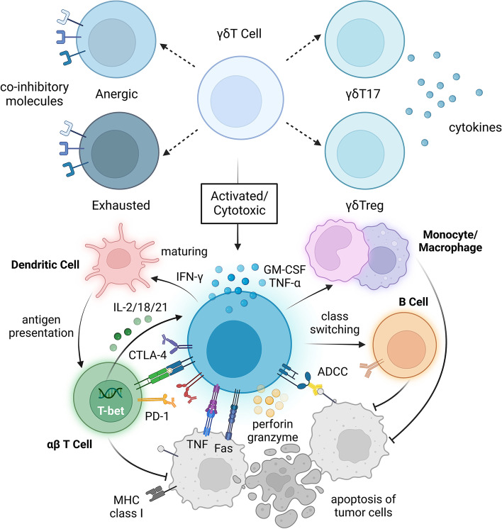
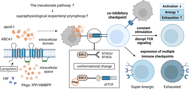
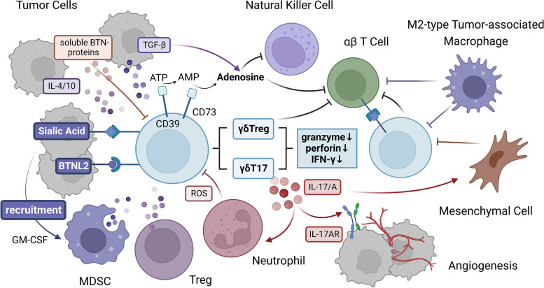
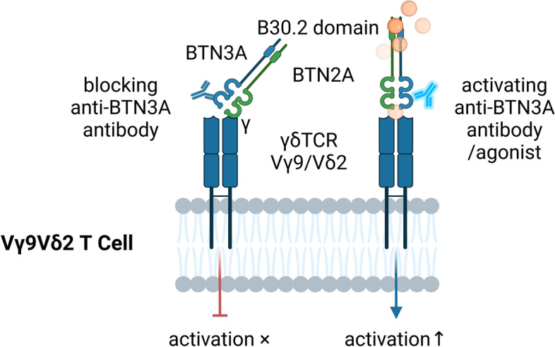
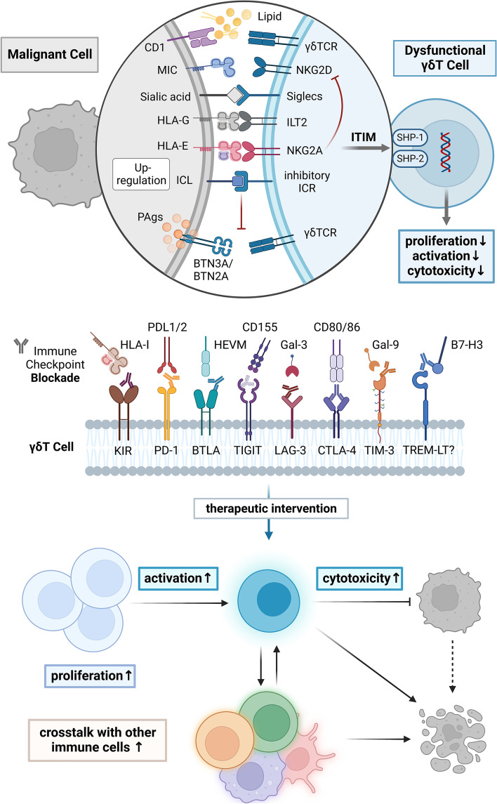
As a nontraditional T-cell subgroup, γδT cells have gained popularity in the field of immunotherapy in recent years. They have extraordinary antitumor potential and prospects for clinical application. Immune checkpoint inhibitors (ICIs), which are efficacious in tumor patients, have become pioneer drugs in the field of tumor immunotherapy since they were incorporated into clinical practice. In addition, γδT cells that have infiltrated into tumor tissues are found to be in a state of exhaustion or anergy, and there is upregulation of many immune checkpoints (ICs) on their surface, suggesting that γδT cells have a similar ability to respond to ICIs as traditional effector T cells. Studies have shown that targeting ICs can reverse the dysfunctional state of γδT cells in the tumor microenvironment (TME) and exert antitumor effects by improving γδT-cell proliferation and activation and enhancing cytotoxicity. Clarification of the functional state of γδT cells in the TME and the mechanisms underlying their interaction with ICs will solidify ICIs combined with γδT cells as a good treatment option.

Keywords: Antitumor immunotherapy; Checkpoint inhibitor (CPI); Immune checkpoint blockade (ICB); Immune checkpoint inhibitors (ICIs); Immune checkpoint molecules; Immune checkpoint therapy (ICT); Tumor microenvironment (TME); γδT cells.


Conflict of interest statement

The authors declare that they have no competing interests.

Figures

Fig. 1
Fig. 1
Under ideal conditions, activated γδT cells can kill tumor cells. Depending on the cytokines secreted by γδT cells and the microenvironment in which they are located, γδT cells can differentiate into different subpopulations, such as γδT17 or γδTregs. To some extent, the expression of checkpoint molecules on the surface of γδT cells can reflect their functional status. Activated effector γδT cells are cytotoxic, and they can express Fas ligand (FasL) and tumor necrosis factor-related apoptosis-induced ligand (TRAIL). Through direct cell–cell contact, apoptosis of tumor cells can be induced through the Fas-FasL and TRAILR-TRAIL death receptor pathways. Proinflammatory cytokines such as IFN-γ and TNF-α can directly inhibit tumor cells, and perforin-granzyme can directly act on the target cell membrane, leading to tumor cell cytolysis. αβT cells can promote the function of γδT cells through cytokines such as IL-2 and the transcription factors T-bet and have a synergistic antitumor effect with γδT cells. Dendritic cells and monocytes-macrophages can also be activated to promote antigen presentation and antibody class switching of B cells, as well as enhance the antibody-dependent cell-mediated cytotoxicity (ADCC) of γδT cells
Fig. 2
Fig. 2
γδT cells gradually become dysfunctional in the tumor microenvironment. During tumorigenesis and tumor development, the metabolism of tumor cells is changed, and the phosphoantigens produced through the mevalonate pathway increase. ABCA1 and apoA-I can act synergistically with BTN proteins to stimulate γδT cells and induce the secretion of supraphysiological levels of IPP. Binding to the B30.2 domain causes the conformation of BTN3A/BTN2A to change, which promotes γδT-cell antigen recognition, proliferation and activation. At the same time, immune checkpoint ligands are highly expressed on the surface of tumor cells, which can interfere with the normal TCR signaling pathway and transmit inhibitory signals to γδT cells by binding to immune checkpoint receptors. Excessive and persistent antigen stimulation and the inhibitory signals transmitted because of the high expression of immune checkpoint molecules eventually cause γδT cells to enter an anergic or exhausted state, resulting in dysfunction and weakened antitumor effects
Fig. 3
Fig. 3
Crosstalk between γδT cells and the TME. Myeloid-derived suppressor cells (MDSCs), regulatory T cells (Tregs), neutrophils with a suppressive phenotype and related immunosuppressive molecules in the tumor microenvironment (TME) can inhibit γδT cells. The binding of glycoimmune checkpoint molecules and BTNL2 to their receptors can lead to the recruitment of MDSCs. Under the action of some cytokines, such as TGF-β, IL-4, and IL-10, in the TME, γδT cells can differentiate into cells with a suppressor phenotype and upregulate the expression of CD39/CD73, which can promote γδT cell exhaustion. γδT cells gradually develop dysfunctional characteristics, such as decreased secretion of perforin, granzyme and IFN-γ; in contrast, γδT cells secrete IL-17/A, which promotes tumor development and even metastasis by increasing angiogenesis and the infiltration of other inhibitory immune cells. At the same time, γδT cells express high levels of immune checkpoint ligands and inhibit other immune effector cells, such as αβT cells, by binding to immune checkpoint receptors on their surface. In addition, CD39/CD73, as the rate-limiting enzyme of the adenosine pathway, can promote the accumulation of adenosine in the TME by acting with soluble molecules such as TGF-β and the sBTN proteins. These interactions generate an immunosuppressive network by inhibiting effector cells, including but not limited to γδT cells, αβT cells and NK cells, through adenosine receptors, which further reduces the antitumor effects of γδT cells
Fig. 4
Fig. 4
The activation of γδT cells can be modulated by anti-BTN3A antibodies. BTN2A can bind to the γ chain of γδTCR and plays an important role with BTN3A in the activation of γδT cells by phosphoantigens. After the binding of an antagonistic monoclonal antibody to BTN3A, the activation of Vγ9Vδ2T cells will be blocked, and the cytotoxicity of Vγ9Vδ2T cells will decrease or even disappear. In contrast, after the use of an agonist targeting BTN3A, Vγ9Vδ2T cells have increased antigen sensitivity and enhanced ability to kill tumor cells, suggesting that targeting BTN family proteins can significantly regulate the immune response of γδT cells against tumor cells
Fig. 5
Fig. 5
Targeting immune checkpoint molecules can revive dysfunctional γδT cells. Dysfunctional γδT cells express high levels of multiple checkpoint molecules on their surface, a phenotype similar to that of anergic or exhausted T cells. Vδ2T cells can recognize phosphoantigens with the assistance of BTN3A/BTN2A, and some subsets of Vδ1T and Vδ3T cells can recognize lipid antigens presented by CD1d and transmit activation signals through γδTCR. Activated γδT cells express NKG2D and/or other similar costimulatory molecules on their surface. Immunoglobulin-like transcripts (ILTs) or leukocyte immunoglobulin-like receptors (LIRs) belong to the Ig superfamily. ILT2 (LIRB1) binds to HLA-G in addition to recognizing other ligands and can inhibit the immune functions of γδT, NK, and B cells. NKG2A can recognize the nonclassical MHC-I molecule HLA-E and inhibit the stimulatory signal of NKG2D. Inhibitory Siglecs are immune regulatory sialic acid-binding receptors that resemble traditional immune checkpoint molecules with one or more ITIM-like motifs in the intracellular segment. Abnormal signaling of immune checkpoint molecules interferes with the normal function of TCRs, affects the level of intracellular protein phosphorylation through ITIM motifs and SHP-1/2, inhibits the proliferation and activation of γδT cells, and ultimately reduces the cytotoxicity of γδT cells. After blocking the inhibitory signals with monoclonal antibodies targeting immune checkpoint molecules, the ability of γδT cells to kill tumor cells and their interactions with other immune effector cells can be enhanced

Similar articles

Cited by

References

    1. Zakeri N, Hall A, Swadling L, Pallett LJ, Schmidt NM, Diniz MO, et al. Characterisation and induction of tissue-resident gamma delta T-cells to target hepatocellular carcinoma. Nat Commun. 2022;13:1372. doi: 10.1038/s41467-022-29012-1. - DOI - PMC - PubMed
    1. Wu Y, Biswas D, Usaite I, Angelova M, Boeing S, Karasaki T, et al. A local human Vδ1 T cell population is associated with survival in nonsmall-cell lung cancer. Nat Cancer. 2022;3:696–709. doi: 10.1038/s43018-022-00376-z. - DOI - PMC - PubMed
    1. Mikulak J, Oriolo F, Bruni E, Roberto A, Colombo FS, Villa A, et al. NKp46-expressing human gut-resident intraepithelial Vδ1 T cell subpopulation exhibits high antitumor activity against colorectal cancer. JCI Insight. 2019;4:125884. doi: 10.1172/jci.insight.125884. - DOI - PMC - PubMed
    1. He W, Hu Y, Chen D, Li Y, Ye D, Zhao Q, et al. Hepatocellular carcinoma-infiltrating γδ T cells are functionally defected and allogenic Vδ2+ γδ T cell can be a promising complement. Clin Transl Med. 2022;12:e800. doi: 10.1002/ctm2.800. - DOI - PMC - PubMed
    1. Bruni E, Cimino MM, Donadon M, Carriero R, Terzoli S, Piazza R, et al. Intrahepatic CD69+Vδ1 T cells re-circulate in the blood of patients with metastatic colorectal cancer and limit tumor progression. J Immunother Cancer. 2022;10:e004579. doi: 10.1136/jitc-2022-004579. - DOI - PMC - PubMed

Publication types