

Lipidomic analysis of brain and hippocampus from mice fed with high-fat diet and treated with fecal microbiota transplantation
- PMID: 36793054
- PMCID: PMC9930259
- DOI: 10.1186/s12986-023-00730-7
Lipidomic analysis of brain and hippocampus from mice fed with high-fat diet and treated with fecal microbiota transplantation
Abstract
Background: Dietary fat intake affects brain composition and function. Different types of dietary fatty acids alter species and abundance of brain lipids in mice. The aim of this study is to explore whether the changes are effective through gut microbiota.
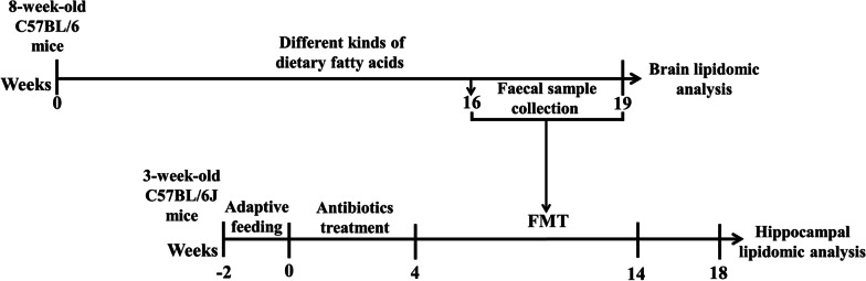
Methods: In our study, 8-week-old male C57BL/6 mice were randomly divided into 7 groups and fed with high-fat diet (HFD) with different fatty acid compositions, control (CON) group, long-chain saturated fatty acid (LCSFA) group, medium-chain saturated fatty acid (MCSFA) group, n-3 polyunsaturated fatty acid (n-3 PUFA) group, n-6 polyunsaturated fatty acid (n-6 PUFA) group, monounsaturated fatty acid (MUFA) group and trans fatty acid (TFA) group. Then, the fecal microbiota transplant (FMT) was performed in other pseudo germ-free mice after antibiotic treatment. The experimental groups were orally perfused with gut microbiota that induced by HFD with different types of dietary fatty acids. The mice were fed with regular fodder before and after FMT. High-performance liquid chromatography-mass spectrometry (LC-MS) was used to analysis the composition of fatty acids in the brain of HFD-fed mice and hippocampus of mice treated with FMT which was collected from HFD-fed mice.
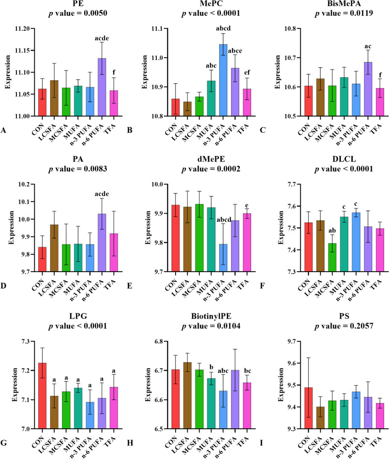
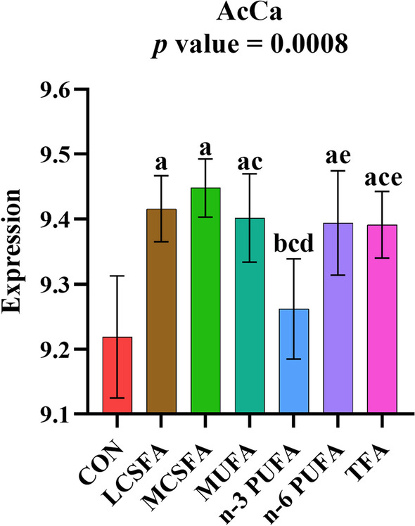
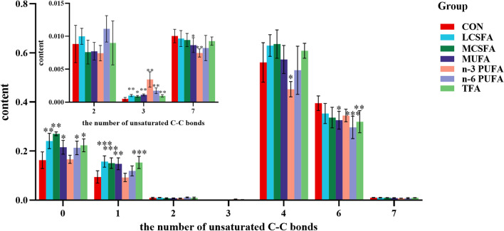
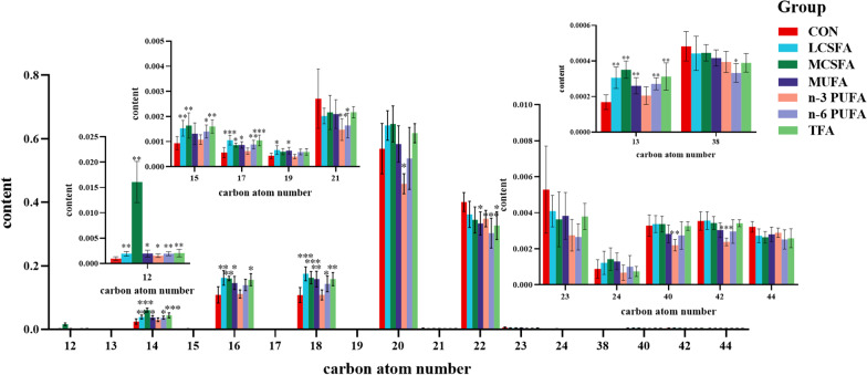
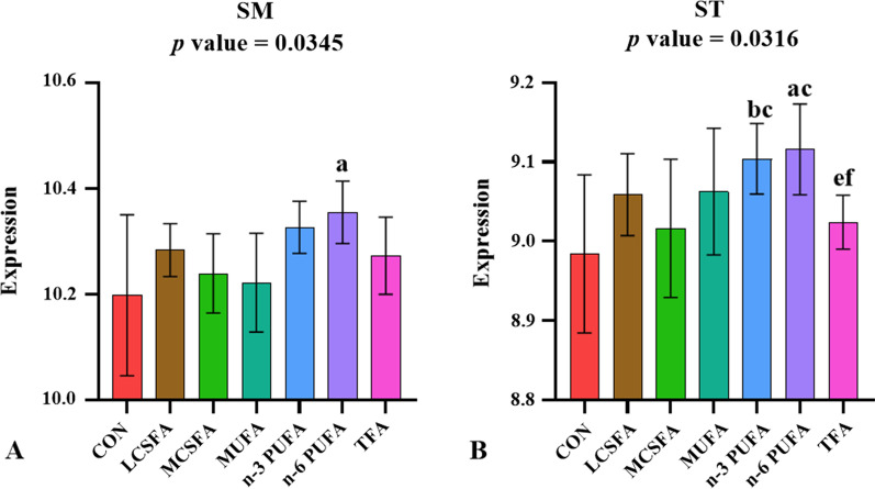
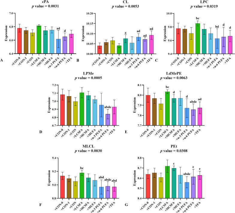
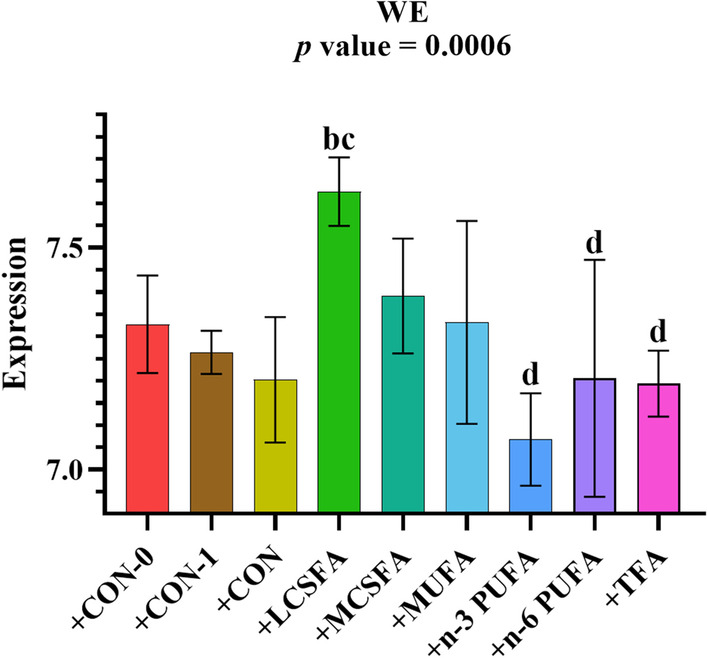
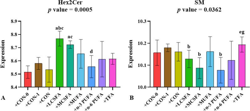
Results: The content of acyl-carnitines (AcCa) increased and lysophosphatidylgylcerol (LPG) decreased in all kinds of HFD groups. phosphatidic acids (PA), phosphatidylethanolamine (PE) and sphingomyelin (SM) contents were significantly increased in the n-6 PUFA-fed HFD group. The HFD elevated the saturation of brain fatty acyl (FA). Lysophosphatidylcholine (LPC), lysodi-methylphosphatidylethanolamine (LdMePE), monolysocardiolipin (MLCL), dihexosylceramides (Hex2Cer), and wax ester (WE) significantly increased after LCSFA-fed FMT. MLCL reduced and cardiolipin (CL) raised significantly after n-3 PUFA-fed FMT.
Conclusions: The study revealed, HFD and FMT in mice had certain effects on the content and composition of fatty acids in the brain, especially on glycerol phospholipid (GP). The change of AcCa content in FA was a good indicator of dietary fatty acid intake. By altering the fecal microbiota, dietary fatty acids might affect brain lipids.
Keywords: Brain lipid; Dietary fatty acid; FMT; High-fat diet; Lipidomics.
© 2023. The Author(s).
Conflict of interest statement
The authors declare no competing interests.
Figures
Similar articles
-
Dietary fatty acids affect learning and memory ability via regulating inflammatory factors in obese mice.J Nutr Biochem. 2022 May;103:108959. doi: 10.1016/j.jnutbio.2022.108959. Epub 2022 Feb 11. J Nutr Biochem. 2022. PMID: 35158028
-
High-fat diets containing different types of fatty acids modulate gut-brain axis in obese mice.Nutr Metab (Lond). 2022 Jun 23;19(1):40. doi: 10.1186/s12986-022-00675-3. Nutr Metab (Lond). 2022. PMID: 35739547 Free PMC article.
-
The relationship between gut microbiota and susceptibility to type 2 diabetes mellitus in rats.Chin Med. 2023 May 5;18(1):49. doi: 10.1186/s13020-023-00717-9. Chin Med. 2023. PMID: 37147692 Free PMC article.
-
Impact of a high-fat diet on the fatty acid composition of the retina.Exp Eye Res. 2020 Jul;196:108059. doi: 10.1016/j.exer.2020.108059. Epub 2020 May 5. Exp Eye Res. 2020. PMID: 32387380
-
Dietary saturated fatty acid type impacts obesity-induced metabolic dysfunction and plasma lipidomic signatures in mice.J Nutr Biochem. 2019 Feb;64:32-44. doi: 10.1016/j.jnutbio.2018.10.005. Epub 2018 Oct 21. J Nutr Biochem. 2019. PMID: 30428423
Cited by
-
Evaluation of Lipid Extraction Protocols for Untargeted Analysis of Mouse Tissue Lipidome.Metabolites. 2023 Sep 9;13(9):1002. doi: 10.3390/metabo13091002. Metabolites. 2023. PMID: 37755282 Free PMC article.
-
Clinical efficacy of washed microbiota transplantation on metabolic syndrome and metabolic profile of donor outer membrane vesicles.Front Nutr. 2024 Nov 19;11:1465499. doi: 10.3389/fnut.2024.1465499. eCollection 2024. Front Nutr. 2024. PMID: 39628464 Free PMC article.
-
Gut microbiota from Mori fructus (Morus alba L.) polyphenols and polysaccharides-dosed mice activates the PPARα/PGC-1α signaling pathway to mitigate HFD-induced metabolic syndrome in mice.Sci Rep. 2025 Aug 1;15(1):28137. doi: 10.1038/s41598-025-13715-8. Sci Rep. 2025. PMID: 40750814 Free PMC article.
-
Short chain fatty acids: the messengers from down below.Front Neurosci. 2023 Jul 6;17:1197759. doi: 10.3389/fnins.2023.1197759. eCollection 2023. Front Neurosci. 2023. PMID: 37483350 Free PMC article.
References
-
- Bascoul-Colombo C, Guschina IA, Maskrey BH, Good M, O'Donnell VB, Harwood JL. Dietary DHA supplementation causes selective changes in phospholipids from different brain regions in both wild type mice and the Tg2576 mouse model of Alzheimer's disease. Biochem Biophys Acta. 2016;1861:524–537. doi: 10.1016/j.bbalip.2016.03.005. - DOI - PMC - PubMed
-
- Zhao Y-C, Zhou M-M, Zhang L-Y, Cong P-X, Xu J, Xue C-H, et al. Recovery of brain DHA-containing phosphatidylserine and ethanolamine plasmalogen after dietary DHA-enriched phosphatidylcholine and phosphatidylserine in SAMP8 mice fed with high-fat diet. Lipids Health Dis. 2020;19:104. doi: 10.1186/s12944-020-01253-3. - DOI - PMC - PubMed
-
- Zhang C, Zhou MM, Zhang TT, Cong PX, Xu J, Xue CH, et al. Effects of dietary supplementation with EPA-enriched phosphatidylcholine and phosphatidylethanolamine on glycerophospholipid profile in cerebral cortex of SAMP8 mice fed with high-fat diet. J Oleo Sci. 2021;70:275–287. doi: 10.5650/jos.ess20212. - DOI - PubMed
LinkOut - more resources
Full Text Sources
Miscellaneous
