

The use of dedicated long-axis views focused on the left atrium improves the accuracy of left atrial volumes and emptying fraction measured by cardiovascular magnetic resonance
- PMID: 36793062
- PMCID: PMC9933380
- DOI: 10.1186/s12968-022-00905-w
The use of dedicated long-axis views focused on the left atrium improves the accuracy of left atrial volumes and emptying fraction measured by cardiovascular magnetic resonance
Abstract
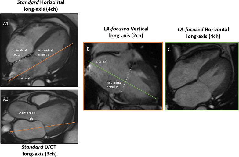
Background: The use of apical views focused on the left atrium (LA) has improved the accuracy of LA volume evaluation by two-dimensional (2D) echocardiography. However, routine cardiovascular magnetic resonance (CMR) evaluation of LA volumes still uses standard 2- and 4-chamber cine images focused on the left ventricle (LV). To investigate the potential of LA-focused CMR cine images, we compared LA maximuml (LAVmax) and minimum (LAVmin) volumes, and emptying fraction (LAEF), calculated on both standard and LA-focused long-axis cine images, with LA volumes and LAEF obtained by short-axis cine stacks covering the LA. LA strain was also calculated and compared between standard and LA-focused images.
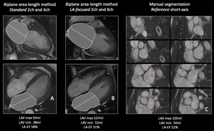
Methods: LA volumes and LAEF were obtained from 108 consecutive patients by applying the biplane area-length algorithm to both standard and LA-focused 2- and 4-chamber cine images. Manual segmentation of a short-axis cine stack covering the LA was used as the reference method. In addition, LA strain reservoir (εs), conduit (εe) and booster pump (εa) were calculated using CMR feature-tracking.
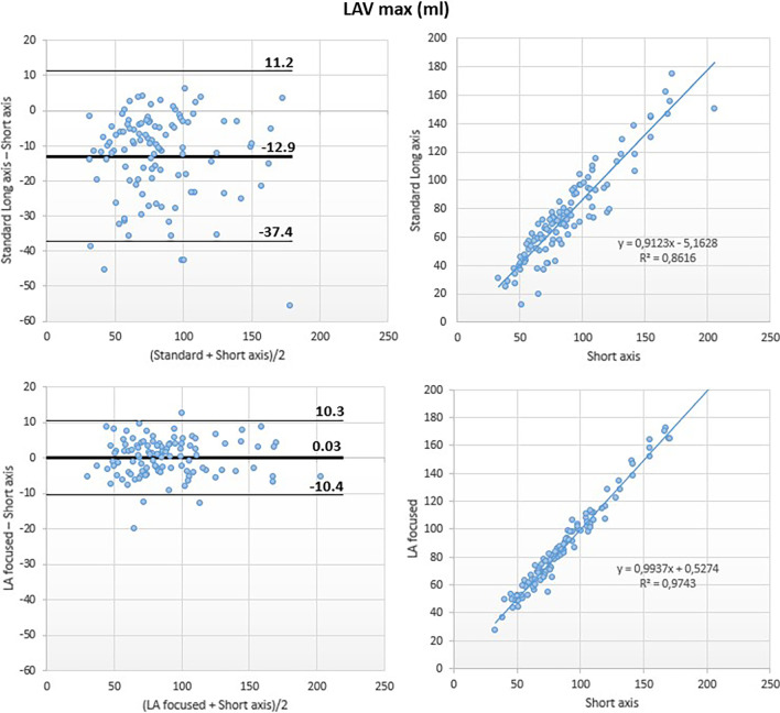
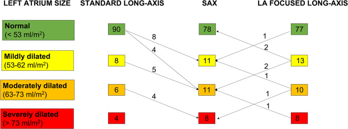
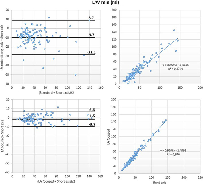
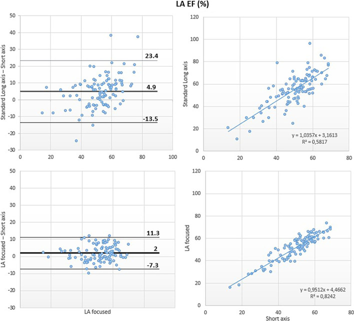
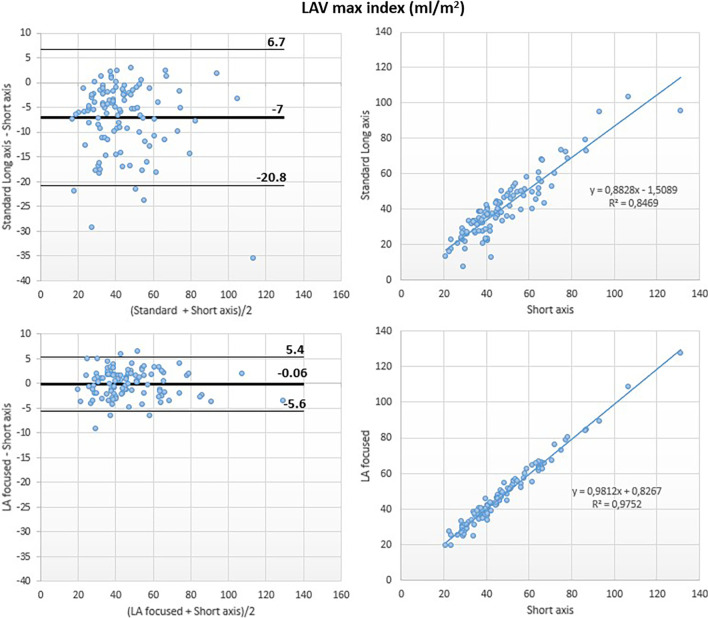
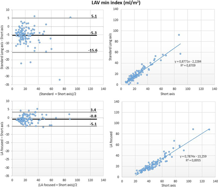
Results: Compared to the reference method, the standard approach significantly underestimated LA volumes (LAVmax: bias - 13 ml; LOA = + 11, - 37 ml; LAVmax i: bias - 7 ml/m2; LOA = + 7, - 21 ml/m2; LAVmin; bias - 10 ml, LOA: + 9, - 28 ml; LAVmin i: bias - 5 ml/m2, LOA: + 5, - 16 ml/m2), and overestimated LA-EF (bias 5%, LOA: + 23, - 14%). Conversely, LA volumes (LAVmax: bias 0 ml; LOA: + 10, - 10 ml; LAVmax i: bias 0 ml/m2; LOA: + 5, - 6 ml/m2; LAVmin: bias - 2 ml; LOA: + 7, - 10 ml; LAVmin i: bias - 1 ml/m2; LOA: + 3, - 5 ml/m2) and LAEF (bias 2%, LOA: + 11, - 7%) by LA-focused cine images were similar to those measured using the reference method. LA volumes by LA-focused images were obtained faster than using the reference method (1.2 vs 4.5 min, p < 0.001). LA strain (εs: bias 7%, LOA = 25, - 11%; εe: bias 4%, LOA = 15, - 8%; εa: bias 3%, LOA = 14, - 8%) was significantly higher in standard vs. LA-focused images (p < 0.001).
Conclusion: LA volumes and LAEF measured using dedicated LA-focused long-axis cine images are more accurate than using standard LV-focused cine images. Moreover, LA strain is significantly lower in LA-focused vs. standard images.
Keywords: Accuracy; Cardiac magnetic resonance; Left atrial emptying fraction; Left atrial strain; Left atrial volume.
© 2023. The Author(s).
Conflict of interest statement
The authors declare that they have no competing interests.
Figures
Similar articles
-
Reproducibility of left atrial function using cardiac magnetic resonance imaging.Eur Radiol. 2021 May;31(5):2788-2797. doi: 10.1007/s00330-020-07399-z. Epub 2020 Oct 30. Eur Radiol. 2021. PMID: 33128187 Free PMC article.
-
Quantification of left atrial function by the area-length method overestimates left atrial emptying fraction.Eur J Radiol. 2023 Mar;160:110705. doi: 10.1016/j.ejrad.2023.110705. Epub 2023 Jan 17. Eur J Radiol. 2023. PMID: 36701824 Free PMC article.
-
Comparing left atrial indices by CMR in association with left ventricular diastolic dysfunction and adverse clinical outcomes.Sci Rep. 2021 Oct 29;11(1):21331. doi: 10.1038/s41598-021-00596-w. Sci Rep. 2021. PMID: 34716361 Free PMC article.
-
Normal Values for Atrial Deformation Measured by Feature-Tracking Cardiac MRI: A Meta-Analysis.J Magn Reson Imaging. 2025 Feb;61(2):882-898. doi: 10.1002/jmri.29465. Epub 2024 May 28. J Magn Reson Imaging. 2025. PMID: 38807354
-
From Left Atrial Dimension to Curved M-Mode Speckle-Tracking Images: Role of Echocardiography in Evaluating Patients with Atrial Fibrillation.Rev Cardiovasc Med. 2022 May 11;23(5):171. doi: 10.31083/j.rcm2305171. eCollection 2022 May. Rev Cardiovasc Med. 2022. PMID: 39077610 Free PMC article. Review.
Cited by
-
The assessment of left ventricular diastolic function: guidance and recommendations from the British Society of Echocardiography.Echo Res Pract. 2024 Jun 3;11(1):16. doi: 10.1186/s44156-024-00051-2. Echo Res Pract. 2024. PMID: 38825710 Free PMC article. Review.
-
CMR to characterize myocardial structure and function in heart failure with preserved left ventricular ejection fraction.Eur Heart J Cardiovasc Imaging. 2024 Oct 30;25(11):1491-1504. doi: 10.1093/ehjci/jeae224. Eur Heart J Cardiovasc Imaging. 2024. PMID: 39205602 Free PMC article. Review.
-
The use of cardiac imaging in patients undergoing atrial fibrillation ablation.J Interv Card Electrophysiol. 2025 Apr 7. doi: 10.1007/s10840-025-02035-6. Online ahead of print. J Interv Card Electrophysiol. 2025. PMID: 40195230 Review.
-
Comparison of right atrial volume measurements using single-plane area-length and stack-of-short-axis methods: A 3.0 T cardiac magnetic resonance study.BMC Med Imaging. 2025 May 14;25(1):160. doi: 10.1186/s12880-025-01708-y. BMC Med Imaging. 2025. PMID: 40369433 Free PMC article.
-
Cardiovascular magnetic resonance imaging for sequential assessment of cardiac fibrosis in mice: technical advancements and reverse translation.Am J Physiol Heart Circ Physiol. 2024 Jan 1;326(1):H1-H24. doi: 10.1152/ajpheart.00437.2023. Epub 2023 Nov 3. Am J Physiol Heart Circ Physiol. 2024. PMID: 37921664 Free PMC article. Review.
References
-
- Gulati A, Ismail TF, Jabbour A, Ismail NA, Morarji K, Ali A, Raza S, et al. Clinical utility and prognostic value of left atrial volume assessment by cardiovascular magnetic resonance in non-ischaemic dilated cardiomyopathy. Eur J Heart Fail. 2013;15:660–670. doi: 10.1093/eurjhf/hft019. - DOI - PubMed
Publication types
MeSH terms
LinkOut - more resources
Full Text Sources
