. 2023 Jan 9;7(2):152-166.
doi: 10.7150/ntno.79050. eCollection 2023.

Gold Nanoparticles Conjugated with Dendrigraft Poly-L-lysine and Folate-Targeted Poly(ethylene glycol) for siRNA Delivery to Prostate cancer

Affiliations

Gold Nanoparticles Conjugated with Dendrigraft Poly-L-lysine and Folate-Targeted Poly(ethylene glycol) for siRNA Delivery to Prostate cancer

Georges Minassian et al. Nanotheranostics. .

Abstract

Dendrigraft Poly-L-Lysine (d-PLL) coated gold nanoparticles (AuNPs) were synthesized by reducing Tetrachloroauric acid with ascorbic acid in the presence of d-PLL. AuNPs-d-PLL formed a stable colloidal solution that absorbs light at a maximum wavelength (λmax) centered at 570 nm as demonstrated by UV-visible (UV-Vis) spectroscopy. From Scanning Electron Microscopy (SEM) analysis, AuNPs-d-PLL were spherical in shape with a mean diameter of 128 ± 47 nm. Dynamic Light scattering (DLS) analysis of the colloidal solution exhibited one size distribution with a hydrodynamic diameter of about 131 nm (size distribution by intensity). Zeta potential (ξ) measurements revealed positively charged AuNPs-d-PLL with ξ about 32 mV, an indicator of high stability in an aqueous solution. The AuNPs-d-PLL was successfully modified with either thiolated poly (ethylene glycol) SH-PEG-OCH3 (Mw 5400 g mol-1) or folic acid-modified thiolated poly (ethylene glycol) SH-PEG-FA of similar molecular weight as demonstrated via DLS and Zeta potential measurements. Complexation of PEGylated AuNPs-d-PLL with siRNA was confirmed by DLS and gel electrophoresis. Finally, we analyzed the functionalization of our nanocomplexes with folic acid via targeted cellular uptake to prostate cancer cells using flow cytometry and LSM imaging. Our findings implicate the broader applicability of folate-PEGylated AuNPs in siRNA-based therapeutics against prostate cancer and perhaps other types of cancer.

Keywords: cancer; gold nanoparticles; polymers; siRNA delivery; stabilization.


Conflict of interest statement

Competing Interests: The authors have declared that no competing interest exists.

Figures

Figure 1
Figure 1
A) Schematic model of the one-pot synthesis of AuNPs-d-PLL solution designed in this study. B) UV-visible spectrum of the obtained AuNPs-d-PLL colloidal solution. C) SEM image of the AuNPs-d-PLL (scale bar 2 µm); and D) Histogram showing the size distribution of the AuNPs-d-PLL from Image J analysis.
Figure 2
Figure 2
A) FTIR analysis of the NHS-activated folic acid (FA-NHS) dot line, SH-PEG-NH2 solid line, and SH-PEG-FA dash line. B) UV-visible spectra of Folic acid (○) and SH-PEG-FA (□).
Figure 3
Figure 3
Size distribution by intensity of AuNPs-d-PLL vs. AuNPs-d-PLL-PEG-OCH3, and AuNPs-d-PLL-PEG-FA. Panel A: An increase of 7nm in size by intensity was noted when comparing AuNPs-d-PLL in the red curve (118.8 ± 1) to AuNPs-d-PLL-PEG-FA in the green curve (125.5 ± 5). An increase of 15 nm in size by intensity was observed when comparing AuNPs-d-PLL (118.8 ± 1) to AuNPs-d-PLL-PEG-OCH3 in the blue curve (133.1 ± 0.9). Panel B: Inset of enlarged peaks to clarify the variation in size (n = 3).
Figure 4
Figure 4
Zeta measurements of AuNPs-d-PLL vs. AuNPs-d-PLL-PEG-OCH3, and AuNPs-d-PLL-PEG-FA. AuNPs-d-PLL-PEG-OCH3 recorded the lowest charge at 21 ± 1 mV when compared to AuNPs-d-PLL 32 ± 1.3 mV, and AuNPs-d-PLL-PEG-FA (30.2 ± 0.9). A 2 mV decrease in zeta potential was observed between AuNPs-d-PLL and AuNPs-d-PLL-PEG-FA. (n = 3).
Scheme 1
Scheme 1
Attachment of SH-PEG-FA onto AuNPs-d-PLL (left) and complexation of siRNA with AuNPs-d-PLL-PEG-FA. A similar method was used for AuNPs-d-PLL-PEG-OCH3.
Figure 5
Figure 5
UV-Vis spectra of AuNPs-d-PLL, AuNPs-d-PLL-PEG, and AuNPs-d-PLL-PEG-FA. A clear redshift of 4 nm from 570 to 574 nm (AuNPs-d-PLL-PEG-FA), and of 6 nm from 570 to 576 nm (AuNPs-d-PLL-PEG-OCH3) is noted upon the addition of SH-PEG-FA and SH-PEG-OCH3 to AuNPs-d-PLL (n = 3).
Figure 6
Figure 6
Size distribution by intensity of AuNPs-d-PLL.siRNA 50nM Vs. AuNPs-d-PLL-PEG-OCH3.siRNA 50nM, and AuNPs-d-PLL-PEG-FA.siRNA 50nM. Panel A: An increase in the size of 21 nm was noted for AuNPs-d-PLL.siRNA, as compared to 10.5 nm for AuNPs-d-PLL-PEG-FA.siRNA. Finally, AuNPs-d-PLL-PEG-OCH3 showed an increase in the size of 1 nm only. Panel B: Inset of enlarged peaks to clarify the variation in size. AuNPs:siRNA was maintained at 25 (n = 3).
Figure 7
Figure 7
WST-1 Cell Proliferation Assay of AuNPs-d-PLL-PEG-FA. The line chart demonstrates the cytotoxicity profile of AuNPs-d-PLL-PEG-FA when incubated with PC-3 PSMA cells over a period of 24 hours. NP concentration was fixed at 50 µg/mL for all time points. Cells were initially seeded onto a 96-well plated, left to reach confluency, and incubated with 50 µg/mL AuNPs-d-PLL-PEG-FA for 4, 6, 8, 12, and 24 hours. Absorbance was read at 450 nm.
Figure 8
Figure 8
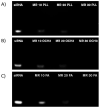
Agarose gel electrophoresis of various AuNPs.siRNA MRs captured under UV light with ChemiDoc™. Free siRNA (0.25 µg/mL) strands were used for the control (first lane in all panels). AuNPs.siRNA complexation was observed on a 1.5 % agarose gel with various coating: AuNPs-d-PLL (A), AuNP-d-PLL-PEG-OCH3 (B), and AuNPs-d-PLL-PEG-FA (C) at MRs 10, 20, and 30, respectively. An evident decrease in band intensity was visible at MR 20. At MR 30, the siRNA band entirely disappeared in all three samples indicating a successful and complete complexation (n = 5).
Figure 9
Figure 9
Mean Percent Fluorescence of AuNPs-d-PLL-PEG OCH3 vs. AuNPs-d-PLL-PEG-FA. PC-3 PSMA cells and PC-3 PSMA.siRNA (50 nM) were used as control. Cells incubated with siRNA.AuNPs-d-PLL-PEG (25 and 50 nM) showed insignificant fluorescence (≤ 5 %). Cells incubated with siRNA.AuNPs-d-PLL-FA (25 nM) presented a mean percent fluorescence of ~ 17% while increasing siRNA.AuNPs-d-PLL-FA to 50 nM had the highest mean percent fluorescence of ~ 30% (n = 3).
Figure 10
Figure 10
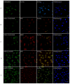
siRNA intracellular localization. PC-3 PSMA cells were incubated for 4 hours with siRNA.NP complexes, tagged with either -OCH3 (B, C) or -FA (D, E) at 37 °C. Panel A serves as a control, where PC-3 PSMA cells were incubated with 50 nM siRNA only. Cells were fixed in 100 % methanol at -20 °C for 5 mins and then stained with Hoechst and anti-early endosomal (EEA1 1/100) or lysosomal (M6PR 1/150) markers. The pattern of siRNA co-localization was visualized by Zeiss Confocal microscopy mainly from the overlay images. Only overlay from panel D showed dot-like structures with partial resemblance to early endosomes in almost 30% of the tested cells with siRNA-NP-FA-50 (D). Scale bar set at 20 µm (n = 5 /panel).
Figure 11
Figure 11
Z-stack imaging of PSMA cells treated with AuNPs-PEG.siRNA (50 nM) vs. AuNPs-FA.siRNA (50 nM). Eight out of 20 sections were chosen as representative sections at a set interval between -10 µm to +10 µm with positive and negative distances relative to the initial focal point of the image. All lasers had the same pinhole. The gain was set at 650 nm for FITC and 550 nm for Hoechst. There is a noticeable decrease in the FITC intensity of AuNPs-PEG.siRNA when compared to AuNPs-FA.siRNA. Furthermore, particles were only visible in sections ranging from -2 µm to + 1 µm in cells incubated with AuNPs-PEG-FA.siRNA (A); particles were absent in cells incubated with AuNPs-PEG.siRNA. The scale bar was set at 20 µm (n = 3).

References

    1. Rahme K, Guo J, Biswas S, O'Driscoll CM, Holmes JD. Branched PEI Capped Gold Nanoparticles in Water for siRNA Delivery to Cancer Cells. Advanced Materials: TechConnect Briefs. 2017. pp. 159–62.
    1. Pernar CH, Ebot EM, Wilson KM, Mucci LA. The Epidemiology of Prostate Cancer. Cold Spring Harbor perspectives in medicine. 2018. 8. - PMC - PubMed
    1. Chanda N, Kan P, Watkinson LD, Shukla R, Zambre A, Carmack TL. et al. Radioactive gold nanoparticles in cancer therapy: therapeutic efficacy studies of GA-198AuNP nanoconstruct in prostate tumor-bearing mice. Nanomedicine: nanotechnology, biology, and medicine. 2010;6:201–9. - PubMed
    1. Cheng Z, Li M, Dey R, Chen Y. Nanomaterials for cancer therapy: current progress and perspectives. Journal of hematology & oncology. 2021;14:85. - PMC - PubMed
    1. Cao L, Zhu Y, Wang W, Wang G, Zhang S, Cheng H. Emerging Nano-Based Strategies Against Drug Resistance in Tumor Chemotherapy. Frontiers in bioengineering and biotechnology. 2021;9:798882. - PMC - PubMed