. 2023 Jan 30:14:1085120.
doi: 10.3389/fneur.2023.1085120. eCollection 2023.

Parent artery occlusion for ruptured aneurysms in moyamoya vessels or on collaterals

Affiliations

Parent artery occlusion for ruptured aneurysms in moyamoya vessels or on collaterals

Zibo Zhou et al. Front Neurol. .

Abstract

Background: Aneurysms in moyamoya vessels or on collaterals are difficult to treat. Parent artery occlusion (PAO) via endovascular treatment (EVT) is often the last resort, but the safety and efficacy of this approach need to be evaluated.

Materials and methods: A retrospective study was performed on patients admitted to our hospital who were diagnosed with unilateral or bilateral moyamoya disease (MMD) associated with ruptured aneurysms in moyamoya vessels or on collaterals. These aneurysms were treated with PAO, and the clinical outcome was recorded.

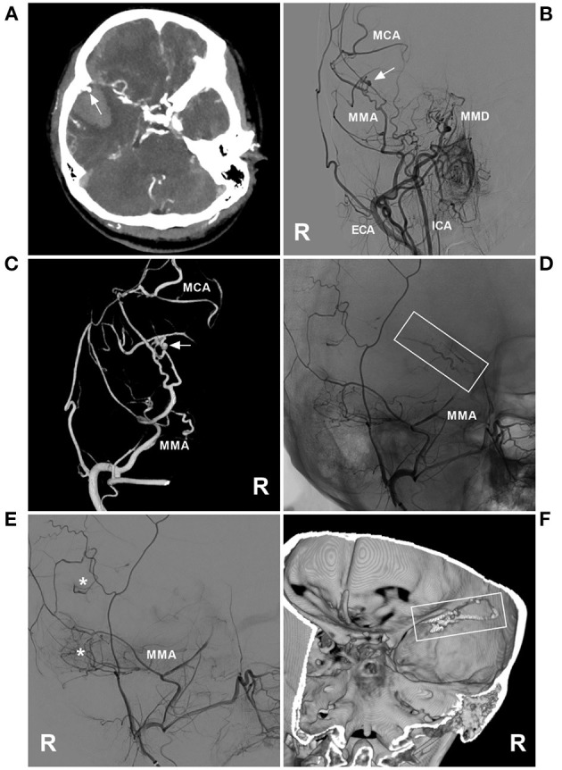
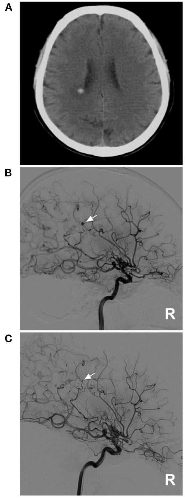
Results: Eleven patients were aged 54.7 ± 10.4 years, and six patients were male (54.5%, 6/11). The aneurysms in 11 patients were single and ruptured, and the average size was 2.7 ± 0.6 mm. Three (27.3%, 3/11) aneurysms were located at the distal anterior choroidal artery, 3 (27.3%, 3/11) were at the distal lenticulostriate artery, 3 (27.3%, 3/11) were at the P2-3 segment of the posterior cerebral artery, 1 (9.1%, 1/11) was at the P4-5 segment of the posterior cerebral artery, and 1 was at the transdural location of the middle meningeal artery. Among the 11 aneurysms, PAO by coiling was performed on 7 (63.6%, 7/11), and Onyx casting was performed on 4 (36.4%, 4/11). Of 11 patients, 2 (18.2%, 2/11) suffered intraoperative hemorrhagic complications. During follow-up, all patients had good outcomes with a modified Rankin scale score of 0-2.

Conclusion: As a last resort, the application of PAO with coiling or casting Onyx for ruptured aneurysms in moyamoya vessels or on collaterals may be safe with an acceptable clinical outcome. However, patients with MMD may not always achieve expected health outcomes, and PAO for the aneurysm can bring only temporary relief.

Keywords: aneurysm; collateral; moyamoya disease; outcome; parent artery occlusion.


Conflict of interest statement

The authors declare that the research was conducted in the absence of any commercial or financial relationships that could be construed as a potential conflict of interest.

Figures

Figure 1
Figure 1
PAO of the PCA aneurysm in MMD in case 4 in Table 1. (A) CT showed subarachnoid hemorrhage focusing on the right ambient cistern. (B) Three-dimensional DSA of the right ICA showed an aneurysm (arrow) at the P2–3 segment of the PCA. (C) Unsubtracted DSA of the right ICA showed that the aneurysm and proximal parent artery underwent PAO by coiling (arrow). (D) Follow-up DSA of the right ICA showed no recurrence of the aneurysm, and the collateral circulation beyond the coils was abundant (circle). CT, computed tomography; DSA, digital subtracted angiography; ICA, internal carotid artery; MMD, moyamoya disease; PAO, parent artery occlusion; PCA, posterior cerebral artery; R, right.
Figure 2
Figure 2
PAO of the PCA aneurysm in case 5 in Table 1. (A) DSA of the right carotid artery showed that the extracranial ICA was thin, and the intracranial ICA was MMD. (B) DSA of the right vertebral artery showed an aneurysm located on the P2–3 segment of the right PCA (arrow). (C) Unsubtracted DSA showed PAO of the P2–3 segment of the PCA by coiling (arrow). (D) DSA showed that the aneurysm (arrow) could be seen by reflux from the collateral circulation in the late artery phase of the arterial angiogram. DSA, digital subtracted angiography; ICA, internal carotid artery; MMD, moyamoya disease; PAO, parent artery occlusion; PCA, posterior cerebral artery; R, right.
Figure 3
Figure 3
PAO of the MMA aneurysm in case 11 in Table 1. (A) Enhanced CT showing hemorrhage of the temporal lobe. An aneurysm (arrow) is indicated at the rim of the hematoma. (B) DSA of the right carotid artery showing MMD in the ICA; an aneurysm (arrow) was at the MMA. (C) Three-dimensional DSA of the right ECA showing an aneurysm (arrow) at the transdural location of the MMA; then, the MCA was continued. (D) Unsubtracted DSA showing the MMA as the parent artery to the aneurysm was occluded with Onyx (frame). (E) DSA of the right ECA showing that the petrous branch of MMD had anastomoses (asterisks) with the MCA. (F) Xper-CT showing the location of Onyx casting (frame). CT, computed tomography; DSA, digital subtracted angiography; ECA, external carotid artery; ICA, internal carotid artery; MCA, middle cerebral artery; MMA, middle meningeal artery; MMD, moyamoya disease; R, right.
Figure 4
Figure 4
Imaging characteristics of cases 1, 2, 3, 7, 8 and 9 in Table 1. (A) Case 1: Left, DSA of the ICA showed an aneurysm on the distal AchA (arrow). Right: Postoperative DSA showing that both the parent artery and aneurysm were occluded with Onyx. (B) Case 2: Left, DSA of the carotid artery showed an aneurysm on the distal AchA (arrow). Right: Postoperative DSA showing that both the parent artery and aneurysm were occluded with coiling (arrow). (C) Case 3: Left, DSA of the ICA showed an aneurysm on the distal AchA (arrow). Right: Postoperative DSA showing that both the parent artery and aneurysm were occluded with coiling (arrow). (D) Case 7: Left, CT showed hemorrhage of the right occipital lobe into the lateral ventricle. Right: DSA of the ICA showing the aneurysm (arrow) located on the P4–5 segment of the PCA. (E) Case 8: Left, DSA of the ICA showed an aneurysm on the distal LSA (arrow). Right: Postoperative DSA showing that both the parent artery and aneurysm were occluded. (F) Case 9: DSA of the ICA in the anterior-posterior view (left) and lateral view (right) showed an aneurysm on the distal LSA (arrow). AchA, anterior choroidal artery; CT, computed tomography; DSA, digital subtracted angiography; ICA, internal carotid artery; LSA, lenticulostriate artery; PAO, parent artery occlusion; PCA, posterior cerebral artery.
Figure 5
Figure 5
A diminishing aneurysm in case 1 in Table 2. (A) CT showed hemorrhage on the lateral ventricle wall. (B) DSA of the right ICA showed an aneurysm on the distal AchA (arrow), and MMD can be seen on this side. (C) One month later, repeat DSA of the right ICA showed that the aneurysm was diminishing, and only a remnant could be seen (arrow). AchA, anterior choroidal artery; CT, computed tomography; DSA, digital subtracted angiography; ICA, internal carotid artery; MMD, moyamoya disease; R, right.
Figure 6
Figure 6
A growing aneurysm in case 2 in Table 2. (A) CT showed hemorrhage (arrow) in the right temporal lobe involving the ventricle. (B) CTA showed an aneurysm (arrow) in the right moyamoya vessels, and bilateral MMD can be seen. (C) One week later, repeated CTA showed that the aneurysm had grown (arrow). (D) Three-dimensional DSA of the right ICA showed that the aneurysm (arrow) vessels affected by MMD were enlarged. (E) Intraoperative image showing the vessels affected by MMD (asterisks); these vessels appeared thin and frail. (F) Postoperative DSA after aneurysm resection showed that the aneurysm had disappeared. CT, computed tomography; CTA, computed tomography angiography; DSA, digital subtracted angiography; ICA, internal carotid artery; MMD, moyamoya disease; R, right.
Figure 7
Figure 7
Fatal hemorrhage in case 5 in Table 2. (A) CT showed hemorrhage (arrow) in the right temporal lobe involving the ventricle. (B) CTA showed an aneurysm (arrow) in the right moyamoya vessels, and bilateral MMD can be seen. CT, computed tomography; CTA, computed tomography angiography; MMD, moyamoya disease.

Similar articles

Cited by

References

    1. Suzuki J, Kodama N. Moyamoya disease: a review. Stroke. (1983) 14:104–9. - PubMed
    1. Lang M, Moore NZ, Witek AM, Kshettry VR, Bain MD. Microsurgical repair of ruptured aneurysms associated with moyamoya-pattern collateral vessels of the middle cerebral artery: a report of two cases. World Neurosurg. (2017) 105:1042.e5–10. 10.1016/j.wneu.2017.06.166 - DOI - PubMed
    1. Sun Y, Zhou G, Feng J, Chen L, Liu G, Wang J, et al. . Incidence and prevalence of moyamoya disease in urban China: a nationwide retrospective cohort study. Stroke Vascul Neurol. (2021) 6:615–23. 10.1136/svn-2021-000909 - DOI - PMC - PubMed
    1. Kim JH, Kwon TH, Kim JH, Chong K, Yoon W. Intracranial aneurysms in adult moyamoya disease. World Neurosurg. (2018) 109:e175–82. 10.1016/j.wneu.2017.09.127 - DOI - PubMed
    1. Lee JK, Lee JH, Kim SH, Lee MC. Distal anterior choroidal artery aneurysm in a patient with moyamoya disease: case report. Neurosurgery. (2001) 48:222–5. 10.1227/00006123-200101000-00043 - DOI - PubMed