

Artificial intelligence method based on multi-feature fusion for automatic macular edema (ME) classification on spectral-domain optical coherence tomography (SD-OCT) images
- PMID: 36793539
- PMCID: PMC9922866
- DOI: 10.3389/fnins.2023.1097291
Artificial intelligence method based on multi-feature fusion for automatic macular edema (ME) classification on spectral-domain optical coherence tomography (SD-OCT) images
Abstract
Purpose: A common ocular manifestation, macular edema (ME) is the primary cause of visual deterioration. In this study, an artificial intelligence method based on multi-feature fusion was introduced to enable automatic ME classification on spectral-domain optical coherence tomography (SD-OCT) images, to provide a convenient method of clinical diagnosis.
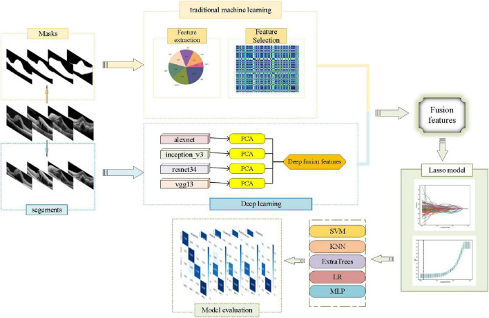
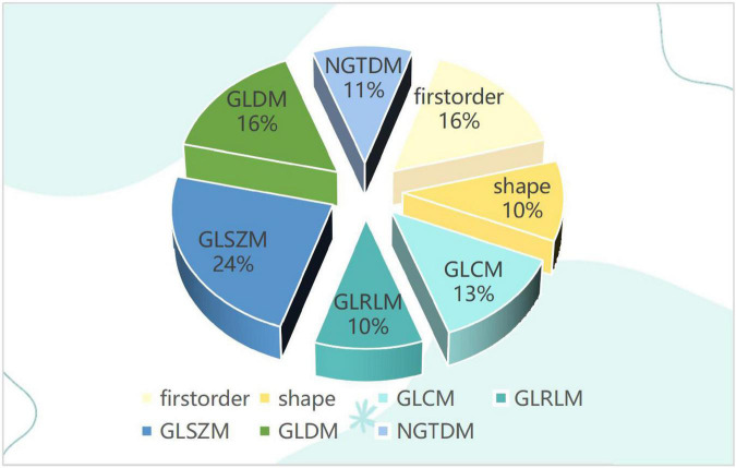
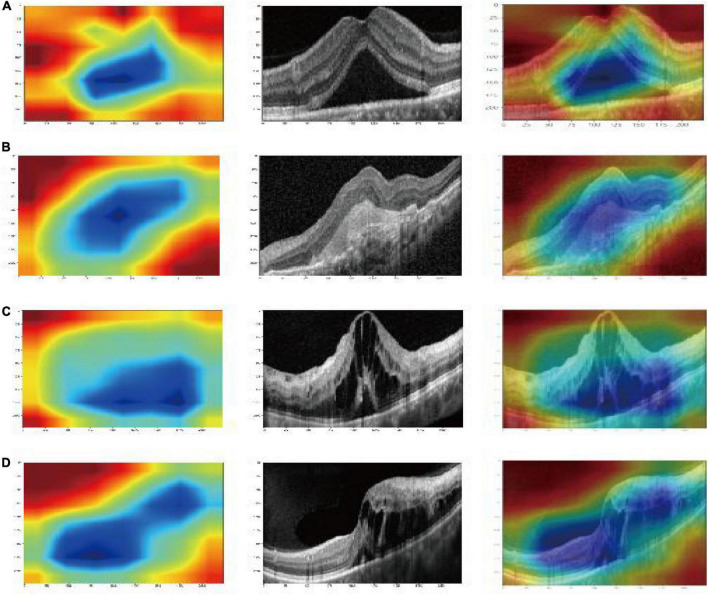
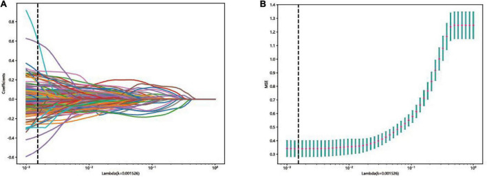
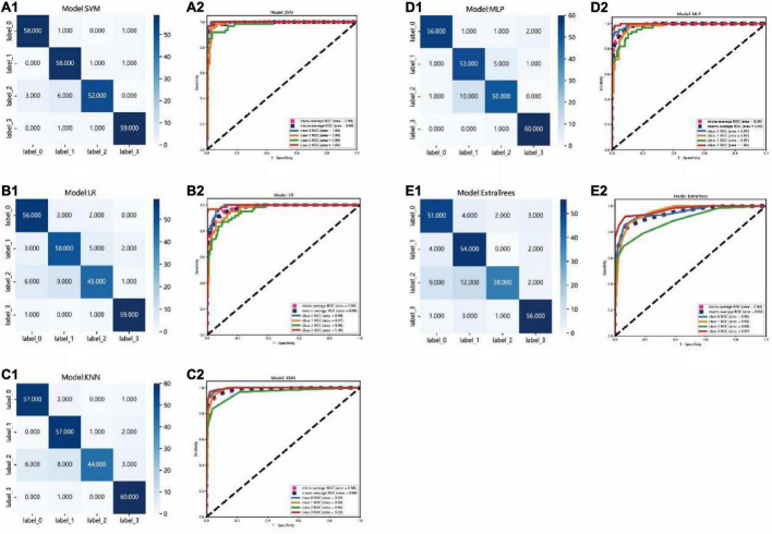
Methods: First, 1,213 two-dimensional (2D) cross-sectional OCT images of ME were collected from the Jiangxi Provincial People's Hospital between 2016 and 2021. According to OCT reports of senior ophthalmologists, there were 300 images with diabetic (DME), 303 images with age-related macular degeneration (AMD), 304 images with retinal-vein occlusion (RVO), and 306 images with central serous chorioretinopathy (CSC). Then, traditional omics features of the images were extracted based on the first-order statistics, shape, size, and texture. After extraction by the alexnet, inception_v3, resnet34, and vgg13 models and selected by dimensionality reduction using principal components analysis (PCA), the deep-learning features were fused. Next, the gradient-weighted class-activation map (Grad-CAM) was used to visualize the-deep-learning process. Finally, the fusion features set, which was fused from the traditional omics features and the deep-fusion features, was used to establish the final classification models. The performance of the final models was evaluated by accuracy, confusion matrix, and the receiver operating characteristic (ROC) curve.
Results: Compared with other classification models, the performance of the support vector machine (SVM) model was best, with an accuracy of 93.8%. The area under curves AUC of micro- and macro-averages were 99%, and the AUC of the AMD, DME, RVO, and CSC groups were 100, 99, 98, and 100%, respectively.
Conclusion: The artificial intelligence model in this study could be used to classify DME, AME, RVO, and CSC accurately from SD-OCT images.
Keywords: SD-OCT images; artificial intelligence; classification models; macular edema; multi-feature fusion.
Copyright © 2023 Gan, Wu and Zhong.
Conflict of interest statement
The authors declare that the research was conducted in the absence of any commercial or financial relationships that could be construed as a potential conflict of interest.
Figures
Similar articles
-
Computerized macular pathology diagnosis in spectral domain optical coherence tomography scans based on multiscale texture and shape features.Invest Ophthalmol Vis Sci. 2011 Oct 21;52(11):8316-22. doi: 10.1167/iovs.10-7012. Invest Ophthalmol Vis Sci. 2011. PMID: 21911579 Free PMC article.
-
Optical Coherence Tomography Image Classification Using Hybrid Deep Learning and Ant Colony Optimization.Sensors (Basel). 2023 Jul 26;23(15):6706. doi: 10.3390/s23156706. Sensors (Basel). 2023. PMID: 37571490 Free PMC article.
-
Evaluation of an Artificial Intelligence-Based Detector of Sub- and Intraretinal Fluid on a Large Set of Optical Coherence Tomography Volumes in Age-Related Macular Degeneration and Diabetic Macular Edema.Ophthalmologica. 2022;245(6):516-527. doi: 10.1159/000527345. Epub 2022 Oct 10. Ophthalmologica. 2022. PMID: 36215958
-
Differentiation of Underlying Pathologies of Macular Edema Using Spectral Domain Optical Coherence Tomography (SD-OCT).Ocul Immunol Inflamm. 2019;27(3):474-483. doi: 10.1080/09273948.2019.1603313. Ocul Immunol Inflamm. 2019. PMID: 31184556 Review.
-
Deep learning algorithms for detection of diabetic macular edema in OCT images: A systematic review and meta-analysis.Eur J Ophthalmol. 2023 Jan;33(1):278-290. doi: 10.1177/11206721221094786. Epub 2022 Apr 27. Eur J Ophthalmol. 2023. PMID: 35473414
Cited by
-
Deep learning-based classification of multiple fundus diseases using ultra-widefield images.Front Cell Dev Biol. 2025 Jul 17;13:1630667. doi: 10.3389/fcell.2025.1630667. eCollection 2025. Front Cell Dev Biol. 2025. PMID: 40746855 Free PMC article.
-
Stitched vision transformer for age-related macular degeneration detection using retinal optical coherence tomography images.PLoS One. 2024 Jun 5;19(6):e0304943. doi: 10.1371/journal.pone.0304943. eCollection 2024. PLoS One. 2024. PMID: 38837967 Free PMC article.
References
-
- Chan G. C., Kamble R., Muller H., Shah S. A., Tang T. B., Meriaudeau F. (2018). “Fusing results of several deep learning architectures for automatic classification of normal and diabetic macular edema in optical coherence tomography,” in Proceedings of the 2018 40th annual international conference of the IEEE engineering in medicine and biology society (EMBC), Vol. 2018 (Honolulu, HI: IEEE; ), 670–673. 10.1109/EMBC.2018.8512371 - DOI - PubMed
LinkOut - more resources
Full Text Sources