

Myopic macular Bruch's membrane defects
- PMID: 36793950
- PMCID: PMC9922809
- DOI: 10.1016/j.heliyon.2023.e13257
Myopic macular Bruch's membrane defects
Abstract
Purpose: To examine histologic characteristics of macular Bruchś membrane defects (BMD) in axially elongated eyes.
Design: Histomorphometric study.
Methods: Using light microscopy, we examined enucleated human globes for BMDs.
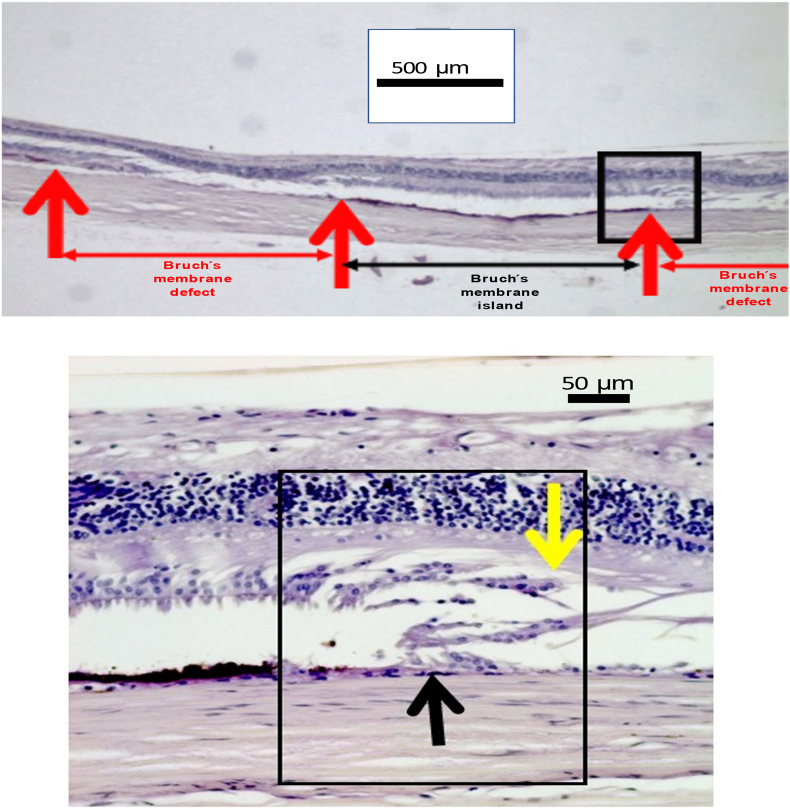
Results: In 247 eyes, BMDs were detected in 15 (6.1%) eyes (axial length:27.0-36.0 mm), in 10 of them in the macular region. Prevalence and size of BMDs (mean:1.93 ± 1.62 mm; range:0.22mm-6.24 mm) correlated with longer axial length (OR:1.52; 95%CI:1.19,1.94; P = 0.001) and higher prevalence of scleral staphylomas (OR:16.3; 95%CI:2.67,99.3; P < 0.001). The BMDs were smaller than corresponding gaps in the retinal pigment epithelium (RPE) (1.93 ± 1.62 mm versus 2.61 mm ± 1.73 mm; P = 0.003), and larger than corresponding gaps in the inner nuclear layer (0.43 ± 0.76 mm; P = 0.008) and inner limiting membrane bridges (0.13 ± 0.33 mm; P = 0.001). Choriocapillaris thickness, BM thickness and RPE cell density did not vary (all P > 0.05) between the BDM border and adjacent areas. In the BMD, choriocapillaris and RPE were absent. The sclera was thinner in the BDM area than in adjacent areas (0.28 ± 0.19 mm versus 0.36 ± 0.13 mm; P = 0.006).
Conclusions: BMDs as hallmarks of myopic macular degeneration are characterized by longer gaps in the RPE and smaller gaps in the outer nuclear layer and inner nuclear layer, by localized scleral thinning, and by a spatial association with scleral staphylomas. Thickness of the choriocapillaris and density of the RPE cell layer, both absent within the BDMs, do not vary between the BMD border and adjacent regions. The results suggest an association between BDMs and absolute scotomas, stretching of the adjacent retinal nerve fiver layer, and an axial elongation-associated stretching effect on BM as etiology of the BDMs.
Keywords: Axial myopia; Bruch’s membrane; Bruch’s membrane defects; Myopia; Myopic macular degeneration; Myopic maculopathy.
© 2023 The Authors.
Figures


References
-
- Ohno-Matsui K., Kawasaki R., Jonas J.B., Cheung C.M., Saw S.M., Verhoeven V.J., Klaver C.C., Moriyama M., Shinohara K., Kawasaki Y., Yamazaki M., Meuer S., Ishibashi T., Yasuda M., Yamashita H., Sugano A., Wang J.J., Mitchell P., Wong T.Y. META-analysis for Pathologic Myopia (META-PM) Study Group. International photographic classification and grading system for myopic maculopathy. Am. J. Ophthalmol. 2015;159(5):877. 83.e7. - PubMed
-
- Ohno-Matsui K., Jonas J.B., Spaide R.F. Macular Bruch membrane holes in highly myopic patchy chorioretinal atrophy. Am. J. Ophthalmol. 2016;166:22–28. - PubMed
-
- Du R., Fang Y., Jonas J.B., Yokoi T., Takahashi H., Uramoto K., Kamoi K., Yoshida T., Ohno-Matsui K. Clinical features of patchy chorioretinal atrophy in pathological myopia. Retina. 2020;40(5):951–959. - PubMed
-
- You Q.S., Peng X.Y., Xu L., Chen C.X., Wei W.B., Wang Y.X., Jonas J.B. Macular Bruch's membrane defects in highly myopic eyes. The Beijing Eye Study. Retina. 2016;36(3):517–523. - PubMed
-
- Jonas J.B., Ohno-Matsui K., Spaide R.F., Holbach L., Panda-Jonas S. Macular Bruch's membrane defects and axial length: association with gamma zone and delta zone in peripapillary region. Invest. Ophthalmol. Vis. Sci. 2013;54(2):1295–1302. - PubMed
LinkOut - more resources
Full Text Sources
