Review
. 2023 Jan 31;12(1):9-22.
doi: 10.21037/acs-2022-urats-23. Epub 2023 Jan 6.

Uniportal fully robotic-assisted sleeve resections: surgical technique and initial experience of 30 cases

Affiliations
Review

Uniportal fully robotic-assisted sleeve resections: surgical technique and initial experience of 30 cases

Diego Gonzalez-Rivas et al. Ann Cardiothorac Surg. .

Abstract

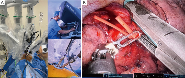
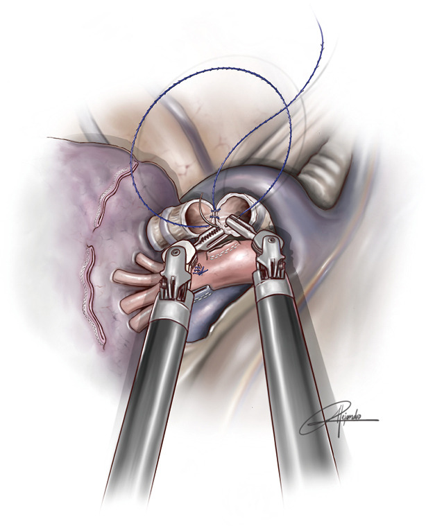
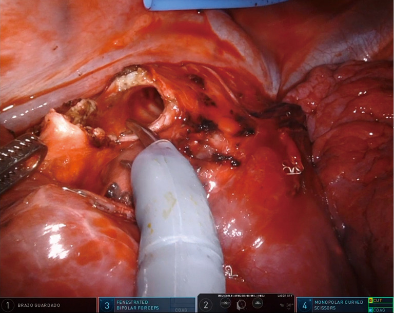
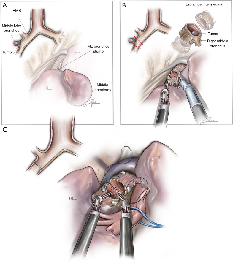
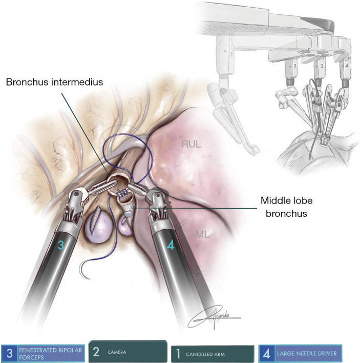
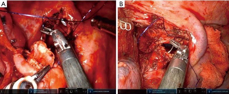
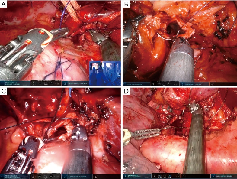
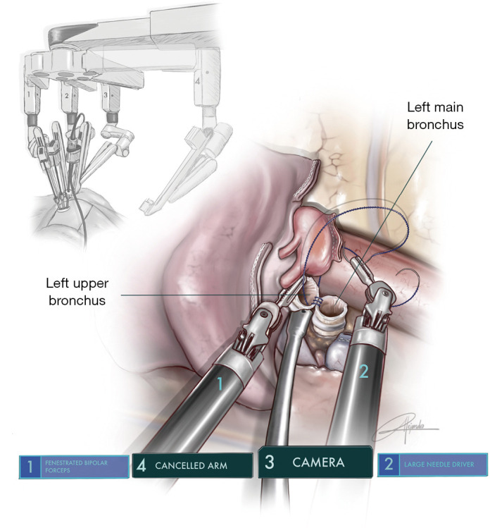
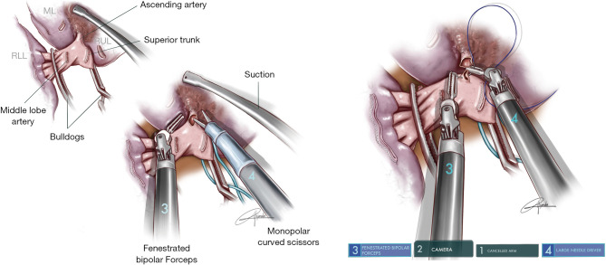
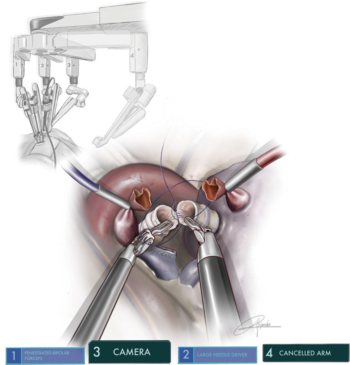
Since the first uniportal video-assisted thoracoscopic surgery (uVATS) performed in 2010, the uniportal approach has evolved up to a point where even the most complex cases can be done. This is thanks to the experience acquired over the years, the specifically designed instruments and improvements in imaging. However, in these last few years, robotic-assisted thoracoscopic surgery (RATS) has also shown progress and distinct advantages compared to the uniportal VATS approach, thanks to advanced maneuverability of the robotic arms as well as the three-dimensional (3D) view. Excellent surgical outcomes have been reported and so too, the ergonomic benefits to the surgeon. The main limitation we find of the robotic systems is that they are designed for a multiport approach, requiring between three to five incisions to be able to perform surgeries. With the aim to offer the least invasive approach, using the robotic technology we decided to adapt the Da Vinci Xi® in September 2021 to develop the uniportal pure RATS approach (uRATS) performed by a single intercostal incision, without rib spreading and using the robotic staplers. We have now reached a point where we perform all type of procedures, including the more complex sleeve resections. Sleeve lobectomy is now widely accepted as a reliable and safe procedure to allow complete resection of centrally located tumors. Although it is a technically challenging surgical technique, it offers better outcomes when compared to pneumonectomy. The intrinsic characteristics of the robot such as the 3D view and improved maneuverability of instruments make the sleeve resections easier compared to thoracoscopic techniques. As in uVATS vs. multiport VATS, the uRATS approach, due to its geometrical characteristics, requires specific instrumentation, different movements and learning curve compared to multiport RATS. In this article we describe the surgical technique and our initial uniportal pure RATS experience with bronchial, vascular sleeves and carinal resections in 30 patients.

Keywords: Sleeve lobectomy; lung cancer; uRATS sleeve; uniportal robotic-assisted thoracoscopic surgery (uRATS).


Conflict of interest statement

Conflicts of Interest: The authors have no conflicts of interest to declare.

Figures

Figure 1
Figure 1
Photo showing the arm placement and location of the 12 mm trocar during a middle sleeve lobectomy (A). Combined photo showing the division of posterior fissure and port placement with the 12 mm trocar through the single incision for stapler insertion during a right lower sleeve lobectomy (B).
Figure 2
Figure 2
Illustration showing a running barbed suture during an upper and middle bilobectomy sleeve (permission provided from medical illustrator Alejandro Garcia, who is also a co-author of this article).
Figure 3
Figure 3
Surgical image showing the use of robotic scissors to divide the intermedius bronchus during a middle sleeve lobectomy.
Figure 4
Figure 4
Illustration showing the procedure of middle sleeve lobectomy (permission provided from medical illustrator Alejandro Garcia, who is also a co-author of this article). (A) Anatomic location of a tumor requiring middle lobe sleeve; (B) division of bronchus intermedius with robotic scissors and tumor at protruding from middle lobe bronchus; (C) running suture of cartilaginous portion between bronchus intermedius and lower lobe bronchus. The lower lobe artery is retracted with a vessel loop. ML, middle lobe; RLL, right lower lobe; RUL, right upper lobe; RMB, right main bronchus.
Figure 5
Figure 5
Illustration showing the running suture for a right lower sleeve lobectomy (reimplantation of middle lobe bronchus to intermedius) (permission provided from medical illustrator Alejandro Garcia, who is also a co-author of this article). ML, middle lobe; RUL, right upper lobe.
Figure 6
Figure 6
Surgical image of the running anastomosis after a right upper and middle sleeve bilobectomy (intermedius to right main bronchus) (A). Surgical image of the running anastomosis after a right lower and middle sleeve bilobectomy (right upper to right main bronchus) (B).
Figure 7
Figure 7
Running barbed suture on left-sided sleeve resections and tracheal anastomosis. (A) Surgical photo showing arm placement and the running barbed anastomosis of left main bronchus to left lower bronchus during a left upper sleeve lobectomy. (B) Surgical image showing the running barbed suture of left main bronchus to trisegmental B1-3 bronchus after a left lower + lingulectomy sleeve lobectomy. (C) Surgical image of second and third carinal sleeve anatomic segmentectomy showing the anastomosis of left main bronchus to left lower bronchus as the initial step. (D) Surgical image showing distal tracheal resection and reconstruction with barbed suture.
Figure 8
Figure 8
Illustration showing the running barbed suture of left main bronchus to left upper lobe bronchus after a left lower sleeve lobectomy (permission provided from medical illustrator Alejandro Garcia, who is also a co-author of this article).
Figure 9
Figure 9
Illustration of vascular reconstruction using the bulldog clamps (permission provided from medical illustrator Alejandro Garcia, who is also a co-author of this article). ML, middle lobe; RUL, right upper lobe; RLL, right lower lobe.
Figure 10
Figure 10
Illustration showing the running barbed suture of left main bronchus to lower lobe bronchus after a double sleeve lobectomy (permission provided from medical illustrator Alejandro Garcia, who is also a co-author of this article).
Video
Video
Uniportal RATS fully robotic sleeve resections - initial experience of 30 cases.

References

    1. Gonzalez D, Paradela M, Garcia J, et al. Single-port video-assisted thoracoscopic lobectomy. Interact Cardiovasc Thorac Surg 2011;12:514-5. 10.1510/icvts.2010.256222 - DOI - PubMed
    1. Gonzalez-Rivas D, Paradela M, Fernandez R, et al. Uniportal video-assisted thoracoscopic lobectomy: two years of experience. Ann Thorac Surg 2013;95:426-32. 10.1016/j.athoracsur.2012.10.070 - DOI - PubMed
    1. Sihoe AD. Uniportal video-assisted thoracic (VATS) lobectomy. Ann Cardiothorac Surg 2016;5:133-44. 10.21037/acs.2016.03.14 - DOI - PMC - PubMed
    1. Gonzalez-Rivas D, Yang Y, Stupnik T, et al. Uniportal video-assisted thoracoscopic bronchovascular, tracheal and carinal sleeve resections†. Eur J Cardiothorac Surg 2016;49 Suppl 1:i6-i16. 10.1093/ejcts/ezv410 - DOI - PubMed
    1. Oh DS, Cho I, Karamian B, et al. Early adoption of robotic pulmonary lobectomy: feasibility and initial outcomes. Am Surg 2013;79:1075-80. - PubMed