. 2022 Jan-Feb:11954:1195408.
doi: 10.1117/12.2609023. Epub 2022 Mar 2.

LED-based Hyperspectral Endoscopic Imaging

Affiliations

LED-based Hyperspectral Endoscopic Imaging

Naeeme Modir et al. Proc SPIE Int Soc Opt Eng. 2022 Jan-Feb.

Abstract

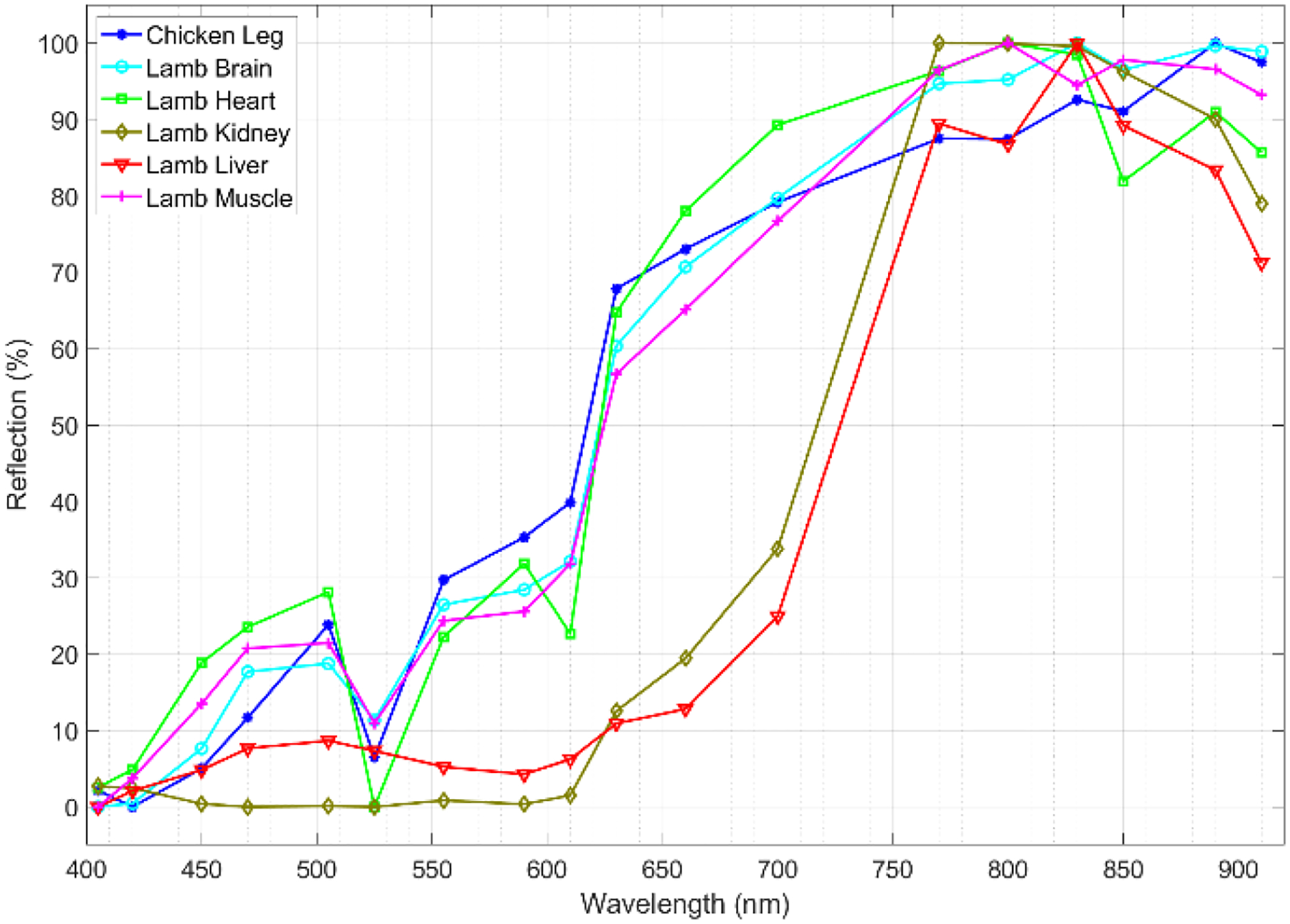
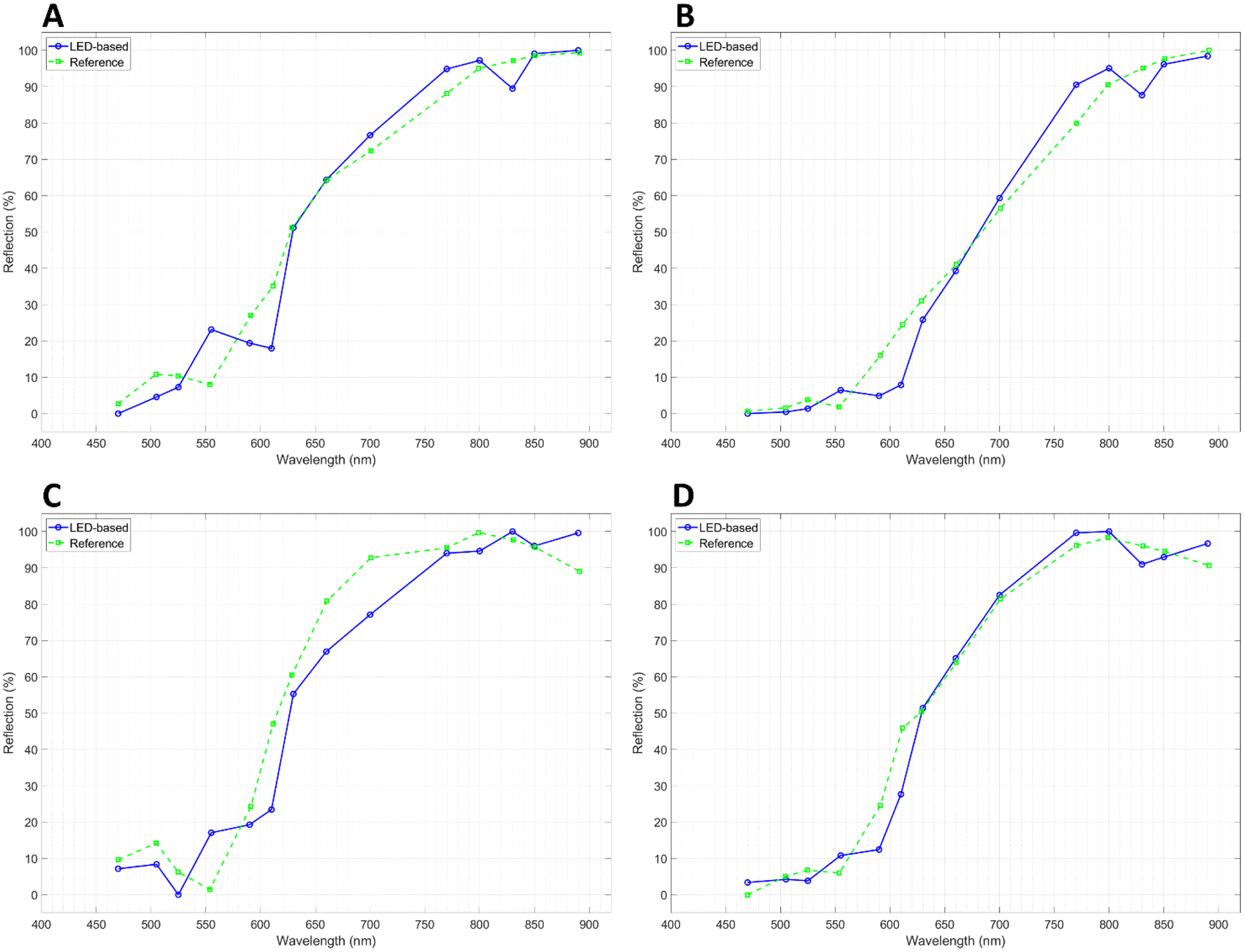
Hyperspectral endoscopy can offer multiple advantages as compared to conventional endoscopy. Our goal is to design and develop a real-time hyperspectral endoscopic imaging system for the diagnosis of gastrointestinal (GI) tract cancers using a micro-LED array as an in-situ illumination source. The wavelengths of the system range from ultraviolet to visible and near infrared. To evaluate the use of the LED array for hyperspectral imaging, we designed a prototype system and conducted ex vivo experiments using normal and cancerous tissues of mice, chicken, and sheep. We compared the results of our LED-based approach with our reference hyperspectral camera system. The results confirm the similarity between the LED-based hyperspectral imaging system and the reference HSI camera. Our LED-based hyperspectral imaging system can be used not only as an endoscope but also as a laparoscopic or handheld devices for cancer detection and surgery.

Keywords: FPGA; Hyperspectral imaging; cancer; endoscopy; handheld device; laparoscope; micro-LED; surgery.


Figures

Figure 1.
Figure 1.
Block diagram of the micro-LED-based hyperspectral endoscopic imaging system.
Figure 2.
Figure 2.
Signals for the LED array driver and control of the system.
Figure 3.
Figure 3.
Experiment setup (A) and the dark box (B) for the ex vivo tissue imaging experiments.
Figure 4.
Figure 4.
Imaging setup using our customized HSI camera system.
Figure 5.
Figure 5.
Spectral images of the white reference before calibration (left) and after calibration (right).
Figure 6.
Figure 6.
Spectral channels of our LED-based HSI prototype system (left images) and the reference HSI camera (right images) from a normal mouse kidney at eight sample bands.
Figure 7.
Figure 7.
Spectral channels of our LED-based HSI prototype system (left images) and the reference HSI camera (right images) from a normal mouse liver at eight sample bands.
Figure 8.
Figure 8.
Spectral channels of our LED-based HSI prototype system (left images) and the reference HSI camera (right images) from a normal lamb brain at eight sample bands.
Figure 9.
Figure 9.
Spectral channels of our LED-based HSI prototype system (left images) and the reference HSI camera (right images) from a neuroblastoma tumor resected from a mouse model at eight sample bands.
Figure 10.
Figure 10.
Spectral signatures of normal lamb and chicken tissues from our LED-based HSI system.
Figure 11.
Figure 11.
Spectral signatures of normal mouse tissues from our LED-based system. The corresponding bands are shown with markers on the graphs.
Figure 12.
Figure 12.
Spectral signature comparison between our LED-based HSI system and the reference system at corresponding wavelengths for the four sample tissues, which are shown in Figures 6 to 9. (A) Mouse kidney, (B) mouse liver, (C) lamb brain, and (D) neuroblastoma tumor.
Figure 13.
Figure 13.
Comparison between spectral signatures of our LED-based HSI system for normal mouse tissues and neuroblastoma tumors. (A) Normal kidney, (B) normal liver, and (C) normal heart vs neuroblastoma tumor. The asterisk symbols show the spectral channels with statistically significant differences between the normal and cancerous tissues (p < 0.05).

References

    1. Siegel RL, Miller KD, Fuchs HE, and Jemal A, “Cancer statistics, 2021,” CA: a cancer journal for clinicians, 71(1), 7–33 (2021). - PubMed
    1. Menon S, and Trudgill N, “How commonly is upper gastrointestinal cancer missed at endoscopy? A metaanalysis,” Endoscopy international open, 2(2), E46 (2014). - PMC - PubMed
    1. Chadwick G, Groene O, Riley S, Hardwick R, Crosby T, Hoare J, Hanna GB, Greenaway K, and Cromwell DA, “Gastric cancers missed during endoscopy in England,” Clinical gastroenterology and hepatology, 13(7), 1264–1270. e1 (2015). - PubMed
    1. Lu G, and Fei B, “Medical hyperspectral imaging: a review,” Journal of biomedical optics, 19(1), 010901 (2014). - PMC - PubMed
    1. Halicek M, Fabelo H, Ortega S, Callico GM, and Fei B, “In-vivo and ex-vivo tissue analysis through hyperspectral imaging techniques: revealing the invisible features of cancer,” Cancers, 11(6), 756 (2019). - PMC - PubMed

LinkOut - more resources