

Pattern of Endodontic Periapical Lesion Extension in Anterior Teeth: A CBCT Study in an Iranian Population
- PMID: 36794107
- PMCID: PMC9923413
- DOI: 10.22037/iej.v14i4.24188
Pattern of Endodontic Periapical Lesion Extension in Anterior Teeth: A CBCT Study in an Iranian Population
Abstract
Introduction: Health of periapical tissues has been considered as an index for the evaluation of endodontic outcomes. The present study sought to assess the pattern of periapical lesion extension in anterior teeth using cone-beam computed tomography (CBCT).
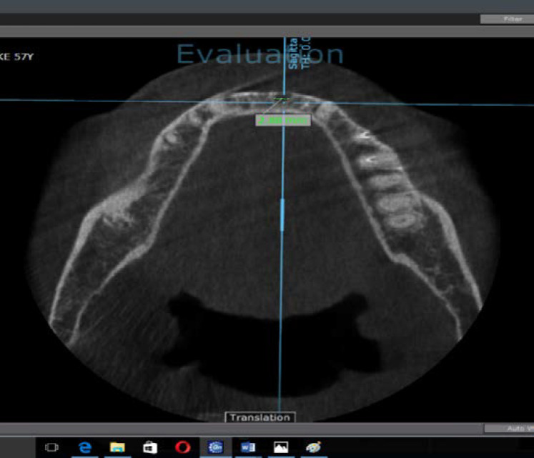
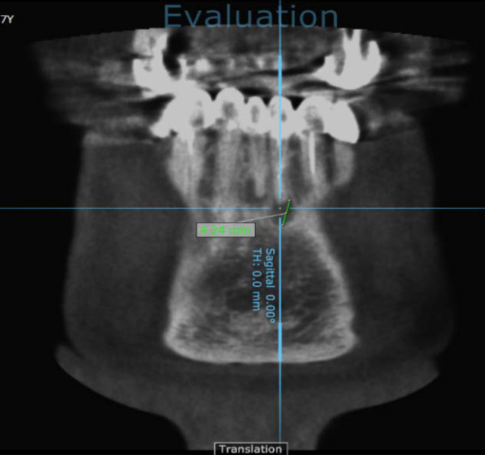
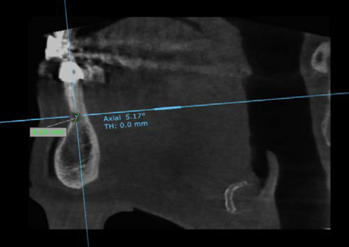
Methods and materials: In this descriptive study' 199 CBCT images belonged to patients aging from 15 to 79 were assessed according to periapical lesion extension in the regions of maxillary and mandibular anterior teeth. Maximum periapical lesion extensions in three orthogonal planes were measured and recorded in millimeters and were assessed according to age' gender' dental arch and tooth type. Statistical analysis was performed using percentages' repeated measure ANOVA and Bonferroni tests. The significant level was set at 0.05.
Results: The highest average of lesion extension, in both maxilla and mandible, was in vertical dimension' followed by horizontal buccolingual and horizontal mesiodistal dimensions' respectively. There were significant differences between the vertical and mesiodistal (P=0.004) and also mesiodistal and buccolingual (P=0.010) periapical lesion extension dimensions. In addition, there were significant differences in maxilla and mandible (P=0.012). In maxilla, there were no significant differences between the three tooth types (P=0.346) but in the mandibular arch, there were significant differences between central-canine (P=0.004) and lateral-canine (P=0.026). According to independent variables, only gender had a significant effect on the lesion extension in anterior regions of maxilla and mandible (P=0.001). The periapical lesion extensions were significantly higher in men compared with women.
Conclusions: The bone destruction_as a consequence of periapical inflammatory process_ was greatest in the vertical, and lowest in the horizontal mesiodistal dimensions. That way, the extension in buccolingual dimension, which could not be detected in the 2-D imaging techniques, was rather high in the present study. Thus CBCT, as a 3-D imaging technique, could be recommended for the precise evaluation of lesion extension in the periapical area.
Keywords: Cone-beam Computed Tomography; Diagnosis; Endodontics; Periapical Disease; Periapical Lesion; Periapical Periodontitis.
Conflict of interest statement
‘None declared’.
Figures
References
-
- Kakehashi S, Stanley HR, Fitzgerald RJ. The effects of surgical exposures of dental pulps in germ-free and conventional laboratory rats. Oral Surg Oral Med Oral Pathol. 1965;20:340–9. - PubMed
-
- Sabeti M, Valles Y, Nowzari H, Simon JH, Kermani-Arab V, Slots J. Cytomegalovirus and Epstein-Barr virus DNA transcription in endodontic symptomatic lesions. Oral Microbiol Immunol. 2003;18(2):104–8. - PubMed
-
- Hahn CL, Liewehr FR. Innate immune responses of the dental pulp to caries. J Endod. 2007;33(6):643–51. - PubMed
-
- Bender IB, Seltzer S. Roentgenographic and direct observation of experimental lesions in bone: II 1961. J Endod. 2003;29(11):707–12; discussion 1. - PubMed
LinkOut - more resources
Full Text Sources