. 2023 Apr 15;25(4):1369-1378.
doi: 10.1093/europace/euad030.

Preclinical assessment of the feasibility, safety and lesion durability of a novel 'single-shot' pulsed field ablation catheter for pulmonary vein isolation

Affiliations

Preclinical assessment of the feasibility, safety and lesion durability of a novel 'single-shot' pulsed field ablation catheter for pulmonary vein isolation

Jacob Koruth et al. Europace. .

Abstract

Aims: Single-shot pulmonary vein isolation can improve procedural efficiency. To assess the capability of a novel, expandable lattice-shaped catheter to rapidly isolate thoracic veins using pulsed field ablation (PFA) in healthy swine.

Methods and results: The study catheter (SpherePVI; Affera Inc) was used to isolate thoracic veins in two cohorts of swine survived for 1 and 5 weeks. In Experiment 1, an initial dose (PULSE2) was used to isolate the superior vena cava (SVC) and the right superior pulmonary vein (RSPV) in six swine and the SVC only in two swine. In Experiment 2, a final dose (PULSE3) was used for SVC, RSPV, and left superior pulmonary vein (LSPV) in five swine. Baseline and follow-up maps, ostial diameters, and phrenic nerve were assessed. Pulsed field ablation was delivered atop the oesophagus in three swine. All tissues were submitted for pathology. In Experiment 1, all 14/14 veins were isolated acutely with durable isolation demonstrated in 6/6 RSPVs and 6/8 SVC. Both reconnections occurred when only one application/vein was used. Fifty-two and 32 sections from the RSPVs and SVC revealed transmural lesions in 100% with a mean depth of 4.0 ± 2.0 mm. In Experiment 2, 15/15 veins were isolated acutely with 14/15 veins (5/5 SVC, 5/5 RSPV, and 4/5 LSPV) durably isolated. Right superior pulmonary vein (31) and SVC (34) sections had 100% transmural, circumferential ablation with minimal inflammation. Viable vessels and nerves were noted without evidence of venous stenosis, phrenic palsy, or oesophageal injury.

Conclusion: This novel expandable lattice PFA catheter can achieve durable isolation with transmurality and safety.

Keywords: Atrial fibrillation; Catheter ablation; Electroporation • Oesophageal injury; Pulmonary vein; Single shot.


Conflict of interest statement

Conflict of interest: Drs Koruth and Reddy have received research grants from Affera, Inc. Drs Koruth and Reddy also hold stock options in Affera, Inc. A comprehensive list of other financial disclosures unrelated to this manuscript is included in Supplement. All other authors have no conflicts of interest relevant to this study.

Figures

Graphical Abstract
Graphical Abstract
Figure 1
Figure 1
Study catheter and electroanatomic visualization. (A) Expandable nitinol-based lattice frame tip (expandable up to 34 mm in diameter) with a 7.5 Fr shaft. (B) The catheter can be visualized within an anatomical map of the left atrium (within the RSPV). (C) RA anatomical shell collected with the study catheter visualized within the chamber. (D) The catheter visualized within a bipolar voltage electroanatomical map. RA, right atrium; RSPV, right superior pulmonary vein. LAA, left atrial appendage; RAA, right atrial appendage; LSPV, left superior pulmonary vein; ICPV, inferior common pulmonary vein; RL, right lateral; PA, postero-anterior.
Figure 2
Figure 2
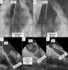
Fluoroscopic views. (A–C) Expanded catheter-tip positioned in SVC, RSPV, LSPV over the wire. Note slight deformation of the catheter as it makes the turn from the transeptal to the RSPV in (B). Coronary sinus catheter is present in all panels. LSPV, left superior pulmonary vein; RSPV, right superior pulmonary vein; SVC, superior vena cava.
Figure 3
Figure 3
Representative electroanatomic maps and electrograms. (A) Baseline, immediate post ablation, and follow-up bipolar voltage maps (0.1–1 mV) of LA demonstrating durably isolated RSPV. (B) Baseline, immediate post ablation and follow-up voltage maps of RA demonstrating durably isolated SVC. (C) Voltage map of LA demonstrating durable isolation of RSPV and LSPV. (D) Changes in electrograms obtained from the study catheter before and immediately after PFA. LA, left atria; LSPV, left superior pulmonary vein; LL, left lateral; PA, posterior anterior; RA, right atria; RSPV, right superior pulmonary vein; SVC, superior vena cava.
Figure 4
Figure 4
Fluoroscopic views and intracardiac echocardiographic views. (A and B) Baseline and 5-week follow-up venous angiograms of the superior vena cava after durable isolation demonstrating no venous stenosis. (C–E) Baseline view of the SVC, catheter-tip positioned in the SVC and the SVC immediately post ablation. Note the slight reduction in the lumen between (C) and (E) (double arrows) post ablation. SVC, superior vena cava.
Figure 5
Figure 5
Gross pathology prior to fixation. ( A and B) The external anterior and lateral view of an ablated SVC. The lesion is outlined with the white dotted line. (C) Internal view of an opened left atrium, demonstrating a circumferential lesion (outlined with the white dotted line) around the RSPV and LSPV. LSPV, left superior pulmonary vein; RSPV, right superior pulmonary vein; - SVC, superior vena cava.
Figure 6
Figure 6
Gross pathology post formalin fixation. (A) The luminal view of an ablated SVC (opened anteriorly) that was shown to have reconnected at follow-up. The lesion (pale region) is outlined with the white dotted line. The white arrow points to an area within the lesion boundary that is partially spared as evident by its darker appearance. The yellow dotted lines are shown to demonstrate where sections, if taken, could miss a region of non-contiguous lesion. (B and C) The lesion is outlined and can be seen to encompass the entire circumference of the SVC and right superior pulmonary vein. SVC, superior vena cava.
Figure 7
Figure 7
Histology: Masson’s trichrome (MT) staining—blue, fibrosis; pink, healthy myocardium. Experiment 1: (A and B) Demonstrates transmural lesions in the SVC and RSPV. (C) Section from SVC in animal with one application/vein that was noted to have reconnected: Small islands of spared myocardium can be seen. (D and E) Experiment 2: Higher magnification sample image of the section of RSPV demonstrating transmural ablation with viable blood vessels and nerves (D) and a transmural wide ablation of the antral wall (E). RSPV, right superior pulmonary vein; SVC, superior vena cava.

References

    1. Koruth J, Kuroki K, Iwasawa J, Enomoto Y, Viswanathan R, Brose Ret al. . Preclinical evaluation of pulsed field ablation: electrophysiological and histological assessment of thoracic vein isolation. Circ Arrhythm Electrophysiol 2019;12:e007781. - PMC - PubMed
    1. Koruth JS, Kuroki K, Kawamura I, Stoffregen WC, Dukkipati SR, Neuzil Pet al. . Focal pulsed field ablation for pulmonary vein isolation and linear atrial lesions: a preclinical assessment of safety and durability. Circ Arrhythm Electrophysiol 2020;13:e008716. - PubMed
    1. Bohnen M, Weber R, Minners J, Jadidi A, Eichenlaub M, Neumann FJet al. . Characterization of circumferential antral pulmonary vein isolation areas resulting from pulsed-field catheter ablation. Europace 2022;24:1248–55. - PMC - PubMed
    1. Stewart MT, Haines DE, Verma A, Kirchhof N, Barka N, Grassl Eet al. . Intracardiac pulsed field ablation: proof of feasibility in a chronic porcine model. Heart Rhythm 2019;16:754–64. - PubMed
    1. Yavin H, Brem E, Zilberman I, Shapira-Daniels A, Datta K, Govari Aet al. . Circular multielectrode pulsed field ablation catheter lasso pulsed field ablation: lesion characteristics, durability, and effect on neighboring structures. Circ Arrhythm Electrophysiol 2021;14:e009229. - PMC - PubMed

Grants and funding