. 2023 Apr 20;8(2):e0001523.
doi: 10.1128/msphere.00015-23. Epub 2023 Feb 16.

The Incompetence of Mosquitoes-Can Zika Virus Be Adapted To Infect Culex tarsalis Cells?

Affiliations

The Incompetence of Mosquitoes-Can Zika Virus Be Adapted To Infect Culex tarsalis Cells?

Emily N Gallichotte et al. mSphere. .

Abstract

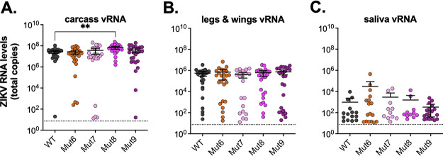
The molecular evolutionary mechanisms underpinning virus-host interactions are increasingly recognized as key drivers of virus emergence, host specificity, and the likelihood that viruses can undergo a host shift that alters epidemiology and transmission biology. Zika virus (ZIKV) is mainly transmitted between humans by Aedes aegypti mosquitoes. However, the 2015 to 2017 outbreak stimulated discussion regarding the role of Culex spp. mosquitoes in transmission. Reports of ZIKV-infected Culex mosquitoes, in nature and under laboratory conditions, resulted in public and scientific confusion. We previously found that Puerto Rican ZIKV does not infect colonized Culex quinquefasciatus, Culex pipiens, or Culex tarsalis, but some studies suggest they may be competent ZIKV vectors. Therefore, we attempted to adapt ZIKV to Cx. tarsalis by serially passaging virus on cocultured Ae. aegypti (Aag2) and Cx. tarsalis (CT) cells to identify viral determinants of species specificity. Increasing fractions of CT cells resulted in decreased overall virus titer and no enhancement of Culex cell or mosquito infection. Next-generation sequencing of cocultured virus passages revealed synonymous and nonsynonymous variants throughout the genome that arose as CT cell fractions increased. We generated nine recombinant ZIKVs containing combinations of the variants of interest. None of these viruses showed increased infection of Culex cells or mosquitoes, demonstrating that variants associated with passaging were not specific to increased Culex infection. These results reveal the challenge of a virus adapting to a new host, even when pushed to adapt artificially. Importantly, they also demonstrate that while ZIKV may occasionally infect Culex mosquitoes, Aedes mosquitoes likely drive transmission and human risk. IMPORTANCE ZIKV is mainly transmitted between humans by Aedes mosquitoes. In nature, ZIKV-infected Culex mosquitoes have been found, and ZIKV infrequently infects Culex mosquitoes under laboratory conditions. Yet, most studies show that Culex mosquitoes are not competent vectors for ZIKV. We attempted to adapt ZIKV to Culex cells to identify viral determinants of species specificity. We sequenced ZIKV after it was passaged on a mixture of Aedes and Culex cells and found that it acquired many variants. We generated recombinant viruses containing combinations of the variants of interest to determine if any of these changes enhance infection in Culex cells or mosquitoes. Recombinant viruses did not show increased infection in Culex cells or mosquitoes, but some variants increased infection in Aedes cells, suggesting adaptation to those cells instead. These results reveal that arbovirus species specificity is complex, and that virus adaptation to a new genus of mosquito vectors likely requires multiple genetic changes.

Keywords: arbovirus; mosquito; species specificity; virology.


Conflict of interest statement

The authors declare no conflict of interest.

Figures

FIG 1
FIG 1
Low level replication of ZIKV in CT cells. Cx. tarsalis CT cells were infected at an MOI of 20 with ZIKV PRVABC59 virus, supernatant collected daily, and analyzed for (A) viral RNA via qRT-PCR and (B) infectious virus via plaque assays. Experiment was performed in biological triplicate (mean ± standard deviation).
FIG 2
FIG 2
ZIKV passaging on Aag2 and CT cells. (A) Schematic of passaging experimental design. Ae. aegypti-derived Aag2 and Cx. tarsalis-derived CT cells were mixed at an initial ratio of 90:10, infected with ZIKV PRVABC59 isolate for 6 days, then culture supernatant was passaged onto new cells. The ratio of Aag2:CT slowly decreased to a final 10:90 ratio over the course of 18 passages. Passaging experiment was performed in biological triplicate. (B) After each passage, supernatant was assayed for infectious virus. (C–D) Cx. tarsalis CT cells were infected at an MOI of 20 with each triplicate of ZIKV passage 18 coculture virus that was passaged once on Vero cells (Co18.1V, Co18.2V and Co18.3V), supernatant collected daily, and analyzed for (C) viral RNA via qRT-PCR and (D) infectious virus via plaque assays. Growth curve experiment was performed in biological triplicate (mean ± standard deviation). Dashed line represents limit of detection. Graphics were generated using BioRender.com.
FIG 3
FIG 3
SNVs associated with ZIKV passaging on Aag2/CT cocultures. (A) Schematic overview of SNVs of interest along the ZIKV PRVABC59 genome. (B–D) Proportion of SNVs out of total virus population (i.e., variant frequencies) for three mutations of interest (B) T1435A, (C) C7460T, (D) and C9800T. Variant frequencies for each passage and each independent passaging replicate are shown. Co18V indicates the Vero propagated virus stock generated by inoculation of Vero cells with Co18 culture supernatant. Dashed line represents 0.5.
FIG 4
FIG 4
Design and characterization of ZIKV recombinant viruses. (A) Schematic of ZIKV genome, with synonymous (blue) and nonsynonymous (red) variants of interest shown as lines. Genome location and type (synonymous versus nonsynonymous) of variants of each of the recombinant viruses is shown (Mut1-9). (B) Recombinant viruses were recovered, stocks generated, and assayed for infectious virus titer (performed in technical duplicate, mean ± 95% confidence intervals). (C) Plaque morphology of ZIKV recombinant viruses on Vero cells.
FIG 5
FIG 5
ZIKV mutants do not infect CT cells. Cx. tarsalis CT cells were infected at an MOI of 5 with ZIKV recombinant mutants, supernatant collected daily, and analyzed for (A) viral RNA via qRT-PCR and (B) infectious virus via plaque assays. Experiment was performed in biological triplicate (mean ± standard deviation). Dashed line represents limit of detection.
FIG 6
FIG 6
Increased replication of ZIKV recombinant viruses in Aag2 cells. Ae. aegypti-derived Aag2 cells were infected at an MOI of 5 with ZIKV recombinant viruses, supernatant collected daily, and analyzed for (A) viral RNA via qRT-PCR and (B) infectious virus via plaque assays. Recombinant viruses are grouped by the general location of their SNVs. Experiment was performed in biological triplicate (mean ± 95% confidence intervals). Two-way ANOVA with Dunnett’s multiple comparison was performed comparing each recombinant virus to WT at each time point. Only statistically significant relationships are shown (*, P < 0.05; **, P < 0.01; ***, P < 0.005).
FIG 7
FIG 7
ZIKV recombinant viruses in Ae. aegypti mosquitoes. Ae. aegypti mosquitoes were infected with ZIKV recombinant viruses, and at day 7 salivated, dissected, and (A) carcass, (B) legs and wings, and (C) saliva were collected. Levels of viral RNA in mosquito tissues and saliva were quantified by qRT-PCR (mean ± 95% confidence intervals). Dashed line represents the limit of detection. Infection was performed in groups of 30 mosquitoes, only samples with detectable levels of virus are shown. One-way ANOVA with Dunnett’s multiple comparison was performed comparing each recombinant virus to WT. Only statistically significant relationships are shown (**, P < 0.005). Dashed line represents limit of detection.

References

    1. Dick GW, Kitchen SF, Haddow AJ. 1952. Zika virus. I. Isolations and serological specificity. Trans R Soc Trop Med Hyg 46:509–520. doi:10.1016/0035-9203(52)90042-4. - DOI - PubMed
    1. Duffy MR, Chen TH, Hancock WT, Powers AM, Kool JL, Lanciotti RS, Pretrick M, Marfel M, Holzbauer S, Dubray C, Guillaumot L, Griggs A, Bel M, Lambert AJ, Laven J, Kosoy O, Panella A, Biggerstaff BJ, Fischer M, Hayes EB. 2009. Zika virus outbreak on Yap Island, Federated States of Micronesia. N Engl J Med 360:2536–2543. doi:10.1056/NEJMoa0805715. - DOI - PubMed
    1. Faye O, Freire CC, Iamarino A, Faye O, de Oliveira JV, Diallo M, Zanotto PM, Sall AA. 2014. Molecular evolution of Zika virus during its emergence in the 20th century. PLoS Negl Trop Dis 8:e2636. doi:10.1371/journal.pntd.0002636. - DOI - PMC - PubMed
    1. Cao-Lormeau VM, Roche C, Teissier A, Robin E, Berry AL, Mallet HP, Sall AA, Musso D. 2014. Zika virus, French Polynesia, South Pacific, 2013. Emerg Infect Dis 20:1085–1086. doi:10.3201/eid2006.140138. - DOI - PMC - PubMed
    1. Petersen LR, Jamieson DJ, Powers AM, Honein MA. 2016. Zika Virus. N Engl J Med 374:1552–1563. doi:10.1056/NEJMra1602113. - DOI - PubMed

Publication types