Review
. 2023 May;78(5):1148-1168.
doi: 10.1111/all.15678. Epub 2023 Feb 26.

Protease allergens as initiators-regulators of allergic inflammation

Affiliations
Review

Protease allergens as initiators-regulators of allergic inflammation

Wai Tuck Soh et al. Allergy. 2023 May.

Abstract

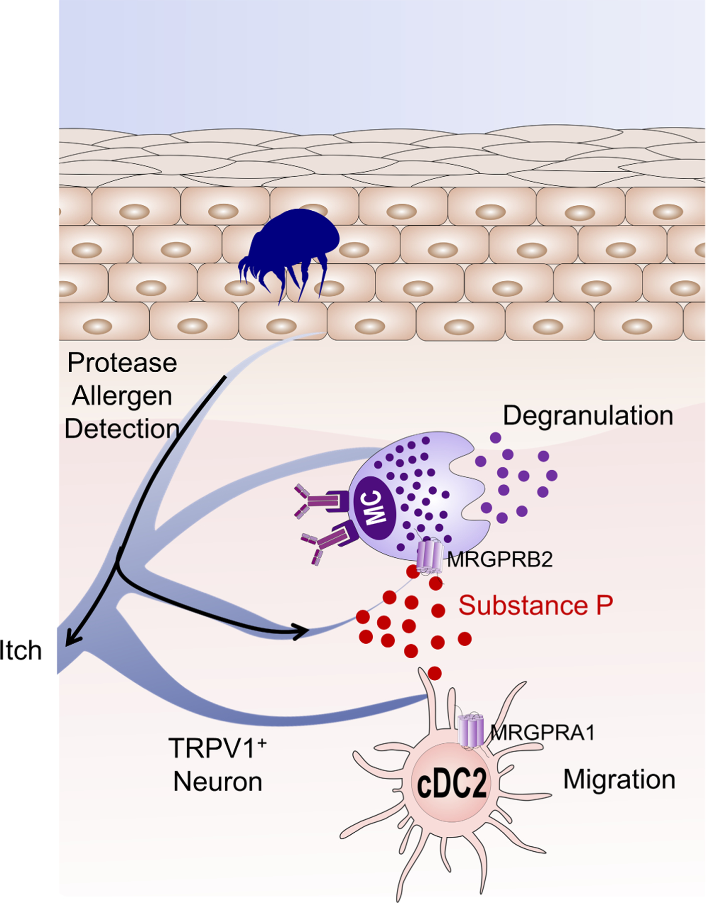
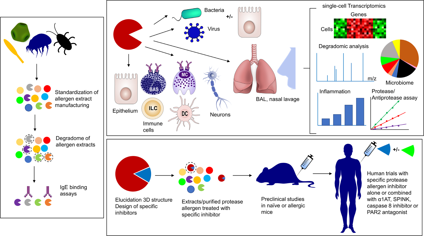
Tremendous progress in the last few years has been made to explain how seemingly harmless environmental proteins from different origins can induce potent Th2-biased inflammatory responses. Convergent findings have shown the key roles of allergens displaying proteolytic activity in the initiation and progression of the allergic response. Through their propensity to activate IgE-independent inflammatory pathways, certain allergenic proteases are now considered as initiators for sensitization to themselves and to non-protease allergens. The protease allergens degrade junctional proteins of keratinocytes or airway epithelium to facilitate allergen delivery across the epithelial barrier and their subsequent uptake by antigen-presenting cells. Epithelial injuries mediated by these proteases together with their sensing by protease-activated receptors (PARs) elicit potent inflammatory responses resulting in the release of pro-Th2 cytokines (IL-6, IL-25, IL-1β, TSLP) and danger-associated molecular patterns (DAMPs; IL-33, ATP, uric acid). Recently, protease allergens were shown to cleave the protease sensor domain of IL-33 to produce a super-active form of the alarmin. At the same time, proteolytic cleavage of fibrinogen can trigger TLR4 signaling, and cleavage of various cell surface receptors further shape the Th2 polarization. Remarkably, the sensing of protease allergens by nociceptive neurons can represent a primary step in the development of the allergic response. The goal of this review is to highlight the multiple innate immune mechanisms triggered by protease allergens that converge to initiate the allergic response.

Keywords: IL-33; protease allergen; epithelial cell; protease-activated receptor; sensory neuron.


Conflict of interest statement

C.L.S is a paid consultant (expert witness) for Bayer and Merck. C.R. and J.Z. are inventors on patents granted in multiple territories concerning allergens, protease inhibitor design and composition of matter, and medical uses thereof. St George’s, University of London is beneficial owner of this patent estate. C.R. is co-founder and holds equity in Aldezon Ltd. M.E.R. is a consultant for Pulm One, Spoon Guru, ClostraBio, Serpin Pharm, Allakos, Celldex, Nextstone One, Bristol Myers Squibb, Astra Zeneca, Ellodi Pharma, GlaxoSmith Kline, Regeneron/Sanofi, Revolo Biotherapeutics, and Guidepoint and has an equity interest in the first seven listed, and royalties from reslizumab (Teva Pharmaceuticals), PEESSv2 (Mapi Research Trust) and UpToDate. M.E.R. is an inventor of patents owned by Cincinnati Children’s Hospital. The remaining authors declare that they have no conflicts of interest.

Figures

FIGURE 1
FIGURE 1. The identified protease allergens inducing innate immune activation are cysteine or serine proteases.
Proteases can be classified according to the location of the cleavage site in their putative substrates (A) and to the nature of their active site residues (B). With the exception of bee venom dipeptidyl peptidase allergens, all the identified protease allergens are endopeptidases (Table S1). The arrows point to the cleavage site on the protein substrate and the red circles the terminal amino acids or dipeptides released by the proteolysis. Most of cysteine and serine protease allergens share structural homologies with Der p 1 and Trypsin, respectively. The insets of panel B shows the key residues of their respective active site (Cysteine–Histidine–Asparagine or Serine–Histidine–Aspartic acid for Der p 1 and trypsin, respectively). Structures taken from the Protein Data Bank (PDB, https://www.rcsb.org/) and figure generated using Pymol.
FIGURE 2
FIGURE 2. General representation of non-IgE mediated innate inflammatory pathways mediated by protease allergens on exposed epithelial and immune cells.
Protease allergens can alter epithelial integrity through degradations of tight (TJ) or adherens junctions (AJ) which facilitates their penetration into submucosal tissues. The junction injuries of bronchiolar club cells are sensed by mechanosensor TRPV4. Epithelial damages are initiated by protease allergens (red-colored enzyme symbol) through their capacity to alter the antiprotease network and to degrade surfactant proteins. Serine protease allergens or thrombin (peach-colored enzyme symbol) trigger protease-activated receptor (PAR) activation. Cleavage of fibrinogen by protease allergens or by thrombin matured by protease allergens elicits TLR4 activation. Activated and/or damaged epithelium produces intracellular ROS leading to the release a large collection of proinflammatory cytokines (IL-6, GM-CSF, TSLP, IL-25), DAMP (IL-1α, IL-33, Uric acid, ATP), and chemokines (CCL2) which activate ILC2 and cDC2 cells. IL-4 produced by activated ILC2s initiates the Th2 differentiation. The production of IL-4, IL-5, IL-13 by Th2 cells and ILC2s induces the production of allergen-specific IgE, the associated sensitization/degranulation of basophils (BAS)/mast cells (MC) and the recruitment of eosinophils. ILC2-derived IL-13 drives goblet cell hyperplasia. The contacts of protease allergens with immune cells, facilitated by the epithelial barrier degradation, allow the cleavage of surface receptors (CD23, CD25, CD40, DC-SIGN/DC-SIGNR) on these target cells which optimizes the Th2-biased inflammatory response. AJ, Adherens Junction; AT, antitrypsin; CCL2, C-C Motif Chemokine Ligand 2; cDC, conventional dendritic cell; cDC2c, type 2 conventional DC; IDO, indoleamine 2,3-dioxygenase; MD2, myeloid differentiation factor 2; PAR, protease-activated receptor; ROS, reactive oxygen species; SLPI, secretory leukocyte protease inhibitor; SP-A/D, surfactant protein-A/D; SPINK 5/7, serine protease inhibitor Kazal type 5 or 7; TJ, tight junction; TLR4, Toll-like receptor 4; TRPV4, Transient receptor potential vanilloid-type 4; TSLP, thymic stromal lymphopoietin.
FIGURE 3
FIGURE 3. Activation of redox-dependent signaling by the prothrombinase activity of Der p 1 and its inhibition by Der p 1 class allergen delivery inhibitors.
Thrombin generation enables the canonical activation of PAR1 and PAR4. In addition to their canonical GPCR signaling, this initiates a signaling network involving epidermal growth factor receptor (EGFR), pannexons and the TLR4-dependent intracellular generation of reactive oxidant species (ROS). This network also receives activating inputs from sensors detecting viral RNA. All these effects can be attenuated by compounds which have been designed to inhibit group 1 allergens potently and selectively. Illustratively, a favored molecular scaffold for inhibitor design is shown. Variation of substituents at positions P1′–P4 (Schechter–Berger nomenclature) tunes the interaction with the binding pockets (S1′–S4) of the protease allergen and confers other essential developability credentials. ADAM10, activation of a disintegrin and metalloprotease 10; EGFR, epidermal growth factor receptor; MDA-5, melanoma differentiation-associated protein 5; PAR, protease-activated receptor; RIG-1, retinoic acid-inducible gene I; ROS, reactive oxygen species; TLR3, Toll-like receptor 3.
FIGURE 4
FIGURE 4. Activation of Protease-activated receptor 2 (PAR2) by trypsin/KLKs or trypsin-like protease allergens.
(A) PAR2 cleavage by trypsin/KLK or serine protease allergens unmasks PAR2’s tethered ligand which interacts with the extracellular loop 2 domain to initiate receptor signaling via the G-proteins, Gq, G12/13 and Gi. Similar effects are triggered by a synthetic tethered ligand-mimicking peptide. (B) Differential cleavage of the N-terminus of rat PAR2 by trypsin and Der p 3, which unmasks the ‘canonical’ tethered receptor-activating ligand versus Der p 1, that cleaves primarily at a distinct site to ‘disarm’ the receptor for trypsin activation. The solid and dotted blue arrows indicate major and minor cleavage sites, respectively, for Der p 1. KLK, kallikrein.
FIGURE 5
FIGURE 5. IL-33 maturation by intracellular RipIL-33 pathway and by the cleavage of IL-33 protease sensor domain exposed to protease allergens or endogenous proteases.
Allergens from different sources trigger the caspase 8–ripoptosome signaling pathway to activate and release mature IL-33 (mIL-33). This RipIL-33 intracellular mechanism is not dependent on PAR2, protease activity of allergens or epithelial damage. In contrast, allergen-induced caspase 8 maturation is required for the downstream cleavage of caspase 3/7. Active caspase 3/7 cleaves the histone-bound full-length IL-33 (FLIL-33) within the IL-1-like domain. The resulting mIL-33 biospecies bound to histone is released and activate the cognate receptor ST2. Both mIL-33 forms had higher bioactivity than FLIL-33. In response to allergens, epithelial cells can release as well histone-bound FLIL-33. Environmental protease allergens from diverse sources (HDM, fungi, pollen, papain) as well as endogenous proteases released from innate effector cells during inflammation (Elastase, Cathepsin G, Granzyme B, Tryptase, chymase) can activate IL-33 maturation through cleavages of the protease sensor domain. Such mIL-33 forms have increased ST2 binding activity compared with FLIL-33.
FIGURE 6
FIGURE 6. Sensing of protease allergens by TRPV1+ nociceptors.
Cysteine and serine protease allergens from papaya, HDM, and Alternaria alternata are detected directly by TRPV1+ sensory neurons in the skin leading to itch and the local release of Substance P. Substance P acts directly on mast cells through their expression of MRGPRB2, leading to mast cell degranulation. Substance P also acts on cDC2 through their expression of MRGPRA1, leading to their migration to the draining lymph node where they initiate Th2 differentiation. cDC2, type 2 conventional DC; MRGPR, Mas-related G protein-coupled receptors; TRPV1, Transient receptor potential vanilloid-type 1.
FIGURE 7
FIGURE 7. Multidisciplinary approaches to tackle important research questions about protease allergens.
Degradome analysis combined with IgE binding assays is needed to elucidate the protease allergen repertoire of allergenic extracts. Signaling pathways activated by protease allergens and their protein substrate repertoire can be fully elucidated by single-cell transcriptomics, degradomics and using different human cell samples (epithelial, immune or neuronal cells). The effects of protease allergens on endosymbiotic or pathogenic bacteria, viruses should be investigated to measure their impact on microbiome and microbial virulence. Proteolytic degradation of the airway antiprotease network can be measured from nasal, bronchoalveolar lavage samples. Specific inhibitors can be designed based on the structure of protease allergens, paving the way for innovative therapeutic intervention. Their preclinical and clinical evaluation, alone or in combination with α1AT/SPINK replacement therapy, caspase 8 inhibitor targeting RipIL-33 pathway or PAR2 antagonist, can pave the way to more effective management of allergic diseases. AT, antitrypsin; SPINK, serine protease inhibitor Kazal.

Similar articles

Cited by

References

    1. Akdis CA, Arkwright PD, Bruggen MC, et al. Type 2 immunity in the skin and lungs. Allergy. 2020;75(7):1582–1605. doi:10.1111/all.14318 - DOI - PubMed
    1. Caraballo L, Valenta R, Puerta L, et al. The allergenic activity and clinical impact of individual IgE-antibody binding molecules from indoor allergen sources. World Allergy Organ J. 2020;13(5):100118. doi:10.1016/j.waojou.2020.100118 - DOI - PMC - PubMed
    1. Sokol CL, Barton GM, Farr AG, Medzhitov R. A mechanism for the initiation of allergen-induced T helper type 2 responses. Nat Immunol. 2008;9(3):310–318. doi:10.1038/ni1558 - DOI - PMC - PubMed
    1. Kheradmand F, Kiss A, Xu J, Lee SH, Kolattukudy PE, Corry DB. A protease-activated pathway underlying Th cell type 2 activation and allergic lung disease. J Immunol. 2002;169(10):5904–5911. doi:10.4049/jimmunol.169.10.5904 - DOI - PubMed
    1. Phipps S, Lam CE, Kaiko GE, et al. Toll/IL-1 signaling is critical for house dust mite-specific helper T cell type 2 and type 17 [corrected] responses. Am J Respir Crit Care Med. 2009;179(10):883–893. doi:10.1164/rccm.200806-974OC - DOI - PubMed

Publication types