Comment
. 2023 Mar 6;222(3):e202211044.
doi: 10.1083/jcb.202211044. Epub 2023 Feb 16.

Mitochondria on the move: Horizontal mitochondrial transfer in disease and health

Affiliations
Comment

Mitochondria on the move: Horizontal mitochondrial transfer in disease and health

Lan-Feng Dong et al. J Cell Biol. .

Abstract

Mammalian genes were long thought to be constrained within somatic cells in most cell types. This concept was challenged recently when cellular organelles including mitochondria were shown to move between mammalian cells in culture via cytoplasmic bridges. Recent research in animals indicates transfer of mitochondria in cancer and during lung injury in vivo, with considerable functional consequences. Since these pioneering discoveries, many studies have confirmed horizontal mitochondrial transfer (HMT) in vivo, and its functional characteristics and consequences have been described. Additional support for this phenomenon has come from phylogenetic studies. Apparently, mitochondrial trafficking between cells occurs more frequently than previously thought and contributes to diverse processes including bioenergetic crosstalk and homeostasis, disease treatment and recovery, and development of resistance to cancer therapy. Here we highlight current knowledge of HMT between cells, focusing primarily on in vivo systems, and contend that this process is not only (patho)physiologically relevant, but also can be exploited for the design of novel therapeutic approaches.


Conflict of interest statement

Disclosures: The authors declare no competing interests exist.

Figures

Figure 1.
Figure 1.
Movement of mitochondria between MSCs and B16 ρ0 melanoma cells. MSCs isolated from a transgenic Su9DsRed mouse expressing RFP in their mitochondria were co-cultured with B16 ρ0 cells labeled with BFP targeted to nuclei and GFP targeted to the plasma membrane. Confocal microscopy shows the presence of RFP mitochondria in a TNT connecting an MSC and a B16 ρ0 cell (Dong et al., 2017).
Figure 2.
Figure 2.
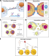
Models of horizontal transfer of mitochondria. (A) Horizontal transfer of mitochondria by TNTs. TNTs are in general formed by F-actin filaments. In case of intercellular transfer of mitochondria, TNTs also contain microtubules and are thicker, thus able to transport bulkier structures. Transport of mitochondria along these cytoskeletal elements is propelled by dynein and kinesin motor complexes consisting of several adaptor proteins, such as Miro1 or Miro2, that are integrated in the outer mitochondrial membrane and facilitate mitochondrial transport not only along microtubules but also along actin filaments (together with Myo19). Formation of TNTs starts either as an actin-driven protrusion of the cell membranes of the two cells involved or as the dislodgement of two previously attached cells that during the partition from each other form the TNT. (B) Horizontal transfer of mitochondria by gap junctions. Gap junctions were shown to involve connexin structures, i.e., protein complexes consisting of six subunits of connexin proteins, such as Cx43. Two juxtapositioned connexon channels form pores connecting two neighboring cells, allowing for bidirectional transport of ions, signaling molecules or whole mitochondria. (C) Horizontal transfer of mitochondria by cell fusion. Cell fusion is a process that is relatively common in cancer progression and comprises several steps. At first two cells are recognized via the so-called fusogenic trigger that could involve different signaling molecules depending on the cell type (TNFα, IL-1, IL-4, and others) or specific conditions (e.g., hypoxia). The cells then approach each other and, using several cell–cell adhesion molecules, such as E-cadherin, syncytin-1 and -2, or ASCT2, which form pore expansions, “fusion” of the two neighboring cells occurs. This yields a cell that shares mitochondria from the two original cells. During this process, several signaling pathways are triggered that result in higher tumorigenesis and increased metastatic potential of the cancer cells. (D) Horizontal transfer of mitochondria by ECVs. Transfer of mitochondria via ECVs involves small double membrane structures that are formed by blebbing of plasma membrane. In contrast, exosomes are of endosomal origin and can transport various cargo including signaling molecules (different metabolites), trans-membrane proteins, nucleic acids, amino acids, and organelle fragments. (E) Inter-organ transport of mitochondria. This mode of mitochondrial horizontal transfer has been recently shown for the organelles moving from adipocytes to the heart tissue. In this particular case, damaged mitochondria from energetically stressed adipocytes of obese patients are transferred via small extracellular vesicles (sEV) to the blood circulation and are taken up by CMs of the heart tissue, triggering small ROS burst. This process results in compensatory antioxidant signaling in the heart muscle, causing metabolic pre-conditioning.
Figure 3.
Figure 3.
Scheme of oxidative phosphorylation and its link to de novo pyrimidine synthesis and tumor formation. In cells with functional mitochondria, electrons are fed into the ETC by CI and CII, and by DHODH, which catalyzes conversion of dihydroorotate (DHO) to orotate in the fourth reaction of de novo pyrimidine synthesis. The electrons are intercepted by the oxidized form of CoQ, which is reduced and carries the electrons to CIII and CIV. CoQ is re-oxidized to accept more electrons from the entry points. This includes DHODH, which allows for de novo pyrimidine synthesis to occur, so that cells can transit the S-phase and eventually undergo cytokinesis, facilitating tumor initiation and progression. In cells devoid of mtDNA (ρ0 cells), respiration is completely inhibited, so that DHODH does not function, de novo pyrimidine synthesis is halted, and tumors cannot develop or progress. DHODHKO cells with functional respiration cannot transit the S-phase of the cell cycle since DHODH is inhibited. Restoration of the function of CIII and CIV, for example, by transfecting ρ0 cells with alternative oxidase (AOX), results in redox-cycling of CoQ, which restores the DHODH activity so that tumors can form and progress. Modified from Bajzikova et al. (2019). OMM, outer mitochondrial membrane; IMS inter-membrane space; IMM, inner mitochondrial space; UMP, uridine monophosphate.

Comment on

References

    1. Abounit, S., Bousset L., Loria F., Zhu S., de Chaumont F., Pieri L., Olivo-Marin J.C., Melki R., and Zurzolo C.. 2016. Tunneling nanotubes spread fibrillar α-synuclein by intercellular trafficking of lysosomes. EMBO J. 35:2120–2138. 10.15252/embj.201593411 - DOI - PMC - PubMed
    1. Acín-Pérez, R., Fernández-Silva P., Peleato M.L., Pérez-Martos A., and Enriquez J.A.. 2008. Respiratory active mitochondrial supercomplexes. Mol. Cell. 32:529–539. 10.1016/j.molcel.2008.10.021 - DOI - PubMed
    1. Acquistapace, A., Bru T., Lesault P.F., Figeac F., Coudert A.E., le Coz O., Christov C., Baudin X., Auber F., Yiou R., et al. . 2011. Human mesenchymal stem cells reprogram adult cardiomyocytes toward a progenitor-like state through partial cell fusion and mitochondria transfer. Stem Cells. 29:812–824. 10.1002/stem.632 - DOI - PMC - PubMed
    1. Aguilar, P.S., Baylies M.K., Fleissner A., Helming L., Inoue N., Podbilewicz B., Wang H., and Wong M.. 2013. Genetic basis of cell-cell fusion mechanisms. Trends Genet. 29:427–437. 10.1016/j.tig.2013.01.011 - DOI - PMC - PubMed
    1. Ahmad, T., Mukherjee S., Pattnaik B., Kumar M., Singh S., Kumar M., Rehman R., Tiwari B.K., Jha K.A., Barhanpurkar A.P., et al. . 2014. Miro1 regulates intercellular mitochondrial transport & enhances mesenchymal stem cell rescue efficacy. EMBO J. 33:994–1010. 10.1002/embj.201386030 - DOI - PMC - PubMed

Publication types