. 2023 Dec:54:293-303.
doi: 10.1016/j.jare.2023.02.006. Epub 2023 Feb 15.

Extracellular vesicles derived from Porphyromonas gingivalis induce trigeminal nerve-mediated cognitive impairment

Affiliations

Extracellular vesicles derived from Porphyromonas gingivalis induce trigeminal nerve-mediated cognitive impairment

Xiaoyang Ma et al. J Adv Res. 2023 Dec.

Abstract

Introduction: Porphyromonas gingivalis (PG)-infected periodontitis is in close connection with the development of Alzheimer's disease (AD). PG-derived extracellular vesicles (pEVs) contain inflammation-inducing virulence factors, including gingipains (GPs) and lipopolysaccharide (LPS).

Objectives: To understand how PG could cause cognitive decline, we investigated the effects of PG and pEVs on the etiology of periodontitis and cognitive impairment in mice.

Methods: Cognitive behaviors were measured in the Y-maze and novel object recognition tasks. Biomarkers were measured using ELISA, qPCR, immunofluorescence assay, and pyrosequencing.

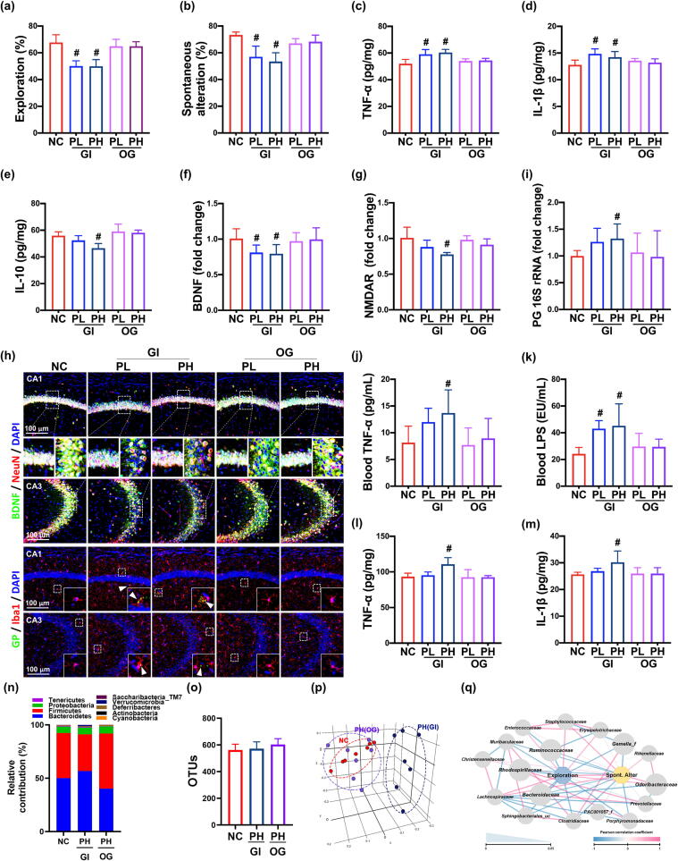
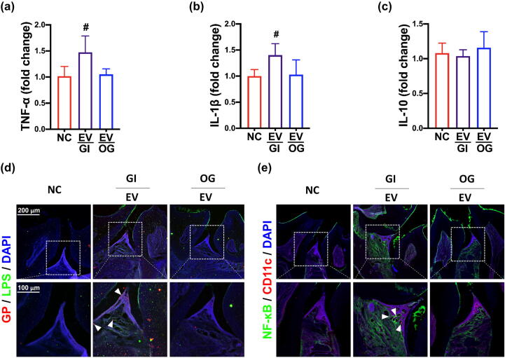
Results: pEVs contained neurotoxic GPs and inflammation-inducible fimbria protein and LPS. Gingivally exposed, but not orally gavaged, PG or pEVs caused periodontitis and induced memory impairment-like behaviors. Gingival exposure to PG or pEVs increased TNF-α expression in the periodontal and hippocampus tissues. They also increased hippocampal GP+Iba1+, LPS+Iba1+, and NF-κB+Iba1+ cell numbers. Gingivally exposed PG or pEVs decreased BDNF, claudin-5, and N-methyl-D-aspartate receptor expression and BDNF+NeuN+ cell number. Gingivally exposed fluorescein-5-isothiocyanate-labeled pEVs (F-pEVs) were detected in the trigeminal ganglia and hippocampus. However, right trigeminal neurectomy inhibited the translocation of gingivally injected F-EVs into the right trigeminal ganglia. Gingivally exposed PG or pEVs increased blood LPS and TNF-α levels. Furthermore, they caused colitis and gut dysbiosis.

Conclusion: Gingivally infected PG, particularly pEVs, may cause cognitive decline with periodontitis. PG products pEVs and LPS may be translocated into the brain through the trigeminal nerve and periodontal blood pathways, respectively, resulting in the cognitive decline, which may cause colitis and gut dysbiosis. Therefore, pEVs may be a remarkable risk factor for dementia.

Keywords: Extracellular vesicle; Memory impairment; Microbiota dysbiosis; Periodontitis; Porphyromonas gingivalis.


Conflict of interest statement

Declaration of Competing Interest The authors declare that they have no known competing financial interests or personal relationships that could have appeared to influence the work reported in this paper.

Figures

None
Graphical abstract
Fig. 1
Fig. 1
Gingivally infected P. gingivalis (PG) caused periodontitis in mice. Effect on TNF-α (a), IL-1β (b), and IL-10 expression (c) in the periodontal tissue. (d) Effct on PG 16S rDNA level in the periodontal tissue. (e) Effect on GP+LPS+ and NF-κB+CD11c+ cell populations in the periodontal tissue. Effects on periodontal microbiota composition: (f) phylum, (g) family levels, (h) OTUs (α-diversity), and (i) β-diversity (principal coordinate analysis plot based on UniFrac). GI, gingivally exposed; OG, orally gavaged; NC, treated with vehicle; PL, treated with 2 × 108 CFU/mouse/day of PG; PH, treated with 1 × 109 CFU/mouse/day. Arrows in (d) indicate positive cells. Data values were described as mean ± SD (n = 7). #p < 0.05 vs. NC.
Fig. 2
Fig. 2
Gingivally infected P. gingivalis (PG) impaired cognitive function in mice. (a) Effects on spontaneous alternation in NORT (a) and YMT (b). Effects on TNF-α (c), IL-1β (d), IL-10 expression (e) in the hippocampus, assessed by ELISA. Effects on BDNF (f) and NMDAR expression (g) in the hippocampus, assessed by qPCR. (h) Effects on BDNF+NeuN+ cells and GP+Iba1+ cell populations in the hippocampus. (i) Effect on PG (PG) 16S rRNA levels in the hippocampus. Effect on TNF-α (j) and LPS levels (k) in the blood. Effect on TNF-α (l) and IL-1β expression (m) in the colon. Effects on fecal microbiota composition: (n) phylum level, (o) OTUs (α-diversity), (p) β-diversity (principal coordinate analysis plot based on UniFrac), and (q) network of differentially enriched gut microbiota in spontaneous alteration in YMT or exploration in NORT scores. GI, gingivally exposed; OG, orally gavaged; NC, treated with vehicle; PL, treated with 2 × 108 CFU/mice/day of PG; PH, treated with 1 × 109 CFU/mice/day. Arrows in (h) indicate positive cells. Data values were described as mean ± SD (n = 7). #p < 0.05 vs. NC.
Fig. 3
Fig. 3
P. gingivalis extracellular vesicles (pEVs) suppressed BDNF expression in neuronal SH-SY5Y cells and induced TNF-α in microphage cells. (a) Transmission electron microscope image of pEVs. (b) Sodium-polyacrylamide gel electrophoresis (SDS-PAGE) of pEVs. (c) The identified components of pEVs: A, Mfa1 family fimbria major subunit partial; B, gingipain R [RpgA]; C, gingipain K [Peptidase C25]; D, fimbria protein; E, gingipain K, assessed by LC-MS-MS. Effect of pEVs on the cytotoxicity (d) and BDNF expression (e) in SH-SY5Y cells. Effects on TNF-α (f) and IL-1β expression (g) in peritoneal macrophages. Effects on TNF-α (h) and IL-1β expression (i) in BV2 cells. N, treated with vehicle; EV, treated with 20, 50, 100, 200, and 400 ng/mL of pEVs. Data values were described as mean ± SD (n = 4). #p < 0.05 vs. NC.
Fig. 4
Fig. 4
Gingivally exposed P. gingivalis extracellular vesicles (pEVs) caused periodontitis in mice. Effects on TNF-α (a), IL-1β (b), and IL-10 (c) in the periodontal tissue. Effects on PG+LPS+ (c) and NF-κB+CD11c+ cell populations (d)in the periodontal tissue. GI, gingivally exposed; OG, orally gavaged; NC, treated with vehicle; EV, treated with 2 μg/mouse/day of pEVs. Arrows in (d) indicate positive cells. Data values were described as mean ± SD (n = 7). #p < 0.05 vs. NC.
Fig. 5
Fig. 5
Gingivally exposed P. gingivalis extracellular vesicles (pEVs) impaired cognitive function in mice. a) Effects on spontaneous alternation in NORT (a) and YMT (b). Effects on TNF-α (c), IL-1β (d), IL-10 expression (e) in the hippocampus, assessed by ELISA. Effects on BDNF (f) and NMDAR expression (g) in the hippocampus, assessed by qPCR. (h) Effects on BDNF+NeuN+, claudin-5+, NF-κB+Iba1+, LPS+Iba1+, TLR4+Iba1+, and GP+Iba1+ cell populations in the hippocampus. (i) Effect on P. gingivalis (PG) 16S rRNA levels in the hippocampus. Effect on TNF-α (j) and LPS levels (k) in the blood. Effect on TNF-α (l) and IL-1β expression (m) in the colon. Effects on periodontal microbiota composition: (n) phylum level, (o) α-diversity (OTUs), (o) β-diversity (principal coordinate analysis plot based on UniFrac), and (q) network of differentially enriched gut microbiota in spontaneous alteration in YMT or exploration in NORT scores. GI, gingivally exposed; OG, orally gavaged; NC, treated with vehicle; EV, treated with 2 μg/mouse/day of pEVs. Arrows in (h) indicate positive cells. Data values were described as mean ± SD (n = 7). #p < 0.05 vs. NC.
Fig. 6
Fig. 6
Trigeminal neurotomy inhibited the translocation of gingivally exposed FITC-labeled pEVs (F-pEVs) into the brain. (a) Effects of administered routes (gingival infection and oral gavage) on the F-pEVs detected in the hippocampus of mice with gingivally exposed to F-pEVs. (b) Effect of right-gingivally injection on the F-pEVs detected in the trigeminal ganglia of mice with gingivally exposed to F-pEVs. (c) Effects of right- and left-gingival injections on the F-pEVs detected on the trigeminal ganglia of mice with right trigeminal neurotomy. NC, treated with vehicle in mice; giEV, gingivally exposed with F-pEVs (2 μg/mouse/day); ogEV, orally gavaged with pEVs (2 μg/mouse/day); EVr, injected with F-pEVs (2 μg/mouse/day) in the right gingiva of mice; EVrl, injected with F-pEVs (2 μg/mouse/day) in the right (R) and left (L) gingiva of mice with the right trigeminal neurotomy. Arrows indicate positive cells.

References

    1. Akiyama H., Barger S., Barnum S., Bradt B., Bauer J., Cole G.M., et al. Inflammation and Alzheimer's disease. Neurobiol Aging. 2000;21(3):383–421. doi: 10.1016/s0197-4580(00)00124-x. - DOI - PMC - PubMed
    1. Peila R, Launer LJ. Inflammation and dementia: epidemiologic evidence. Acta Neurol Scand Suppl. 2006;185:102–6. doi:10.1111/j.1600-0404.2006.00693.x. - PubMed
    1. Zhang Y., Geng R., Tu Q. Gut microbial involvement in Alzheimer's disease pathogenesis. Aging (Albany NY) 2021;13(9):13359–13371. doi: 10.18632/aging.202994. - DOI - PMC - PubMed
    1. Lotz SK, Blackhurst BM, Reagin KL, Funk KE. Microbial infections are a risk factor for neurodegenerative diseases. Front Cell Neurosci. 2021;15:691136. doi:10.3389/fncel.2021.691136. - PMC - PubMed
    1. Zhao J., Bi W., Xiao S., Lan X., Cheng X., Zhang J., et al. Neuroinflammation induced by lipopolysaccharide causes cognitive impairment in mice. Sci Rep. 2019;9(1):5790. doi: 10.1038/s41598-019-42286-8. - DOI - PMC - PubMed

Publication types

Substances

LinkOut - more resources