

ACTA1 H40Y mutant iPSC-derived skeletal myocytes display mitochondrial defects in an in vitro model of nemaline myopathy
- PMID: 36796746
- PMCID: PMC9993434
- DOI: 10.1016/j.yexcr.2023.113507
ACTA1 H40Y mutant iPSC-derived skeletal myocytes display mitochondrial defects in an in vitro model of nemaline myopathy
Abstract
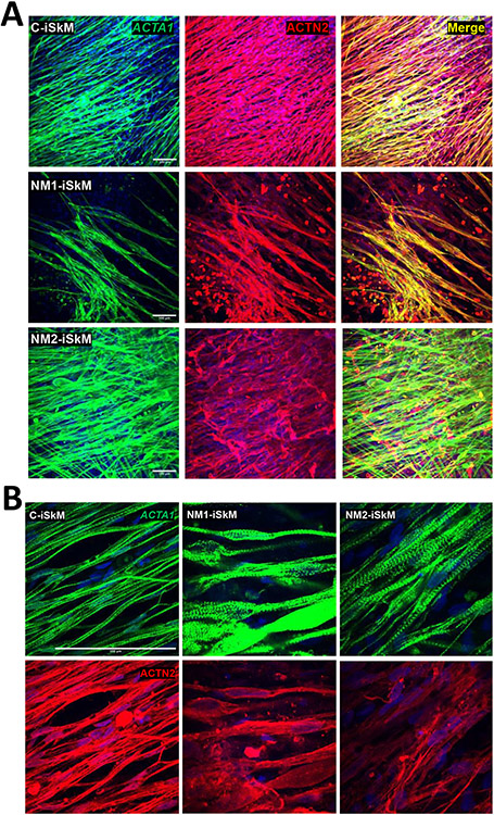
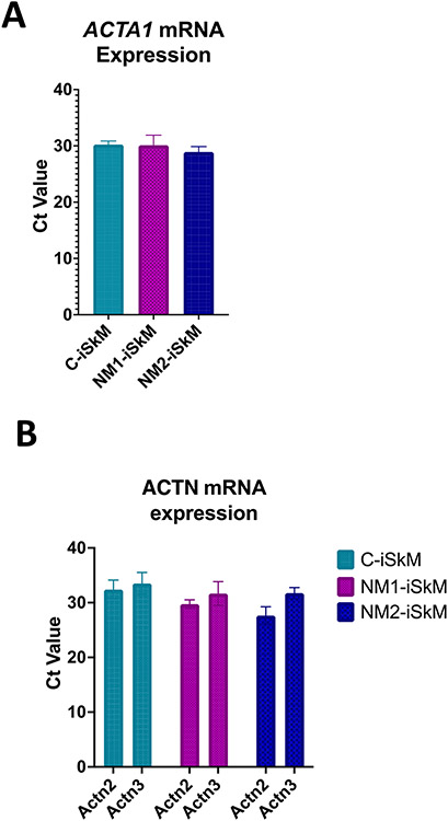
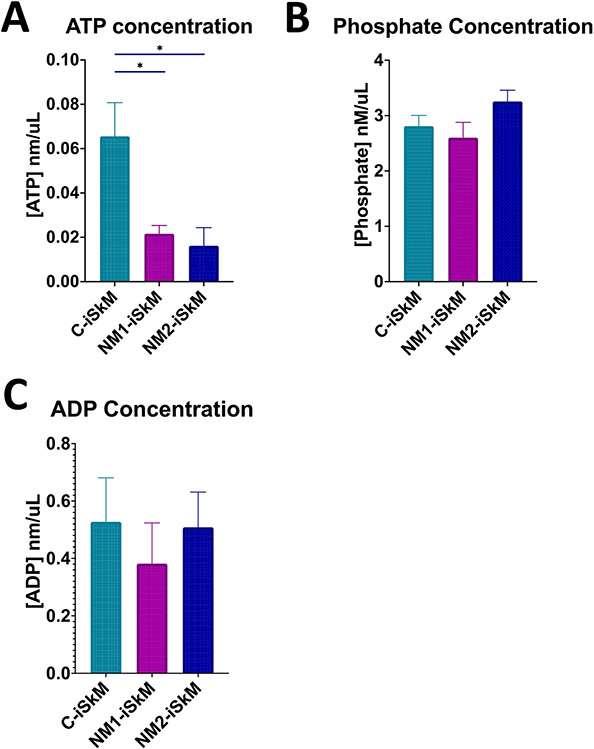
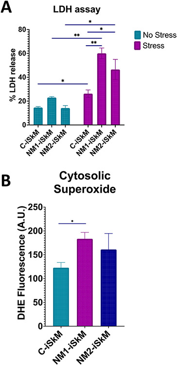
Nemaline myopathies (NM) are a group of congenital myopathies that lead to muscle weakness and dysfunction. While 13 genes have been identified to cause NM, over 50% of these genetic defects are due to mutations in nebulin (NEB) and skeletal muscle actin (ACTA1), which are genes required for normal assembly and function of the thin filament. NM can be distinguished on muscle biopsies due to the presence of nemaline rods, which are thought to be aggregates of the dysfunctional protein. Mutations in ACTA1 have been associated with more severe clinical disease and muscle weakness. However, the cellular pathogenesis linking ACTA1 gene mutations to muscle weakness are unclear To evaluate cellular disease phenotypes, iPSC-derived skeletal myocytes (iSkM) harboring an ACTA1 H40Y point mutation were used to model NM in skeletal muscle. These were generated by Crispr-Cas9, and include one non-affected healthy control (C) and 2 NM iPSC clone lines, therefore representing isogenic controls. Fully differentiated iSkM were characterized to confirm myogenic status and subject to assays to evaluate nemaline rod formation, mitochondrial membrane potential, mitochondrial permeability transition pore (mPTP) formation, superoxide production, ATP/ADP/phosphate levels and lactate dehydrogenase release. C- and NM-iSkM demonstrated myogenic commitment as evidenced by mRNA expression of Pax3, Pax7, MyoD, Myf5 and Myogenin; and protein expression of Pax4, Pax7, MyoD and MF20. No nemaline rods were observed with immunofluorescent staining of NM-iSkM for ACTA1 or ACTN2, and these mRNA transcript and protein levels were comparable to C-iSkM. Mitochondrial function was altered in NM, as evidenced by decreased cellular ATP levels and altered mitochondrial membrane potential. Oxidative stress induction revealed the mitochondrial phenotype, as evidenced by collapsed mitochondrial membrane potential, early formation of the mPTP and increased superoxide production. Early mPTP formation was rescued with the addition of ATP to media. Together, these findings suggest that mitochondrial dysfunction and oxidative stress are disease phenotypes in the in vitro model of ACTA1 nemaline myopathy, and that modulation of ATP levels was sufficient to protect NM-iSkM mitochondria from stress-induced injury. Importantly, the nemaline rod phenotype was absent in our in vitro model of NM. We conclude that this in vitro model has the potential to recapitulate human NM disease phenotypes, and warrants further study.
Keywords: Mitochondria; Nemaline myopathy; Skeletal myotube; Stress injury; iPSC.
Copyright © 2023 Elsevier Inc. All rights reserved.
Conflict of interest statement
Declaration of competing interest M.W.L. is the founder, CEO, and owner of Diverge Translational Science Laboratory. M.W.L. is or has recently been a member of advisory boards for Solid Biosciences, Taysha Gene Therapies, Astellas Gene Therapies (formerly Audentes Therapeutics), and Ichorion Therapeutics. M.W.L. is also a consultant for Astellas Gene Therapies (formerly Audentes Therapeutics), Encoded Therapeutics, Modis Therapeutics, Lacerta Therapeutics, Dynacure, AGADA Biosciences, Affinia Therapeutics, Biomarin, Locanabio, and Vertex Pharmaceuticals. M.W.L. receives research support from Astellas Gene Therapies, Solid Biosciences, Kate Therapeutics, Prothelia, Cure Rare Disease, and Rocket Pharma, Ultragenyx. S.M.L. is now a Sr. Application Scientist at Curi Bio and D.L.M is on Curi Bio's SAB. All other authors have no disclosures to declare.
Figures

References
-
- Sanoudou D, Beggs AH. Clinical and genetic heterogeneity in nemaline myopathy–a disease of skeletal muscle thin filaments. Trends in molecular medicine. 2001;7(8):362–8. - PubMed
Publication types
MeSH terms
Substances
Grants and funding
LinkOut - more resources
Full Text Sources
