Review
. 2023 Feb 17;8(1):70.
doi: 10.1038/s41392-023-01332-8.

Hypoxic microenvironment in cancer: molecular mechanisms and therapeutic interventions

Affiliations
Review

Hypoxic microenvironment in cancer: molecular mechanisms and therapeutic interventions

Zhou Chen et al. Signal Transduct Target Ther. .

Abstract

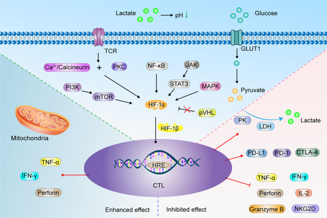
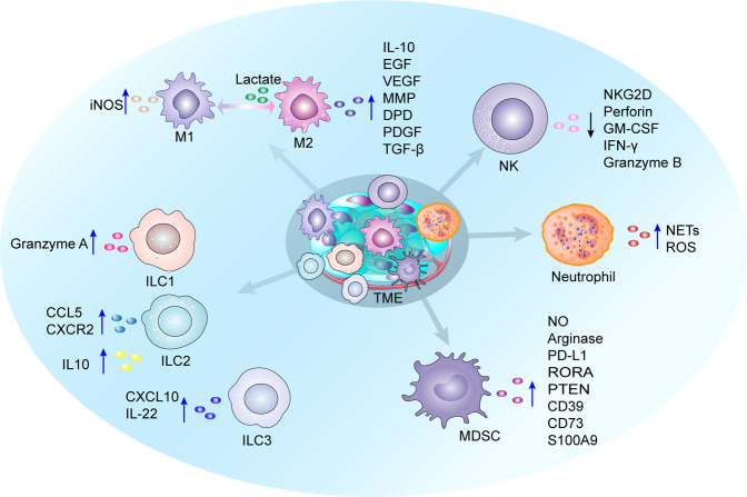
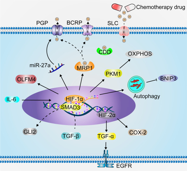
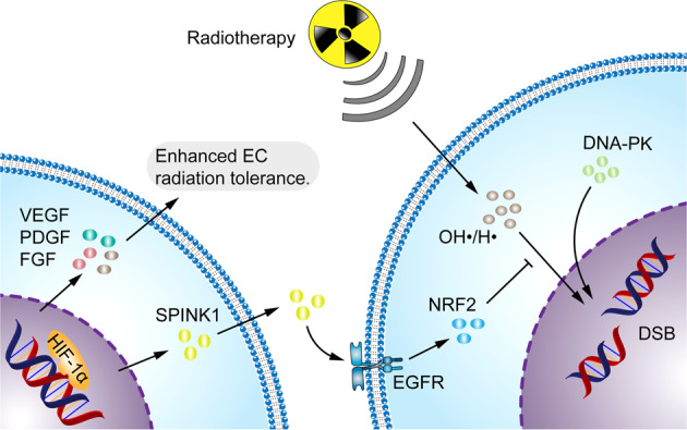
Having a hypoxic microenvironment is a common and salient feature of most solid tumors. Hypoxia has a profound effect on the biological behavior and malignant phenotype of cancer cells, mediates the effects of cancer chemotherapy, radiotherapy, and immunotherapy through complex mechanisms, and is closely associated with poor prognosis in various cancer patients. Accumulating studies have demonstrated that through normalization of the tumor vasculature, nanoparticle carriers and biocarriers can effectively increase the oxygen concentration in the tumor microenvironment, improve drug delivery and the efficacy of radiotherapy. They also increase infiltration of innate and adaptive anti-tumor immune cells to enhance the efficacy of immunotherapy. Furthermore, drugs targeting key genes associated with hypoxia, including hypoxia tracers, hypoxia-activated prodrugs, and drugs targeting hypoxia-inducible factors and downstream targets, can be used for visualization and quantitative analysis of tumor hypoxia and antitumor activity. However, the relationship between hypoxia and cancer is an area of research that requires further exploration. Here, we investigated the potential factors in the development of hypoxia in cancer, changes in signaling pathways that occur in cancer cells to adapt to hypoxic environments, the mechanisms of hypoxia-induced cancer immune tolerance, chemotherapeutic tolerance, and enhanced radiation tolerance, as well as the insights and applications of hypoxia in cancer therapy.


Conflict of interest statement

The authors declare no competing interests.

Figures

Fig. 1
Fig. 1
A historical and chronological figure of major events in tumor hypoxia research
Fig. 2
Fig. 2
Potential factors contributing to tumor hypoxia. Carcinogenic factors, such as drug, carcinogen, and microbiota dysbiosis, impair EC shape and function in the vascular system. TME is remodeled by tumor cells, stromal cells and stromal components (e.g., fibrin), resulting in vascular deformation due to pressure. High metabolism in cancer cells, such as increased nucleic acid synthesis and increased protein anabolism, leads to relative hypoxia. Dysregulated proliferation and alignment of vascular ECs result in the formation of non-functional blood vessels. With the increased distance between tumor cells and blood vessels, O2 diffusion decreases and leads to hypoxia
Fig. 3
Fig. 3
Biological changes in cancer cells adapt to hypoxia. Hypoxia promotes carcinogenesis by inducing DNA strand breaks, including DNA DSB and SSB, and by weakening DNA repair pathways, such as HR and MMR. HIF-1α is upgraded by PI3K-mTOR, JAK-STAT3, NF-κB, MAPK, Wnt/β-catenin, and Notch pathway. Deletion of tumor suppressor genes, such as p53, PTEN, and ROS production, also contributes to the upregulation of HIF-1α. The loss of pVHL function under hypoxic conditions indirectly leads to HIF-1α accumulation. HIF-1α dimerizes with HIF-1β and enters the nucleus to bind to HRE, which regulates various downstream target genes (Table 1) to promote cancer cell proliferation, migration, invasion, EMT and angiogenesis
Fig. 4
Fig. 4
Glucose and lipid metabolism in cancer cells under hypoxic conditions. Glucose is taken up by cancer cells via GLUT1 and glycolysed to pyruvate via PKM, PGK1 and PGAM1. Hypoxic cancer cells promote pyruvate glycolysis to lactate by upregulating LDHA, LDHC and LDH-5, and the lactate produced is excreted outside the cell via MCT1/4. In addition, HIF-1α inactivates PDH by activating PDK1, which in turn fails to convert pyruvate to acetyl-CoA, preventing the entry of pyruvate into the TCA cycle. Cytoplasmic citrate is catalyzed by ALCY to acetyl-CoA, and acetyl-CoA catalyzed by ACC to malonyl-CoA. Acetyl-CoA and malonyl-CoA are catalyzed to FA via FASN upregulated by SREBP-1. SCD1 upregulated by SREBP-1 catalyzes the formation of MUFA from saturated FA. PHD3 loss reduces ACC2 hydroxylation and promotes FAO to provide energy. α-KG as a product of glutamine is reduced and carboxylated to isocitrate by IDH, and then oxidation to citrate. PLD hydrolyzes PC to produce PA. PLA2 catalyzes the hydrolysis of GPL to produce Lyso-PL
Fig. 5
Fig. 5
Hypoxia remodels CTL immune effect. HIF-1α in CTL is upgraded by TCR-PKC and Ca2+/calcineurin, PI3K/mTOR, NF-κB, NF-κB, JAK-STAT3, MAPK pathway. Deletion of pVHL impaired HIF-1α degradation. HIF-1α and HIF-1β dimerization stimulates downstream factor expression, such as perforin, IFN-γ, TNF-α, which can enhance the antitumor efficacy of CTL. However, some experiments elaborate the opposite results. Hypoxia inhibits the expression of IFN-γ, TNF-α, granzyme B, IL-2, perforin and NKG2D, while promoting PD-L1,PD-1 and CTLA-4 expression. CTL increases glucose uptake via GLUT1, which is metabolized to lactate by glycolytic enzymes PK and LDH. Lactate acidifies the TME, which inhibits CTL activation
Fig. 6
Fig. 6
Changes in cytokine secretion by innate immune cells under hypoxic conditions. M1 microphage secretes iNOS, and M2 macrophages secrete IL-10, EGF, VEGF, MMP, DPD, PDGF and TGF-β to suppress immune responses. Lactate induces M2-like polarization. HIF-1α inhibits NK cell expression of perforin, NKG2D, GM-CSF, granzyme B and IFN-γ. Hypoxia induces neutrophils to produce ROS and NETs, which promote tumorigenesis and metastasis. HIF-1α induces MDSCs to secrete cytokines NO, arginase, PD-L1, RORA, PTEN, CD39, CD73 and S100A9, which inhibit immune response and promote the tumor cell stemness and growth. ILC1 secretes granzyme A and maintains antitumor efficacy. ILC2 enhances immune response through selective expression of CCL5, CXCR2. Hypoxia induces IL-10 expression in ILC2 and suppresses immunity. ILC3 secretes CXCL10 and IL-22, improving antitumor immune response
Fig. 7
Fig. 7
Hypoxia enhances tumor chemotherapy resistance. IL-6 stimulates HIF-1α expression, which increases PGP expression through upregulation of miR-27a. Chemotherapeutic agents are transported by HIF-1α-induced intracellular MRP1 and excreted from cells via PGP and BCRP. HIF-1α induces OLFM4 to enhance cancer cell chemoresistance. HIF-1α enhances chemotherapeutic resistance in cancer cells by promoting PKM1, enhancing mitochondrial OXPHOS. Under hypoxic conditions, cancer cells induce enhanced autophagy and inhibit the chemotherapeutic drug-induced BNIP3 death pathway to resist drug toxicity. HIF-1α synergizes with TGF-β to promote GLI2 expression through SMAD3, inducing cancer cell stemness and chemoresistance. HIF-2α upregulation promotes the ability of hypoxic cancer cells to resist drug toxicity by activating the TGF-α/EGFR pathway and COX-2. In addition, CDD and SLC play important roles in the drug resistance of cancer cells, the effect of hypoxic conditions on CDD and SLC is unclear
Fig. 8
Fig. 8
Hypoxia enhances the ability of the tumor to resist radiotherapy. Radiotherapy induces cancer cell death by directly damaging DNA through the production of free radicals such as OH• and H•. SPINK1, secreted by hypoxic cancer cells, upregulates EGFR and NRF2 antioxidant response in adjacent cancer cells to reduce radiation-induced DNA damage, thereby inducing cancer radioresistance. VEGF, FGF and PDGF secreted by hypoxic cancer cells enhanced the radiation resistance of ECs. HIF-1α stimulates DNA-PK expression and repairs DNA DSB

References

    1. Fane M, Weeraratna AT. How the ageing microenvironment influences tumour progression. Nat. Rev. Cancer. 2020;20:89–106. doi: 10.1038/s41568-019-0222-9. - DOI - PMC - PubMed
    1. Siegel RL, Miller KD, Fuchs HE, Jemal A. Cancer statistics, 2022. CA Cancer J. Clin. 2022;72:7–33. doi: 10.3322/caac.21708. - DOI - PubMed
    1. Tannock IF. Population kinetics of carcinoma cells, capillary endothelial cells, and fibroblasts in a transplanted mouse mammary tumor. Cancer Res. 1970;30:2470–2476. - PubMed
    1. Gray LH, et al. The concentration of oxygen dissolved in tissues at the time of irradiation as a factor in radiotherapy. Br. J. Radio. 1953;26:638–648. doi: 10.1259/0007-1285-26-312-638. - DOI - PubMed
    1. Semenza GL, Wang GL. A nuclear factor induced by hypoxia via de novo protein synthesis binds to the human erythropoietin gene enhancer at a site required for transcriptional activation. Mol. Cell Biol. 1992;12:5447–5454. - PMC - PubMed

Publication types