. 2023 Feb 17;8(1):66.
doi: 10.1038/s41392-022-01268-5.

B-lymphoid tyrosine kinase-mediated FAM83A phosphorylation elevates pancreatic tumorigenesis through interacting with β-catenin

Affiliations

B-lymphoid tyrosine kinase-mediated FAM83A phosphorylation elevates pancreatic tumorigenesis through interacting with β-catenin

Cefan Zhou et al. Signal Transduct Target Ther. .

Abstract

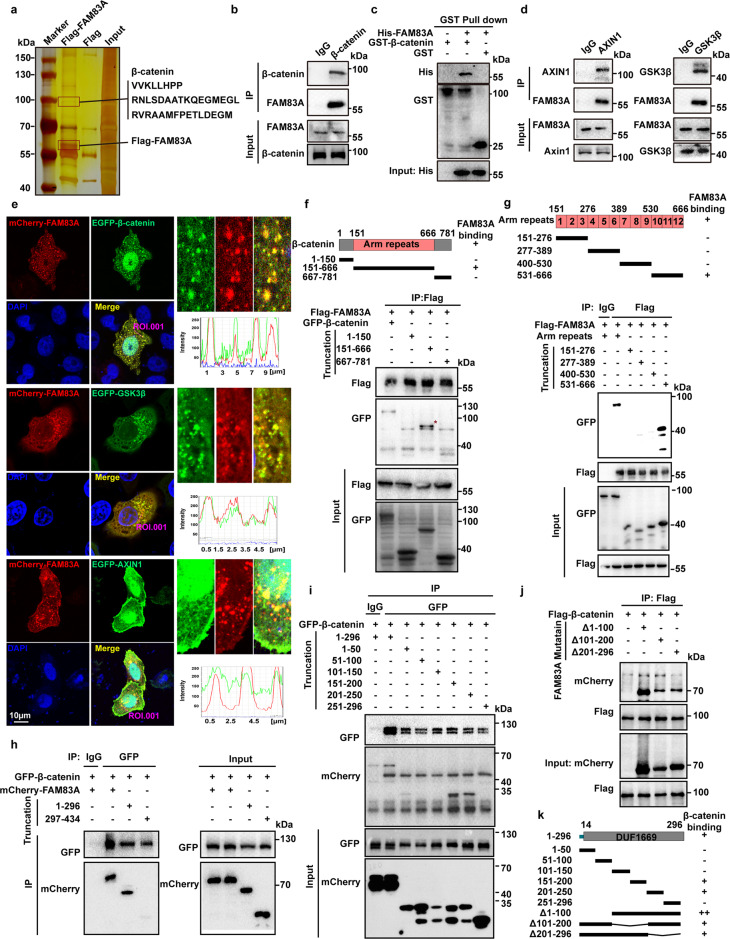
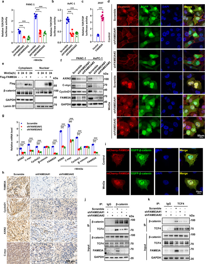
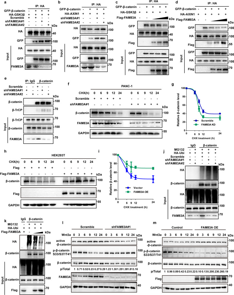
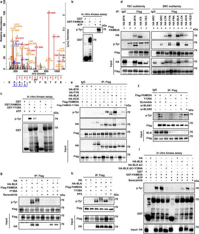
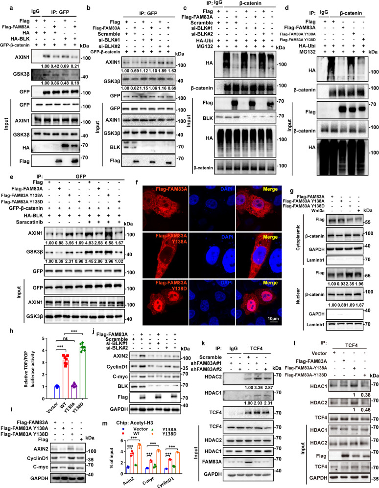
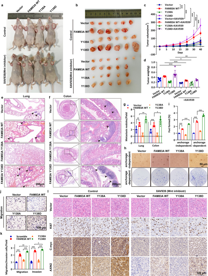
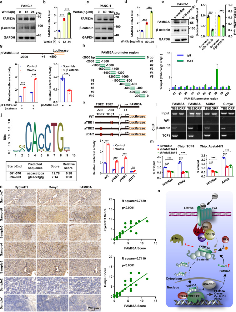
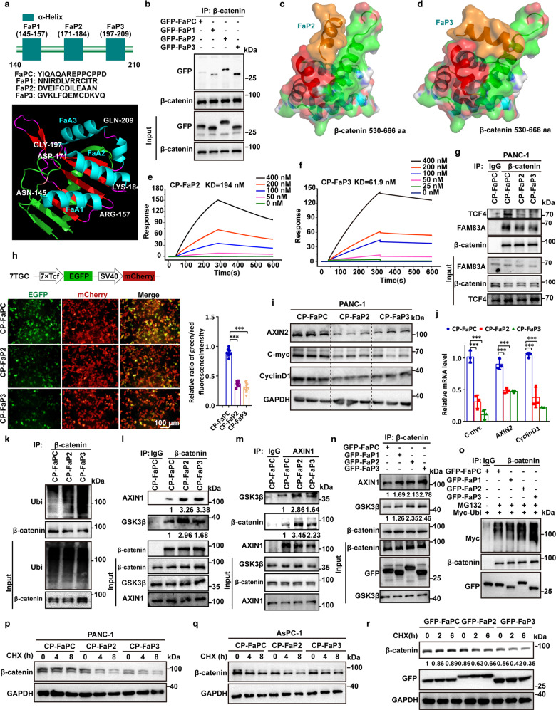
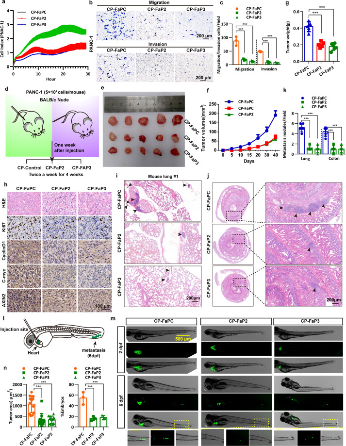
Abnormal activation of Wnt/β-catenin-mediated transcription is closely associated with the malignancy of pancreatic cancer. Family with sequence similarity 83 member A (FAM83A) was shown recently to have oncogenic effects in a variety of cancer types, but the biological roles and molecular mechanisms of FAM83A in pancreatic cancer need further investigation. Here, we newly discovered that FAM83A binds directly to β-catenin and inhibits the assembly of the cytoplasmic destruction complex thus inhibiting the subsequent phosphorylation and degradation. FAM83A is mainly phosphorylated by the SRC non-receptor kinase family member BLK (B-lymphoid tyrosine kinase) at tyrosine 138 residue within the DUF1669 domain that mediates the FAM83A-β-catenin interaction. Moreover, FAM83A tyrosine 138 phosphorylation enhances oncogenic Wnt/β-catenin-mediated transcription through promoting β-catenin-TCF4 interaction and showed an elevated nucleus translocation, which inhibits the recruitment of histone deacetylases by TCF4. We also showed that FAM83A is a direct downstream target of Wnt/β-catenin signaling and correlates with the levels of Wnt target genes in human clinical pancreatic cancer tissues. Notably, the inhibitory peptides that target the FAM83A-β-catenin interaction significantly suppressed pancreatic cancer growth and metastasis in vitro and in vivo. Our results revealed that blocking the FAM83A cascade signaling defines a therapeutic target in human pancreatic cancer.


Conflict of interest statement

The authors declare no competing interests.

Figures

Fig. 1
Fig. 1
FAM83A is identified as a new component of the cytoplasmic β-catenin destruction complex. a Silver staining of immunoprecipitate using Flag antibody from HEK293T cell lysates transfected with Flag-tagged FAM83A. b The interaction between endogenous FAM83A and β-catenin in pancreatic cancer PANC-1 cells. Protein interactions were analyzed by western blotting. c Externally purified GST-tagged full-length β-catenin was incubated with externally purified His-tagged FAM83A, the mixture was pulled down by GST beads and then the precipitates were subjected to western blotting. d The interaction between endogenous FAM83A and the components of β-catenin destructive complex AXIN1 and GSK3β in PANC-1 cells. Protein interactions were analyzed by western blotting. e The co-localization of mCherry-tagged FAM83A and EGFP-tagged β-catenin, GSK3β, and AXIN1 in HeLa cells were analyzed with confocal microscopy. f, g HEK293T cells were transfected with the β-catenin truncation mutations indicated for 48 h and then harvested for IP and western blotting analysis. The schematic diagram of β-catenin truncation generation and FAM83A-β-catenin mutants interacting domain were shown. hk HEK293T cells were transfected with the FAM83A truncation or deletion mutations indicated for 48 h and then harvested for IP and western blotting analysis. The schematic diagram of FAM83A truncation and deletion generation and β-catenin-FAM83A mutants interacting domain were shown. Scale bars: 10 µm
Fig. 2
Fig. 2
FAM83A promotes canonical β-catenin transcriptional activity in human pancreatic cancer. ac Wnt reporter luciferase activity (TOP Flash) in PANC-1, AsPC-1, and HEK293T cells with or without FAM83A knockdown or overexpression and Wnt3a treatment (100 ng/mL for 4 hours) (n = 7). d The localization of β-catenin with or without FAM83A knockdown (cells expressing FAM83A shRNA co-expressed GFP) and Wnt3a treatment (100 ng/mL for 4 hours) were analyzed with confocal microscopy in PANC-1 cells. e The protein level of cytoplasmic and nuclear β-catenin in HEK293T cells with or without Wnt3a treatment (100 ng/mL) were analyzed with western blotting (n = 3). f (n = 3). g mRNA level of Wnt target genes AXIN2, C-myc, and CyclinD1 in PANC-1 cell lysates with or without FAM83A knockdown and Wnt3a treatment (100 ng/mL) (n = 6). h Representative immunohistochemical images of FAM83A and Wnt target genes AXIN2, C-myc, and CyclinD1 expression in excised xenograft tumor tissues with or without FAM83A knockdown. i The localization of mCherry-tagged FAM83A and GFP-tagged β-catenin with or without Wnt3a treatment (100 ng/mL) in PANC-1 cells was analyzed with confocal microscopy. j, k FAM83A knockdown decreased the interaction between endogenous TCF4 and β-catenin in PANC-1 cell lysates. Protein interactions were analyzed by western blotting (n = 3). Scale bars: 10 µm. *P < 0.05; **P < 0.01; ***P < 0.001. Data were presented as mean ± SD
Fig. 3
Fig. 3
FAM83A inhibits the destruction complex formation and stabilizes β-catenin. a, b FAM83A knockdown using two specific small interfering RNAs (shFAM83A#1 and shFAM83A#2) enhanced the interaction between exogenous HA-tagged GSK3β, AXIN1 and GFP-tagged β-catenin in PANC-1 cell lysates. Protein interactions were analyzed by western blotting (n = 3). c, d The exogenous Flag-FAM83A dose-dependently decreased β-catenin-GSK3β and β-catenin-AXIN1 interactions in PANC-1 cell lysates. Protein interactions were analyzed by western blotting (n = 3). e FAM83A knockdown enhanced the interaction between endogenous β-TrCP and β-catenin in PANC-1 cell lysates. Protein interactions were analyzed by western blotting (n = 3). f, g FAM83A knockdown promoted the degradation of β-catenin protein upon cycloheximide (CHX, 20 μM) treatment for indicated times in PANC-1 cell lysates. Representative western blotting images and quantification data were shown (n = 3). h, i FAM83A overexpression inhibited the degradation of β-catenin protein upon cycloheximide (CHX, 20 μM) treatment for indicated times in PANC-1 cell lysates. Representative western blotting images and quantification data were shown (n = 3). j FAM83A knockdown increased the level of β-catenin ubiquitination upon MG132 (10 μM) treatment in PANC-1 cell lysates. Representative western blotting images were shown (n = 3). k FAM83A overexpression dose-dependently decreased the level of β-catenin ubiquitination upon MG132 (10 μM) treatment in PANC-1 cell lysates. Representative western blotting images were shown (n = 3). l, m Representative western blotting images of active-β-catenin, p-β-catenin S33/S37/T41 were shown after FAM83A knockdown or FAM83A overexpression upon Wnt3a (100 ng/mL) treatment for indicated times (n = 3). *P < 0.05; **P < 0.01; ***P < 0.001. Data were presented as mean ± SD
Fig. 4
Fig. 4
FAM83A is phosphorylated by BLK kinase at Y138. a The mass spectrum showed that FAM83A extracted from HEK293T cell lysates was phosphorylated at tyrosine 138 residue. b In vitro purified GST-FAM83A were incubated with HEK293T cell lysates with or without ATP addition, and the reaction products were subjected to western blotting. c In vitro purified GST-tagged FAM83A and GST-tagged FAM83A Y138A mutant were incubated with HEK293T cell lysates with or without ATP addition, and the reaction products were subjected to western blotting (n = 3). d The in vivo interaction between Flag-tagged FAM83A and the components of non-receptor TEC kinase subfamily or SRC kinase subfamily were analyzed in HEK293T cells by western blotting (n = 3). e Level of total tyrosine phosphorylation of Flag-tagged FAM83A and its Y138A mutant in HEK293T cells after HA-tagged BTK, FYN, BLK, and HCK transfection. Cell lysates were used for IP and western blotting with the indicated antibodies (n = 3). f Level of total tyrosine phosphorylation of Flag-tagged FAM83A and its Y138A mutant in HEK293T cells after BLK kinase knockdown using two specific small interfering RNAs (siBLK#1 and siBLK#2). Cell lysates were used for IP and western blotting with the indicated antibodies (n = 3). g, h Level of total tyrosine phosphorylation of Flag-tagged FAM83A and its Y138A mutant in HEK293T cells with or without BLK kinase overexpression and saracatinib (BLK inhibitor, 10 μM)/PP2 (LCK or FYN inhibitor, 10 μM) treatment. Cell lysates were used for IP and western blotting with the indicated antibodies (n = 3). i The BLK kinases was obtained from the immunoprecipitate of HEK293T cell lysates that transfected with either HA-tagged BLK wild-type, BLK kinase-dead mutant (Y389A) or BLK constitutive activated mutant with the deletion of the C-terminal auto-inhibitory domain (BLK ΔC) using HA tag antibody. Then the BLK kinases were incubated with the in vitro purified GST-tagged FAM83A protein at 30 °C for 30 min with or without BLK inhibitor saracatinib, and then the reaction products were subjected to western blotting assay (n = 3)
Fig. 5
Fig. 5
Phosphorylated FAM83A promotes β-catenin destruction complex assembly and inhibits HDAC recruitment by TCF4. a, b The interaction between GFP-tagged β-catenin and endogenous GSK3β and AXIN1 after BLK kinase overexpression or knockdown in PANC-1 cells. Protein interactions were analyzed by western blotting (n = 3). c The level of β-catenin ubiquitination with or without Flag-tagged FAM83A overexpression or BLK kinase knockdown after MG132 treatment in HEK293T cells. Cell lysates were used for IP and western blotting with the indicated antibodies (n = 3). d The level of β-catenin ubiquitination with or without Flag-tagged FAM83A, Y138A, or Y138D mutants overexpression after MG132 treatment in HEK293T cells. Cell lysates were used for IP and western blotting with the indicated antibodies (n = 3). e The interaction between GFP-tagged β-catenin and endogenous GSK3β and AXIN1 after BLK kinase overexpression with or without Flag-tagged FAM83A, Y138A, or Y138D mutants transfection or saracatinib treatment in HEK293T cells. Cell lysates were used for IP and western blotting with the indicated antibodies (n = 3). f The localization of Flag-tagged FAM83A and its Y138A, Y138D mutants in HEK293T cells were analyzed with confocal microscopy. g The protein level of cytoplasmic and nuclear β-catenin in HEK293T cells with or without Flag-tagged FAM83A, Y138A, or Y138D mutants transfection and Wnt3a treatment. Cell lysates were used for western blotting with the indicated antibodies (n = 3). h, i Wnt reporter luciferase activity (TOP Flash, n = 7) and the level of Wnt target genes AXIN2, C-myc, and CyclinD1 in HEK293T cells with or without Flag-tagged FAM83A, Y138A, or Y138D mutants transfection (n = 3). j Western blotting analysis of Wnt target genes AXIN2, C-myc, and CyclinD1 in PANC-1 cell lysates with or without FAM83A transfection and BLK kinase knockdown (n = 3). k, l The interaction between HDAC1, HDAC2 and TCF4 after FAM83A knockdown in PANC-1 cells or Flag-tagged FAM83A, Y138A or Y138D mutants transfection in HEK293T cells (n = 3). Cell lysates were used for IP and western blotting with the indicated antibodies. m The level of Wnt target genes AXIN2, C-myc and CyclinD1 in the PANC-1 cells stably expressed Flag-tagged FAM83A, Y138A or Y138D mutants in chromatin immunoprecipitation using histone H3 acetylation antibodies (n = 3). *P < 0.05; **P < 0.01; ***P < 0.001. Data were presented as mean ± SD
Fig. 6
Fig. 6
FAM83A Y138 phosphorylation enhances Wnt/β-catenin signaling and pancreatic cancer cell proliferation and metastasis. a, b Representative images of nude mice with two sides of axilla subcutaneous injected with PANC-1 cells stably expressed Flag-tagged wild-type FAM83A, Y138A, or Y138D mutants and representative images of excised tumors in different groups of nude mice were shown. The Wnt inhibitor XAV939 (20 mg/kg) was intravenously injected into the caudal vein of the nude mice one week later after xenograft formation. c Growth curve showing the changes in the tumor volume in mice in different groups from the injection (n = 6 or 5). d Weight of the excised tumors in each group (n = 6 or 5). e, f Representative images of lung and colon metastasis of the nude mice model that tail intravenous injected with PANC-1 cells stably expressed Flag-tagged wild-type FAM83A, Y138A, or Y138D mutants were shown. g The number of lung or colon metastasis was quantified per field (n = 5). h, i Representative images and quantification of the soft agarose and plate colony formation assays of PANC-1 cells stably expressed Flag-tagged wild-type FAM83A, Y138A, or Y138D mutants (n = 3). j, k Representative images and quantification of the transwell assays without matrigel of PANC-1 cells stably expressed Flag-tagged wild-type FAM83A, Y138A or Y138D mutants (n = 3). l Representative H&E staining images and immunohistochemical images of Ki67, C-myc, and AXIN2 expression in excised tumors tissues. *P < 0.05; **P < 0.01; ***P < 0.001. Data were presented as mean ± SD
Fig. 7
Fig. 7
FAM83A is a direct downstream target of Wnt/β-catenin signaling and forms positive feedback in pancreatic cancer cells. ad Representative western blotting images of the protein and mRNA levels of FAM83A after Wnt3a treatment for indicated concentrations and times in PANC-1 cells (n = 3). e, f The protein and mRNA levels of FAM83A after β-catenin knockdown in PANC-1 cells. Cell lysates were used for western blotting with the indicated antibodies (n = 3). g FAM83A gene promoter-reporter luciferase activity in HEK293T cells with or without β-catenin overexpression (left) and PANC-1 cells with or without β-catenin knockdown (right) (n = 6). h The schematic diagram of FAM83A gene promoter truncation and deletion generation. i PACN-1 cells were subjected to chromatin immunoprecipitation using antibodies against TCF4 and IgG, and the fold change of TCF/IgG was quantified (n = 3). j Predicted TCF4/LEF-binding sequence in the FAM83A gene promoter from the JASPAR website. k Schematic diagram of the generated luciferase report plasmids and the relative luciferase activity after transfection with the plasmids in HEK293T cells with or without Wnt3a treatment (n = 6). l A ChIP experiment was used to detect binding between the β-catenin-TCF4 complex and the predicted TCF/LEF-binding element in the FAM83A promoter (TBE1 and TBE2). C-myc and AXIN2 were used as a positive control (n = 3). m The level of FAM83A and Wnt target gene AXIN2 in the PANC-1 cells with FAM83A depletion in chromatin immunoprecipitation using TCF4 and histone H3 acetylation antibodies (n = 3). n Representative IHC images of FAM83A, CyclinD1, and C-myc in 44 human pancreatic cancer patient tissues. The correlation between the level of CyclinD1, C-myc, and FAM83A in 44 human pancreatic cancer patient tissues was shown. o Schematic diagram of the biological role of FAM83A in cytoplasmic β-catenin destruction complex and nuclear TCF4/β-catenin-mediated transcription in pancreatic cancer cells. *P < 0.05; **P < 0.01; ***P < 0.001. Data were presented as mean ± SD
Fig. 8
Fig. 8
Peptides disrupting FAM83A-β-catenin interaction inhibit Wnt/β-catenin signaling. a Identification of β-catenin binding α-helical peptide(s) in FAM83A DUF1669 (140aa–210aa) region. The secondary structure of the FAM83A DUF1669 (140aa–210aa) region was analyzed based on the PDB data (4urj). b The FaPC, FaP1, FaP2, and FaP3 fragments were constructed into pEGFP-C1 expression vector connection with a flexible linker ((GGGGS)n). The interactions between β-catenin and the four fragments were evaluated with Co-IP assays in HEK293T cells (n = 3). c, d The predicted docking model of FaP2, FaP3 with β-catenin 530–666 aa region. e, f Kinetic interactions of cell-penetrating peptide TAT-linked peptides with β-catenin was determined by surface plasmon resonance analyses. g Effect of CP-FaP2 and CP-FaP3 on β-catenin-FAM83A and β-catenin-TCF4 interactions in PANC-1 cells. Cells were treated with control (CP-FaPC), CP-FaP2, and CP-FaP3 (5 μM) for 24 hours. Cell lysates were used for IP and western blotting with the indicated antibodies (n = 3). h The schematic diagram of 7TGC plasmid construction and the fluorescence images of PANC-1 cells with control, CP-FaP2, and CP-FaP3 treatment. The relative ratio of green/red fluorescence intensity was quantified (n = 9). i, j Protein and mRNA levels of Wnt target genes AXIN2, C-myc, and CyclinD1 in PANC-1 cells with control, CP-FaP2, and CP-FaP3 treatment (n = 3). k The level of β-catenin ubiquitination in PANC-1 cells after control, CP-FaP2 and CP-FaP3 treatment in PANC-1 cells (n = 3). Cell lysates were used for IP and western blotting with the indicated antibodies. ln The interaction between β-catenin and AXIN1, GSK3β in PANC-1 cells after control, CP-FaP2 and CP-FaP3 treatment or GFP-tagged FaP1, FaP2, and FaP3 transfection in PANC-1 cells (n = 3). Cell lysates were used for IP and western blotting with the indicated antibodies. o The level of β-catenin ubiquitination in HEK293T cells after GFP-tagged FaP1, FaP2, and FaP3 transfection (n = 3). Cell lysates were used for IP and western blotting with the indicated antibodies. pr The stability of β-catenin protein upon CHX treatment for indicated times in PANC-1 cells after control, CP-FaP2 and CP-FaP3 treatment or GFP-tagged FaP1, FaP2, and FaP3 transfection (n = 3). Cell lysates were used for IP and western blotting with the indicated antibodies
Fig. 9
Fig. 9
FAM83A-β-catenin interaction inhibitory peptides restrain pancreatic cancer development. a Real-time cell analysis (cell index, CI) showing the differences in growth profile for PANC-1 cells treated with control (CP-FaPC, 2 mg/kg/2d), CP-FaP2 (2 mg/kg/2d), and CP-FaP3 (2 mg/kg/2d) peptides measured in a 16-well E-plate. b, c Representative images and quantification of the transwell assays with or without matrigel of PANC-1 cells treated with control, CP-FaP2, and CP-FaP3 peptides (n = 3). d Schematic diagram of the experimental procedure of the treatment of the peptides to nude mice xenograft formation and tail vein injection. e Representative images of excised tumors in the groups with respective peptides treatment were shown. f The growth curve shows the changes in the tumor volume in mice in different groups (n = 5). g Weight of the excised tumors in each group (n = 5). h Representative H&E staining images and immunohistochemical images of Ki67, C-myc CyclinD1, and AXIN2 expression in excised tumors tissues. ik Representative images and quantification of lung and colon metastasis of each group was shown (n = 5). l Schematic diagram of the zebrafish embryo xenograft assay. m Representative overview images at two days and six days post-implantation of zebrafish injected with EGFP-tagged PANC-1 cells. Scale bar: 500 μm. n Quantification of the area of metastatic cells in individual zebrafish embryos (n = 15) and the percentage of the zebrafish embryos displaying tumor progression to the tail fin (n = 3)

References

    1. Siegel RL, Miller KD, Fuchs HE, Jemal A. Cancer statistics, 2021. CA Cancer J. Clin. 2021;71:7–33. doi: 10.3322/caac.21654. - DOI - PubMed
    1. Zhou C, et al. TSPAN1 promotes autophagy flux and mediates cooperation between WNT-CTNNB1 signaling and autophagy via the MIR454-FAM83A-TSPAN1 axis in pancreatic cancer. Autophagy. 2021;17:3175–3195. doi: 10.1080/15548627.2020.1826689. - DOI - PMC - PubMed
    1. Park W, Chawla A, O’Reilly EM. Pancreatic cancer: a review. JAMA. 2021;326:851–862. doi: 10.1001/jama.2021.13027. - DOI - PMC - PubMed
    1. Morris JPT, Wang SC, Hebrok M. KRAS, Hedgehog, Wnt and the twisted developmental biology of pancreatic ductal adenocarcinoma. Nat. Rev. Cancer. 2010;10:683–695. doi: 10.1038/nrc2899. - DOI - PMC - PubMed
    1. Kim SE, et al. Wnt stabilization of β-catenin reveals principles for morphogen receptor-scaffold assemblies. Science. 2013;340:867–870. doi: 10.1126/science.1232389. - DOI - PMC - PubMed

Publication types

MeSH terms