. 2023 Feb 17;14(2):135.
doi: 10.1038/s41419-023-05666-7.

VE-Cadherin modulates β-catenin/TCF-4 to enhance Vasculogenic Mimicry

Affiliations

VE-Cadherin modulates β-catenin/TCF-4 to enhance Vasculogenic Mimicry

Daniel Delgado-Bellido et al. Cell Death Dis. .

Abstract

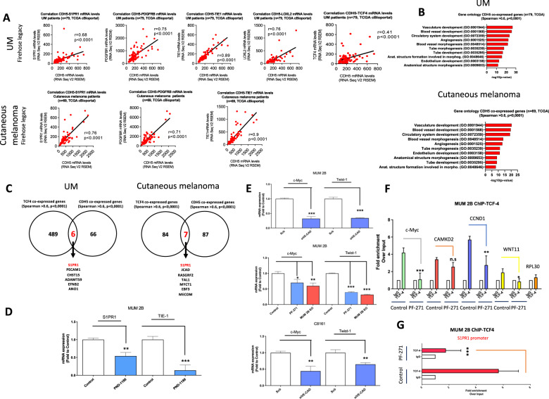
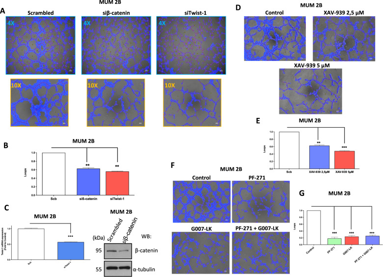
Vasculogenic Mimicry (VM) refers to the capacity to form a blood network from aggressive cancer cells in an independent way of endothelial cells, to provide nutrients and oxygen leading to enhanced microenvironment complexity and treatment failure. In a previous study, we demonstrated that VE-Cadherin and its phosphorylation at Y658 modulated kaiso-dependent gene expression (CCND1 and Wnt 11) through a pathway involving Focal Adhesion kinase (FAK). In the present research, using a proteomic approach, we have found that β-catenin/TCF-4 is associated with nuclear VE-cadherin and enhances the capacity of malignant melanoma cells to undergo VM in cooperation with VE-Cadherin; in addition, preventing the phosphorylation of Y658 of VE-cadherin upon FAK disabling resulted in VE-Cadherin/β-catenin complex dissociation, increased β-catenin degradation while reducing TCF-4-dependent genes transcription (C-Myc and Twist-1). Uveal melanoma cells knockout for VE-Cadherin loses β-catenin expression while the rescue of VE-Cadherin (but not of the phosphorylation defective VE-Cadherin Y658F mutant) permits stabilization of β-catenin and tumor growth reduction in vivo experiments. In vivo, the concomitant treatment with the FAK inhibitor PF-271 and the anti-angiogenic agent bevacizumab leads to a strong reduction in tumor growth concerning the single treatment. In conclusion, the anomalous expression of VE-Cadherin in metastatic melanoma cells (from both uveal and cutaneous origins), together with its permanent phosphorylation at Y658, favors the induction of the aggressive VM phenotype through the cooperation of β-catenin with VE-Cadherin and by enhancing TCF-4 genes-dependent transcription.


Conflict of interest statement

The authors declare no competing interests.

Figures

Fig. 1
Fig. 1. LC-MS experiments showed an elevated coupling of β-catenin/VE-cadherin complex in VM.
A Immunoprecipitation of VE-cadherin experiments shows union with β-catenin in MUM 2B cells, B representative union catenin to VE-cadherin graph of LC-MS proteomics IP-VE-cadherin CE (cytosol extract), NE (nuclear extract), score sequest HT in the X axis and #peptides sequest HT in Y axis. C String graph union protein of VE-cadherin.
Fig. 2
Fig. 2. Elevated expression of active β-catenin and Y397 phosphorylation of FAK in aggressive melanoma cells.
A Total expression of VE-cadherin, Y658 VE-cadherin, of β-catenin or non-phospho β-catenin (active β-catenin), focal adhesion kinase (FAK) or the active form phospho FAK (Y397) were measured in aggressive melanoma cells (MUM 2B, C8161) and poorly melanoma cells (MUM 2C, C81-61), and B in a subfractionation experiment. C We confirm these results in an immunofluorescent experiment (active β-catenin: red, VE-cadherin: green and DAPI (nuclear stain, blue). Bars 15 μm. D Immunochemistry analysis is performed to observe nuclear Y658 VE-Cadherin expression in melanoma in situ, metastasis patients. Consecutive sections are shown. Bars: 58 μm.
Fig. 3
Fig. 3. VE-Cadherin/β-catenin forms a complex in FAK-dependent manner to enhance TCF-4 transcription activity.
A Immunoprecipitation after cytosol-nucleus fractionation of VE-Cadherin or β-catenin B in MUM 2B cells, input was used to protein expression controls, α-tubulin cytosol control or lamin B1 nucleus control. C Immunoprecipitation after cytosol-nucleus fractionation of VE-Cadherin or β-catenin D in C8161 cells, input was used for protein expression controls, α-tubulin cytosol control or lamin B1 nucleus control.
Fig. 4
Fig. 4. VE-Cadherin/β-catenin forms a complex in FAK-dependent manner enhance TCF-4 transcription activity.
A Evaluation of the correlation between CDH5 and S1PR1, PDGFRB, TIE1, LOXL2, and TCF4 mRNA levels in a cohort of uveal melanoma (n = 79, TCGA Firehose legacy) and cutaneous melanoma patients (n = 69, TCGA, Firehose legacy) using cBioPortal. Correlation analysis was done using the Spearman test. B Top 10 gene ontology terms of CDH5 positive correlated genes (r > 0.6, p < 0.0001) from previous cohorts. C Venn diagram comparing CDH5 and TCF4 positive correlated genes was done using the Spearman test (r > 0.6, p < 0.0001). D, E qPCR experiments with FAK-inhibitors, silencing VE-Cadherin (50 nM during 48 h) in MUM 2B and C8161 cells or MUM 2B ko cells showed strong downregulation of TCF-4-dependent genes (c-Myc, Twist1, S1PR1) and VM implications gen TIE-1. Statistical analyses were conducted using Graph Pad Prism software. Statistical significance was calculated using a Student’s t-test (unpaired, two-tailed) with measurements from at least three independent trials. F ChIP-TCF4 assay in MUM 2B cells at the Wnt11 promoter, CCND1 promoter, c-Myc promoter, and S1PR1 with and without PF-271 treatment. CAMK2D promoter is used as a positive control and RPL30 as an irrelevant sequence for TCF-4 binding (negative control). Results are represented as fold enrichment over input. Asterisks denote significance in an unpaired t-test (p < 0.05, p < 0.01, p < 0.001), and error bars denote SD. G ChIP-TCF4 assay in MUM 2B cells at the S1PR1 promoter with and without PF-271 treatment.
Fig. 5
Fig. 5. Abrogation of VM through siRNA of TCF-4 target genes and β-catenin silencing/TNKs inhibitors.
A In vitro angiogenesis assay with Matrigel in MUM 2B showed the effect of siβ-catenin or siTwist1, images were acquired using an Olympus CKX41 microscope (10× or 4× lens) (bars 50μm) and the formation of tube-like structures was then quantified by Wimasis program. B Each treatment was performed in triplicate, and the experiment was independently repeated at least three times. C siRNA of Twist1 was confirmed by qPCR and siβ-catenin by western blot experiments. D Angiogenesis assay with TNKS inhibitor (XAV-939 2,5 µM and 5 µM during 24 h) and quantification by wimasis program represented in E Bars: 100 µm. F In vitro angiogenesis assay with Matrigel in MUM 2B showed the effect of PF-271 (1 µM during 24 h) or G007-LK (5 µM during 24 h), images were acquired using an Olympus CKX41 microscope (10× lens) (bars 50 μm) and the formation of tube-like structures was then quantified by G Wimasis program. Each treatment was performed in triplicate, and the experiment was independently repeated at least three times. Results are represented as fold enrichment. Asterisks denote significance in an unpaired t-test (p < 0.05, p < 0.01, p < 0.001), and error bars denote SD.
Fig. 6
Fig. 6. Y658 phosphorylation of VE-cadherin is essential in the ability to form VM by stabilization of β-catenin and TCF-4 gene transcription.
AC Western blot assay, VE-Cad K.O, VE-Cad K.O WT VE-Cad, VE-Cad K.O Y658F construct (1µgr during 48 h) in MUM 2B showed that VE-cadherin/Y658 is essential for β-catenin stabilization. D Immunoprecipitation after cytosol-nucleus fractionation of VE-Cadherin in MUM 2B cells, VE-Cad K.O, VE-Cad K.O WT VE-Cad, VE-Cad K.O Y658F construct (1 µgr during 48 h), input was used to protein expression controls, α-tubulin cytosol control or PARP-1 nucleus control. E Tumor growth progression graphic representation on different days, the annotations of the tumor were every two days, being the day 4 post-injection of MUM 2B cells, VE-Cad K.O, VE-Cad K.O WT VE-Cad, VE-Cad K.O Y658F construct (1 µgr during 48 h) before the beginning the injection of the cells (N = 6 per group). Results are represented with asterisks that denote significance in an unpaired t-test (p < 0.05, p < 0.01, p < 0.001), and error bars denote SD.
Fig. 7
Fig. 7. FAK inhibition in combination prime line anti-angiogenesis Bevacizumab reduces the tumor growth in MUM 2B xenograft approach.
A Tumor growth progression graphic representation on different days, the annotations of the tumor were every two days, being the day 12 post-injection of MUM 2B cells the beginning of different treatments. B Graphic representing final tumor growth with the treatment’s groups (N = 6). C Representative pictures of different tumors from treatment groups.
Fig. 8
Fig. 8. Proposed model for the mechanism by which elevated Y658 of VE-Cadherin/ β-catenin/TCF-4 axis leads to increased VM in malignant melanoma cells.
VM VE-Cadherin positive cells express a basal activate FAK Y397 enhance Y658 VE-Cadherin implication to different transcription factors (Kaiso, TCF-4) as well as target genes (CCDN1, Wnt11, C-Myc, Twist-1), VEGFi, FAKi, TNKSi, and VE-PTPi finally inhibit VM formation in cutaneous/uveal melanoma cells. Further details are explained in the “Discussion” section.

References

    1. McLean IW, Keefe KS, Burnier MN. Uveal melanoma: comparison of the prognostic value of fibrovascular loops, mean of the ten largest nucleoli, cell type, and tumor size. Ophthalmology. 1997;104:777–80. doi: 10.1016/S0161-6420(97)30234-6. - DOI - PubMed
    1. Warso MA, Maniotis AJ, Chen X, Majumdar D, Patel MK, Shilkaitis A, et al. Prognostic significance of periodic acid-schiff-positive patterns in primary cutaneous melanoma. Clin Cancer Res. 2001;7:473–7. - PubMed
    1. Paradis JS, Acosta M, Saddawi-Konefka R, Kishore A, Lubrano S, Gomes FG, et al. Synthetic lethal screens reveal co-targeting FAK and MEK as a multimodal precision therapy for GNAQ-driven uveal melanoma. Clin Cancer Res. 2021;27:4664–4664. doi: 10.1158/1078-0432.CCR-21-2433. - DOI - PubMed
    1. Maniotis AJ, Folberg R, Hess A, Seftor EA, Gardner LMG, Pe’er J, et al. Vascular channel formation by human melanoma cells in vivo and in vitro: Vasculogenic mimicry. Am J Pathol. 1999;155:739–52. doi: 10.1016/S0002-9440(10)65173-5. - DOI - PMC - PubMed
    1. Hendrix MJC, Seftor EA, Meltzer PS, Gardner LMG, Hess AR, Kirschmann DA, et al. Expression and functional significance of VE-cadherin in aggressive human melanoma cells: Role in vasculogenic mimicry. Proc Natl Acad Sci USA. 2001;98:8018–23. doi: 10.1073/pnas.131209798. - DOI - PMC - PubMed

Publication types

LinkOut - more resources