

doi: 10.1038/s41375-023-01836-w.
Epub 2023 Feb 16.
CMV serostatus and T-cell repertoire diversity 5 years after allogeneic hematopoietic stem cell transplantation
Affiliations
- PMID: 36797415
- PMCID: PMC10079542
- DOI: 10.1038/s41375-023-01836-w
Item in Clipboard
CMV serostatus and T-cell repertoire diversity 5 years after allogeneic hematopoietic stem cell transplantation
Leukemia.
2023 Apr.
No abstract available
Conflict of interest statement
YC: consulting fees from MSD, Novartis, Incyte, BMS, Pfizer, Abbvie, Roche, Jazz, Gilead, Amgen, Astra-Zeneca, Servier; Travel support from MSD, Roche, Gilead, Amgen, Incyte, Abbvie, Janssen, Astra-Zeneca, Jazz. The other authors declare no competing interests.
Figures
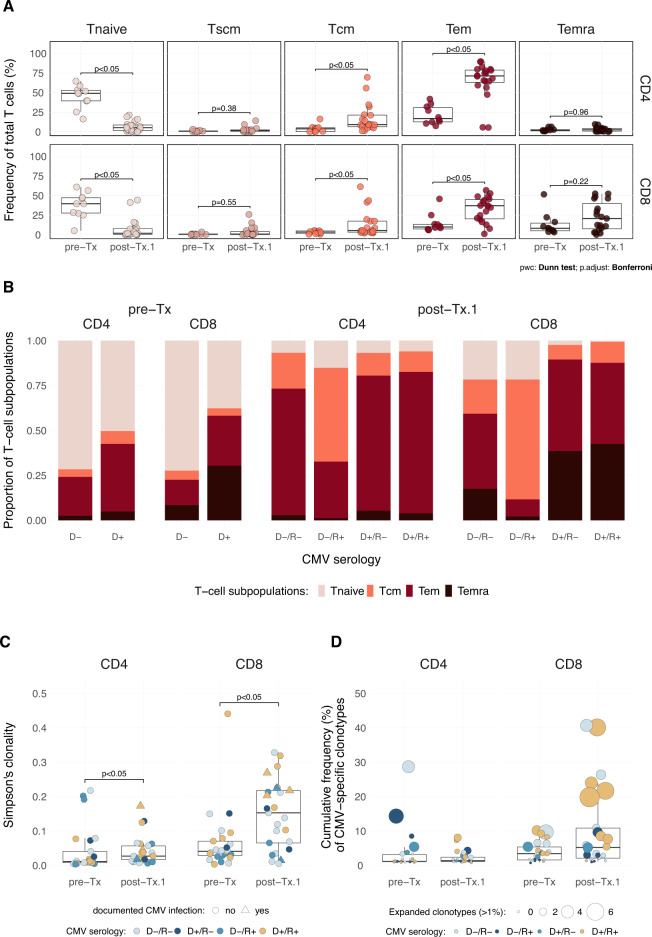
A Frequency of the Tnaive, Tscm, Tcm, Tem, and Temra cells in the CD4+ and CD8+ T-cell populations. Box plots show the median frequency per group, and dots correspond to one subject. B Proportion of Tnaive, Tcm, Tem, and Temra subsets within four sets of donor/recipient pairs (D/R) classified on their CMV serotype (D-/R-, D+/R-, D-/R+ and D+/R+). Only the donor status is reported for pre-Tx data. Each bar plot displays the four T cell subsets distribution in CD4+ and CD8+ compartments at both time points. C Simpson’s clonality of the TCR CDR3β repertoire from CD4+ and CD8+ fractions. Each dot corresponds to a subject and is color-coded according to the CMV serologic status (see B). D Clonal expansion of CMV-specific clonotypes. Box plots show the cumulative frequency of CMV-specific clonotypes. The dot’s size denotes the number of the most abundant CMV-specific clonotypes (i.e., expanded clones with a frequency above 1%). D: donor, R: recipient, (−) CMV negative, (+) CMV positive. Boxes show median and interquartile ranges, and dots correspond to individuals. Dunn’s test was used for multiple comparisons between groups for each T cell fraction. P values below 0.05 were considered statistically significant.

A Three time points were considered and compare the repertoire in 10 related and 16 unrelated donors prior to transplantation (pre-Tx), at 1 year post-HSCT in 26 full chimeric recipients (post-Tx.1) and their follow-up at 5 or 6 years (post-Tx.5). At the latter time point, 23 recipients were still fully chimeric, while 3 had mixed chimerism (i.e., recipient chimerism estimated between 7% and 15% in peripheral blood mononuclear cells according to an analysis performed with microsatellite markers). Clonality and Morisita’s values are shown along the y axis with lines connecting donors and recipients. In B, Simpson’s clonality and Morisita’s index are plotted according to CMV serologic status and a documented CMV reactivation or infection (i.e., defined as CMV DNA in plasma above the limit of detection, currently 2.1E + 1 UI/ml, in patients with or without clinical symptoms) within the first year post-transplantation. Of note, no CMV reactivation or infection was documented in the last year prior to the follow-up at 5 or 6 years. The clonal expansion of the repertoire across the three time points is presented in C according to CMV serologic status. The cumulative frequency of clonotypes for a given category is plotted with color representing the level of expansion considered based on the number of sequenced reads. D provides a detailed view of the expansion of the repertoire in the nine D+/R+ pairs. The clonotypes are categorized as “private” (i.e., observed in only one donor/recipient pair of the current cohort) or “public” (i.e., observed in two or more donor/recipient pairs) and the data are stratified according to occurrence of CMV reactivation or infection within the first year post-transplantation. D: donor CMV negative (−) or positive (+), R: recipient CMV negative (−) or positive (+), Inf: CMV reactivation or infection within the first year post transplantation, Tx: allogeneic HSCT.
References
-
- Huttunen P, Taskinen M, Siitonen S, Saarinen-Pihkala UM. Impact of very early CD4(+) /CD8(+) T cell counts on the occurrence of acute graft-versus-host disease and NK cell counts on outcome after pediatric allogeneic hematopoietic stem cell transplantation. Pediatr Blood Cancer. 2015;62:522–8. doi: 10.1002/pbc.25347. - DOI - PubMed
