. 2023 Feb;29(2):412-421.
doi: 10.1038/s41591-022-02190-7. Epub 2023 Feb 16.

Efficient in vivo genome editing prevents hypertrophic cardiomyopathy in mice

Affiliations

Efficient in vivo genome editing prevents hypertrophic cardiomyopathy in mice

Daniel Reichart et al. Nat Med. 2023 Feb.

Abstract

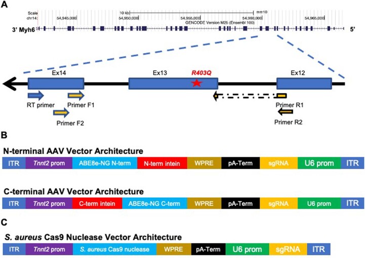
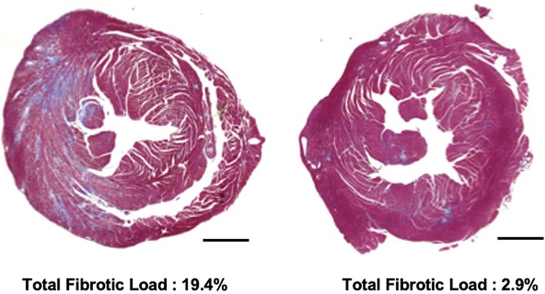
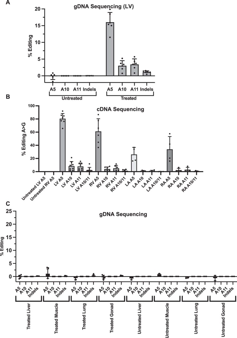
Dominant missense pathogenic variants in cardiac myosin heavy chain cause hypertrophic cardiomyopathy (HCM), a currently incurable disorder that increases risk for stroke, heart failure and sudden cardiac death. In this study, we assessed two different genetic therapies-an adenine base editor (ABE8e) and a potent Cas9 nuclease delivered by AAV9-to prevent disease in mice carrying the heterozygous HCM pathogenic variant myosin R403Q. One dose of dual-AAV9 vectors, each carrying one half of RNA-guided ABE8e, corrected the pathogenic variant in ≥70% of ventricular cardiomyocytes and maintained durable, normal cardiac structure and function. An additional dose provided more editing in the atria but also increased bystander editing. AAV9 delivery of RNA-guided Cas9 nuclease effectively inactivated the pathogenic allele, albeit with dose-dependent toxicities, necessitating a narrow therapeutic window to maintain health. These preclinical studies demonstrate considerable potential for single-dose genetic therapies to correct or silence pathogenic variants and prevent the development of HCM.


Conflict of interest statement

L.H.V. holds equity and serves on the Board of Directors for Affinia Therapeutics and Ciendias Bio, where he is employed, and holds equity in Akouos. He has licensed technology to Affinia, Akouos and Novartis. D.R.L. is a consultant and equity owner of Beam Therapeutics, Prime Medicine, Pairwise Plants and Chroma Medicine, companies that use genome editing or genome engineering. J.G.S. and C.E.S are founders of Myokardia (a Bristol Myers Squibb subsidiary) and are consultants for Maze and BridgeBio. C.E.S serves on the Merck Board of Directors and the Burroughs Wellcome Fund. None of these companies had any input into the design, execution, analyses or writing of this manuscript. Other authors declare no competing interests.

Figures

Fig. 1
Fig. 1. Base editing the pathogenic variant R403Q mediated by injection of dual-AAV9 ABE8e.
a, Schematic of the genomic sequence surrounding the HCM R403Q pathogenic variant. The ABE protospacer (green line) and PAM (purple line) are underlined. The pathogenic variant R403Q (numbered according to the human MYH7 amino acid residue) is shown with flanking amino acid residues (N, amino terminus; C, carboxyl terminus). R403Q is located at position A5 (blue), and potential bystander edits are at positions 10 (brown) and 11 (orange), numbered from the 5′ end of the protospacer. Bystander editing at each position (arrows) would encode missense amino acids. b, Schematic of the experimental design depicting AAV9 delivery and subsequent evaluation of disease. Dual-AAV9 ABE8e or single AAV9-Cas9 were injected (solid filled arrow, first dose; unfilled arrow, second dose) into R403Q-129SvEv or R403Q-129SvEv/S4 mice at postnatal weeks 2–3. All R403Q-129SvEv/S4 mice consistently develop LVH by 8–10 weeks (gray box), whereas only male R403Q-129SvEv mice show LVH at 20–25 weeks (blue box). Cardiac morphology and function were assessed by echocardiography at 2–4-week intervals. c, Editing efficiency (%) of the targeted pathogenic variant R403Q was based on high-throughput sequencing gDNA from all LV cells, including cardiomyocytes, fibroblasts, macrophages and endothelial cells, after a single dose of dual-AAV9 ABE8e injection (n = 6 males) or in untreated R403Q mice (n = 5 males), quantified by CRISPResso2. gDNA editing efficiency was calculated as: WT nucleotide percentage minus 50% (to measure the editing beyond the heterozygous baseline) and then divided by 50% (to determine the percentage of observed editing out of the theoretical maximum). d, Editing efficiency of the targeted R403Q allele after a single AAV9 ABE8e injection was assessed by sequencing Myh6 cDNA derived from RNA extracted from the LV (n = 5 males, 3 females), RV (n = 4 males, 2 females), LA (n = 2 males, 2 females) and RA (n = 2 males, 2 females) and in five untreated LVs and RVs from male mice. RNA editing efficiency was calculated as: WT nucleotide percentage minus 50% and then divided by 50%. Data are presented as mean values ± s.d. Source data
Fig. 2
Fig. 2. A single injection of dual-AAV9 ABE8e prevents hypertrophy and fibrosis in two strains of HCM mice.
a,b, Echocardiographic measurements of the LVPW thickness (left panels), ratio of LVPW and LVDd (middle panels) and %FS (right panels) in studied mice. Untreated R403Q-129SvEv and R403Q-129SvEv/S4 developed hypertrophy (increased LVPW thickness, reduced LV volumes and increased LVPW/LVDd ratios). R403Q-129SvEv/S4 has earlier onset of hypertrophy and hypercontractility (increased FS). a, Longitudinal echocardiographic measurements in WT 129SvEv (n = 4 males; black line), untreated R403Q-129SvEv (n = 10 males, red line) and R403Q-129SvEv (n = 6 males, blue line) mice treated with a single dose of dual-AAV9 ABE8e. b, Longitudinal echocardiographic measurements in WT R403Q-129SvEv/S4 (n = 5 males, 6 females; black line), untreated R403Q-129SvEv/S4 (n = 6 males, 6 females; red line) and R403Q-129SvEv/S4 (n = 3 males, 7 females; blue line) treated with a single dose of dual-AAV9 ABE8e. c, Left: representative Masson-trichrome-stained LV and RV histological sections from a single dose of dual-AAV9 ABE8e-treated (upper panel) and untreated (lower panel) male R403Q-129SvEv mice. Collagen deposition (blue staining) within regions of myocardial fibrosis was prominent in the untreated mouse, whereas the histology of the treated mouse was similar to WT hearts (Fig. 3b). Scale bars, 1 mm (low magnification) and 50 μm (high magnification). Right panel: quantification of fibrosis from Masson trichrome staining in 29 ventricular sections derived from six treated male mice and 17 sections from four untreated male mice. The fibrotic load in untreated mice ranged from 2.9% to 19.4% (Extended Data Fig. 3). The untreated section shown here has a mean fibrotic load of 3.4%. Data are presented as mean values ± s.d. Significance was assessed by two-tailed t-test (Supplementary Information). Source data
Fig. 3
Fig. 3. Two doses of dual-AAV9 ABE8e increase atrial but not ventricular base editing.
a, Longitudinal echocardiographic measurements (defined in Fig. 2) of the LVPW (left panel), LVPW/LVDd ratio (middle panel) and %FS (right panel) in WT 129SvEv/S4 (n = 5 males, black line), untreated R403Q-129SvEv/S4 (n = 6 males, red line) and R403Q-129SvEv/S4 mice treated with two doses of dual-AAV9 ABE8e (n = 6 males, blue line). Untreated but not treated R403Q-129SvEv/S4 developed early-onset hypertrophy (increased LVPW thickness and increased LVPW/LVDd) and hypercontractility (increased FS). b, Left: representative Masson-trichrome-stained LV and RV histological sections from WT 129SvEv/S4 male (left), treated (two doses of AAV9 ABE8e) male R403Q-129SvEv/S4 (middle) and untreated R403Q-129SvEv/S4 (right) male mice. Collagen deposition (blue staining) within regions of myocardial fibrosis was prominent in the untreated but minimal in the treated mouse and WT mice. (Scale bar, 1 mm). Right panel: quantification of fibrosis from Masson-trichrome-stained ventricular sections (five per mouse) from untreated (n = 5 males) and treated (n = 3 males; two doses) of AAV9 ABE8e R403Q-129SvEv/S4 mice. c, Editing efficiency of the targeted R403Q allele and indels was assessed in LV gDNA derived from five treated and five untreated male mice. d, Editing efficiency was assessed by sequencing Myh6 cDNA derived from RNAs extracted from the LVs, RVs, LAs and RAs from three male mice. Atrial editing was increased after two doses. e, The mean percent of bystander edits was detected in pooled LV cDNAs from R403Q-129SvEv/S4 mice (n = 3 males) treated with two doses. Data are presented as mean values ± s.d., and significance was assessed by two-tailed t-test (Supplementary Information). Source data
Fig. 4
Fig. 4. AAV9-Cas9 silencing of the pathogenic variant R403Q and cardiac function in R403Q-129SvEv mice.
a, Schematic of the genomic sequence surrounding the HCM R403Q pathogenic variant, showing the pathogenic variant R403Q (numbered according to the human MYH7 amino acid residues) and flanking amino acid residues (N, amino terminus; C, carboxyl terminus). The S. aureus Cas9 nuclease protospacer (green line), PAM site (purple line) and double-stranded cleavage position of the nuclease (dotted orange line) are shown. b, The percent of inactivation of the R403Q allele after 1 × 1013 vg kg−1 of AAV9-Tnnt2-S. aureus-Cas9 (designated AAV9-Cas9) was assessed by sequencing Myh6 cDNA, amplified from RNA extracted from each cardiac chamber of surviving R403Q-129SvEv mice (n = 4 males) at 33 weeks. The percentage of inactivation of the pathogenic variant R403Q was calculated as: (1 − (total number of R403Q reads, divided by total number of WT reads)) multiplied by 100. c, Longitudinal echocardiographic measurements (defined in Fig. 2) of the LVPW (left panel), LVPW/LVDd ratio (middle panel) and %FS (right panels) in WT R403Q-129SvEv (n = 4 males; black line), untreated R403Q-129SvEv (n = 10 males, red line) and treated (AAV9-Cas9) R403Q-129SvEv (n = 5 males; blue line) mice. Note impaired contractile function (FS <40%) in treated mice. Data are presented as mean values ± s.d., and significance was assessed by two-tailed t-test (Supplementary Information). Source data
Fig. 5
Fig. 5. Assessment of R403Q-allele inactivation and echocardiographic findings in R403Q-129SvEv/S4 mice treated with low, medium and high doses of AAV9-Cas9.
a, Percent of R403Q alleles with indels in three untreated male or AAV9-Cas9 treated (low dose: n = 1 male, 2 females; medium dose, n = 3 females and high dose, n = 4 females) R403Q-129SvEv/S4 mice was assessed by next-generation sequencing of PCR-amplified LV gDNA and analyzed using CRISPResso2. b, Myh6 cDNAs, amplified from RNAs extracted from each cardiac chamber of R403Q-129SvEv/S4 mice, treated with low (1.1 × 1012 vg kg−1; n = 1 male, 2 females), medium (5.4 × 1012 vg kg−1; n = 3 females) and high (2.2 × 1013 vg kg−1; n = 2 females) doses of AAV9-Cas9 for 18–33 weeks. The percentage of inactivation of the pathogenic variant R403Q was calculated as: (1 – (total number of R403Q reads, divided by total number of WT reads)) multiplied by 100. c, Longitudinal echocardiographic measurements LVPW, LVPW/LVDd and FS (defined in Fig. 2) in WT 129SvEv/S4 (n = 5 males, 5 females, black line) and untreated R403Q-129SvEv/S4 (n = 5 males, 6 females, red line) and treated (AAV9-Cas9 2.2 × 1013 vg kg−1) R403Q-129SvEv/S4 (n = 6 females, blue line). Note impaired contractile function (FS <40%) in high-dose-treated mice. d, Echocardiographic measurements of LVPW thickness, LVPW/LVDd ratio and FS (defined in Fig. 2) in R403Q-129SvEv/S4 mice at 20 weeks of age treated with low (1.1 × 1012 vg kg−1, n = 1 male, 2 females), medium (5.4 × 1012 vg kg−1, n = 3 females) and high (2.2 × 1013 vg kg−1, n = 6 females) doses of AAV9-Cas9, compared to untreated WT 129SvEv/S4 (n = 3 females) and untreated R403Q-129SvEv/S4 (n = 4 females) mice. Note that one mouse treated with the low dose had increased LVPW and increased LVPW/LVDd, indicating a non-therapeutic response. Data are presented as mean values ± s.d., and significance was assessed by two-tailed t-test (Supplementary Information). Source data
Extended Data Fig. 1
Extended Data Fig. 1. AAV9 vectors used to assess cardiotropic co-expression of fluorescent marker genes.
A) Schematics of AAV9 genomes containing fluorescent reporter genes (not to scale) used to assess dual transduction. ITR: AAV9 inverted terminal repeats. Tnnt2 prom: cardiomyocyte specific troponin T promoter. CMV prom: Cytomegalovirus promoter, a strong, constitutive promoter. WPRE: Woodchuck hepatitis virus post-transcriptional regulatory element, used to stabilize transcripts. pA-Term: polyadenylation signal and transcriptional terminator. B) Fluorescent micrographs of ventricular section (short axis view) and liver from a WT mouse injected with 3×10¹³ vg/kg AAV2/9-Tnnt2-eGFP.RBG (Tnnt2-eGFP.RBG) and AAV2/9-CMV-mScarlet.WPRE.bGH (CMV-mScarlet.WPRE.bGH) and imaged with a fluorescence microscope. Scale bar: 1 mm. Imaging revealed co-expression of green fluorescent protein (top panel) delivered by AAV9-Tnnt2-eGFP.RBG and red fluorescent protein (middle panel) delivered by AAV9-CMV-mScarlet.WPRE.bGH are co-localized (yellow signal, lower panel) in cardiac cells. Representative images are shown from two technical replicates.
Extended Data Fig. 2
Extended Data Fig. 2. Schematic of Myh6 gene and AAV vector genomes.
A) Schematic of Myh6 gene indicating the location of the pathogenic variant R403Q (numbered according to the human amino acid residues) in exon 13 and the targeted Myh6 mRNA region that was amplified to assess editing (Methods). The locations of reverse transcriptase primer (RT-primer) and PCR primer pairs 1 and 2 are indicated. B) Schematics of dual AAV vector genomes (not to scale) that encode an amino-terminal fragment (N-term) and carboxyl-terminal fragment (C-term) that form base editors after protein splicing. ITR: inverted terminal repeats. Tnnt2 Prom: the cardiomyocyte-specific troponin T promoter. WPRE: Woodchuck hepatitis virus post-transcriptional regulatory element, used to stabilize transcripts. pA-Term: polyadenylation signal and transcriptional terminator. sgRNA: single guide RNA. U6 prom: the human Pol III promoter that efficiently transcribes guide RNAs. C) Schematic of the AAV vector genome used for Cas9 nuclease delivery (not to scale). ITR: Inverted terminal repeats. Tnnt2 prom: the cardiomyocyte specific troponin T promoter. WPRE: Woodchuck hepatitis virus post-transcriptional regulatory element, used to stabilize transcripts. pA-Term: polyadenylation signal and transcriptional terminator. U6 prom: the human Pol III promoter that efficiently transcribes guide RNAs. sgRNA: single guide RNA.
Extended Data Fig. 3
Extended Data Fig. 3. Cardiac histopathology in untreated R403Q-SvEV mice.
Representative Masson-trichrome stained ventricular sections (short axis view) from two untreated R403Q-129SvEv mice. Across all sections of untreated mice the extent of collagen deposition (blue staining), indicative of regions of myocardial fibrosis, was assessed. Shown here are sections from two mice with a mean percentage of myocardial fibrosis equaling 2.9% (right) and 19.4% (left). Scale bars: 1 mm.
Extended Data Fig. 4
Extended Data Fig. 4. Bystander edits assessed after single injection of AAV9-ABE8e.
A) Editing efficiencies (%) measured after a single injection of AAV9-ABE8e based on high-throughput sequencing of PCR product amplified from LV gDNA (untreated n = 5, treated n = 6), quantified by CRISPResso2 (https://github.com/pinellolab/CRISPResso2). Editing efficiency at the on-target site A5 is compared to editing at bystander nucleotides A10 and A11. Indel rates were also assessed. B) Editing efficiencies (%) in untreated mice and mice treated with a single dose of AAV9-ABE8e. High-throughput sequencing of PCR product amplified from Myh6 cDNA derived from untreated LV and treated LV (n = 8), RV (n = 6), LA (n = 4) and RA (n = 4) identified bystander nucleotides A10, A11 or both (A10/11). C) Editing efficiency (%) assessed by sequencing PCR amplified gDNA extracted from non-targeted tissues: liver, skeletal muscle, lung and gonads from 6 treated and 3 untreated mice. Note low levels of on target Myh6 editing of the pathogenic variant R403Q (A5) in these non-targeted tissues. Data are presented as mean values ±SD. Source data
Extended Data Fig. 5
Extended Data Fig. 5. Liver and lung histology after AAV9-ABE8e injection.
Representative sections of liver (left) and lung (right) obtained from R403Q-129SvEv mice (n = 3) injected with 1.1×10¹³ AAV9-ABE8e vg/kg showed normal architecture and no infiltrates of inflammatory cells.
Extended Data Fig. 6
Extended Data Fig. 6. Bystander editing analysis after two injections of AAV9-ABE8e in each cardiac chamber.
A) Editing efficiency (%) of the targeted pathogenic variant R403Q based on high-throughput sequencing of PCR product amplified from gDNA extracted from LV tissues from untreated (n = 5) and treated (n = 5; two doses of AAV9-ABE8e) mice. Editing efficiency at the dominant heterozygous R403Q pathogenic variant is calculated as: (wild type nucleotide percentage – 50%) divided by 50%. B) Editing efficiencies (%) after two injections based on high-throughput sequencing of PCR products amplified from Myh6 cDNA derived from RNA extracted from the LV (n = 3), RV (n = 3), LA (n = 3), and RA (n = 3) at bystander nucleotides A10, A11 or both (A10/11). Data are presented as mean values ±SD. These data are summarized in Fig. 3. Source data
Extended Data Fig. 7
Extended Data Fig. 7. LV RNA expression in WT, treated R403Q and untreated R403Q mice.
A) Principal component analyses of WT (n = 12, black triangles), R403Q-129SvEv/S4 (n = 6, red circles) and treated (2 doses of AAV9-ABE8e) R403Q-129SvEv/S4 (n = 3, blue squares) mice. Principal component 1 (PC1) provided the best discrimination of phenotype and treatment, accounting for 25% of the variance. PC3 accounted for 12% of the variance. B) and C) Expression levels of genes involved in development of LV hypertrophy, sarcomere structure, cytoskeletal function, calcium handling, extracellular matrix and metabolism were assessed in untreated (n = 6) and treated (two doses of AAV9-ABE8e; n = 3) R403Q-129SvEv/S4 mice. * denotes RNAs with significantly (p < 0.05) altered expression in treated compared to untreated R403Q-129SvEv/S4. Data are presented as mean values ±SD and significance assessed by two-sided T-test (provided in Supplementary Table S3). Source data
Extended Data Fig. 8
Extended Data Fig. 8. Off-target editing analysis in AAV9-ABE8e treated mice.
Candidate genomic loci identified by CIRCLE-seq (see Methods) were PCR-amplified and sequenced by high throughput DNA sequencing (Illumina MiSeq). Adenine base editing at the nucleotide closest to the window center was quantified by CRISPResso2 (https://github.com/pinellolab/CRISPResso2). A) Detected Off-target editing in untreated mice (n = 3) compared to mice receiving two doses of base editor AAVs sacrificed at 5 weeks of age (n = 2). B) Detected off-target editing in untreated mice (n = 3) compared to mice receiving two doses of base editor AAVs sacrificed at 30 weeks of age (n = 3). C) Assessment of RNA editing (A-to-I nucleotide changes) by ABE8e in RNA sequencing data from LV tissues of treated (n = 3) and untreated (n = 4) R403Q mice. Data are presented as mean values ±SD and significance assessed by two-sided T-test (provided in Source Data). Source data
Extended Data Fig. 9
Extended Data Fig. 9. Genomic DNA sequencing of mice treated with AAV9-Cas9 nuclease.
A) The frequency of unmodified WT alleles measured from high-throughput sequencing of PCR products amplified from the targeted locus in gDNA from the LV of WT mice treated with high dose (1.1×10¹³) AAV9-Cas9 with sgRNA (n = 2) or without sgRNA (n = 2). B) and C) The frequency of unmodified WT and R403Q alleles measured from high-throughput sequencing of the targeted genomic locus in gDNA from LVs of 3-4 untreated mice and mice treated with low, medium, and high doses of AAV9-Cas9 mice after 5 weeks (B) and 30 weeks (C). Data are presented as mean values ±SD and significance was assessed by two-sided T-test. Source data
Extended Data Fig. 10
Extended Data Fig. 10. Assessment of Indels from extracardiac tissues derived from mice treated with AAV9-Cas9 nuclease.
Indels were measured by high-throughput sequencing of PCR products amplified from the targeted Myh6 region in gDNA extracted from the liver, lung, gonad and skeletal muscle (quadricep) of mice treated with AAV9-Cas9 nuclease (n = 2-3 per dose) and from untreated mice (n = 3). Reads were aligned to the target site by CRISPResso2. CRISPResso2 quantification of indels is plotted for each sample. Source data

Comment in

Similar articles

Cited by

References

    1. Garfinkel AC, Seidman JG, Seidman CE. Genetic pathogenesis of hypertrophic and dilated cardiomyopathy. Heart Fail Clin. 2018;14:139–146. doi: 10.1016/j.hfc.2017.12.004. - DOI - PMC - PubMed
    1. Yotti R, Seidman CE, Seidman JG. Advances in the genetic basis and pathogenesis of sarcomere cardiomyopathies. Annu. Rev. Genomics Hum. Genet. 2019;20:129–153. doi: 10.1146/annurev-genom-083118-015306. - DOI - PubMed
    1. Walsh R, et al. Reassessment of Mendelian gene pathogenicity using 7,855 cardiomyopathy cases and 60,706 reference samples. Genet. Med. 2017;19:192–203. doi: 10.1038/gim.2016.90. - DOI - PMC - PubMed
    1. Ho CY, et al. Genotype and lifetime burden of disease in hypertrophic cardiomyopathy: insights from the Sarcomeric Human Cardiomyopathy Registry (SHaRe) Circulation. 2018;138:1387–1398. doi: 10.1161/CIRCULATIONAHA.117.033200. - DOI - PMC - PubMed
    1. Ho CY, et al. Assessment of diastolic function with Doppler tissue imaging to predict genotype in preclinical hypertrophic cardiomyopathy. Circulation. 2002;105:2992–2997. doi: 10.1161/01.CIR.0000019070.70491.6D. - DOI - PubMed

Publication types