

The clinicopathological significance of thymic epithelial markers expression in thymoma and thymic carcinoma
- PMID: 36797681
- PMCID: PMC9936685
- DOI: 10.1186/s12885-023-10619-6
The clinicopathological significance of thymic epithelial markers expression in thymoma and thymic carcinoma
Abstract
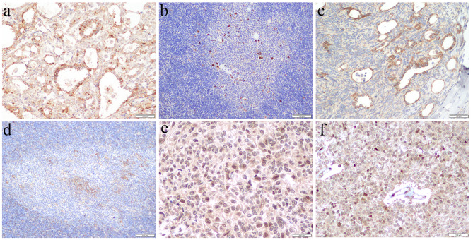
Background: The classification of thymomas is based on the morphology of epithelial tumor cells and the proportion of lymphocytes. Type A thymomas are composed of the spindle or oval tumor epithelial cells. Tumor cells of B thymomas are epithelioid-shaped with increasing atypia. Type AB thymomas have the features of epithelial tumor cells of A and B thymomas. The diagnosis can be difficult because of the complex morphology. Some novel thymic epithelial markers have been reported in several preclinical studies, but they have not been applied to clinical practice. Here, we investigated the expression of 3 cortical and 3 medullary markers, which are thymoproteasome-specific subunit β5t (β5t), thymus-specific serine protease 16 (PRSS16), cathepsin V, autoimmune regulator (AIRE), CD40 and claudin-4.
Methods: Immunohistochemistry was used to analyze 53 cases of thymomas and thymic squamous cell carcinomas (TSCC), aiming to explore the expression of cortical and medullary epithelial markers and their correlation with histological classification, Masaoka-Koga stage, and prognosis.
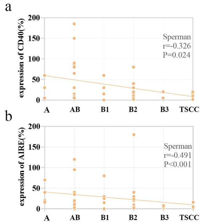
Results: Our results found that for cortical epithelial markers the expression of β5t, PRSS16, and cathepsin V was higher in type AB and B thymomas than in micronodular thymoma with lymphoid stroma (MNT), and we observed a dramatic increase of β5t and PRSS16 expression in type AB compared to type A thymomas. In medullary epithelial markers, the expression of AIRE was higher in type A than in B3 thymomas. CD40 and β5t expression were associated with the Masaoka-Koga stage. High cathepsin V expression was related to a good prognosis and a longer progression-free survival.
Conclusion: This is the first comprehensive analysis of the role of thymic cortical and medullary epithelial markers as biomarkers for differential diagnosis and prognosis in thymic tumors. Thymic medullary epithelial immunophenotype was found to exhibit in type A, MNT, and TSCC. Type B thymomas primarily exhibited a cortical epithelial immunophenotype. Type AB thymomas showed cortical, medullary, or mixed corticomedullary epithelial immunophenotype. Our results demonstrated that thymic cortical and medullary epithelial markers including β5t, PRSS16, cathepsin V, and AIRE could be used as ancillary markers in the diagnosis and prognosis of thymic epithelial tumors.
Keywords: Cortex; Differentiation; Immunohistochemical staining; Medulla; Thymic epithelial markers; Thymic squamous cell carcinoma; Thymoma.
© 2023. The Author(s).
Conflict of interest statement
The authors declare that they have no competing interests.
Figures


Similar articles
-
Corticomedullary differentiation and maturational arrest in thymomas.Histopathology. 2014 Mar;64(4):557-66. doi: 10.1111/his.12279. Epub 2013 Nov 18. Histopathology. 2014. PMID: 24236644
-
Expression of proteasome subunit β5t in thymic epithelial tumors.Am J Surg Pathol. 2011 Sep;35(9):1296-304. doi: 10.1097/PAS.0b013e3182237f5d. Am J Surg Pathol. 2011. PMID: 21836487
-
Expression of thymoproteasome subunit β5t in type AB thymoma.J Clin Pathol. 2014 Mar;67(3):276-8. doi: 10.1136/jclinpath-2013-201930. Epub 2013 Nov 29. J Clin Pathol. 2014. PMID: 24293611
-
Histogenetic and disease-relevant phenotypes in thymic epithelial tumors (TETs): The potential significance for future TET classification.Pathol Int. 2023 Jul;73(7):265-280. doi: 10.1111/pin.13343. Epub 2023 Jun 6. Pathol Int. 2023. PMID: 37278579 Review.
-
Pathological aspects of malignant and benign thymic disorders.Ann Med. 1999 Oct;31 Suppl 2:5-14. Ann Med. 1999. PMID: 10574149 Review.
Cited by
-
Thymic Carcinoma: Unraveling Neuroendocrine Differentiation and Epithelial Cell Identity Loss.Cancers (Basel). 2023 Dec 25;16(1):115. doi: 10.3390/cancers16010115. Cancers (Basel). 2023. PMID: 38201543 Free PMC article.
-
A rare case of tumor-to-tumor metastasis from thymic carcinoma to an ovarian mature teratoma.Thorac Cancer. 2024 Apr;15(11):934-937. doi: 10.1111/1759-7714.15277. Epub 2024 Mar 11. Thorac Cancer. 2024. PMID: 38468427 Free PMC article.
-
Synchronous HPV-related Tonsillar Squamous Cell Carcinoma and Type AB Thymoma: Successful Cancer Treatment Complicated by Fatal COVID-19 Infection.In Vivo. 2025 Jul-Aug;39(4):2085-2090. doi: 10.21873/invivo.14003. In Vivo. 2025. PMID: 40578983 Free PMC article.
-
Hypofractionated Radiotherapy as a Standalone Treatment Modality for Locally Advanced Type B2 Thymoma in an Octogenarian Patient: 45 Gy in 15 Fractions.Cureus. 2024 Jan 2;16(1):e51528. doi: 10.7759/cureus.51528. eCollection 2024 Jan. Cureus. 2024. PMID: 38304685 Free PMC article.
-
Pathological snapshots of thymic epithelial tumors with invasion into neighboring structures: preparing for the forthcoming revision of the TNM classification.Mediastinum. 2023 Sep 19;7:36. doi: 10.21037/med-23-28. eCollection 2023. Mediastinum. 2023. PMID: 38090038 Free PMC article. Review.
References
-
- WHO Classification of Tumours Editorial Board . Thoracic tumours. WHO classification of Tumours. 5. Lyon, France: International Agency for Research on Cancer; 2021.
-
- Valavanis C, Stanc GM, Baltayiannis N. Classification, histopathology and molecular pathology of thymic epithelial tumors: a review. J BUON: official J Balkan Union Oncol. 2021;26(4):1198–207. - PubMed
MeSH terms
Substances
Grants and funding
LinkOut - more resources
Full Text Sources
Medical
Research Materials
