

Ectopic sphenoidal ACTH-secreting adenoma revealed by 11C Methionine PET scan: case report
- PMID: 36797716
- PMCID: PMC9933249
- DOI: 10.1186/s12902-023-01298-2
Ectopic sphenoidal ACTH-secreting adenoma revealed by 11C Methionine PET scan: case report
Abstract
Background: Ectopic ACTH pituitary adenomas (EAPA), located outside the sella turcica and deriving from cellular remnants of Rathke's pouch are a very rare cause of Cushing's syndrome (CS). The diagnosis is often difficult and delayed, even after comprehensive work-up. To our knowledge, we report for the first time an ectopic corticotroph tumor of the posterior wall of the sphenoid sinus, leading to false positive results of bilateral inferior petrosal sinus sampling (BIPPS) and which was finally localized by a co-registered11 C Methionine PET/MR imaging.
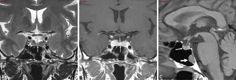
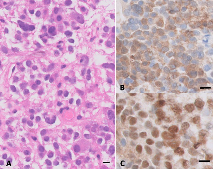
Case presentation: A 48-year-old woman was referred for a high clinical suspicion of ACTH-dependent CS. Biological testing comprising low dose dexamethasone suppression and CRH stimulation tests were indicative of pituitary Cushing's disease, but comprehensive pituitary MRI did not reveal any pituitary adenoma. BIPSS confirmed however a central origin of ACTH secretion (central-to-peripheral ACTH ratio > 100) and revealed a significant right-to-left gradient (6.2), leading to a first right-sided exploratory hypophysectomy, that did not cure the patient. BIPSS images were reviewed and revealed preferential drainage of the left pituitary to the right petrosal sinus, leading us to a left sided exploratory hypophysectomy, which was again unsuccessful. A11 C Methionine PET/MRI was performed and revealed a hypermetabolic lesion adjacent to the posterior wall of the sphenoidal sinus. After surgical resection, this polypoid mass was identified as an ectopic ATCH-secreting pituitary adenoma expressing ACTH and T-Pit and complete remission of hypercortisolism was observed.
Conclusions: In conclusion, we report a case of ACTH-dependent Cushing's syndrome, caused by an ectopic corticotroph adenoma located in the sphenoidal sinus, which perfectly mimicked the biological features of a classical pituitary ACTH adenoma on a comprehensive hormonal evaluation including BIPPS, and the features of a benign naso-sinusal polyp at MRI. We report for the first time a key role of11 C Methionine PET co-registered to high resolution MRI for localizing ectopic adenomas, efficiently guiding surgical removal and leading to complete remission of hypercortisolism.
Keywords: 11C Methionine PET/MRI; ACTH-secreting adenoma; Cushing’s syndrome; Ectopic pituitary adenoma.
© 2023. The Author(s).
Conflict of interest statement
The authors declare that they have no competing interests.
Figures

References
-
- Pecori Giraldi F, Cavallo LM, Tortora F, et al. The role of inferior petrosal sinus sampling in ACTH-dependent Cushing’s syndrome: review and joint opinion statement by members of the Italian Society for Endocrinology, Italian Society for Neurosurgery, and Italian Society for Neuroradiology. Neurosurg Focus. 2015;38(2):E5. doi: 10.3171/2014.11.FOCUS14766. - DOI - PubMed
-
- Young J, Deneux C, Grino M, Oliver C, Chanson P, Schaison G. Pitfall of petrosal sinus sampling in a cushing’s syndrome secondary to ectopic Adrenocorticotropin-Corticotropin Releasing Hormone (ACTH-CRH) Secretion. J Clin Endocrinol Metab. 1998;83(2):305–308. doi: 10.1210/jcem.83.2.4549. - DOI - PubMed
Publication types
MeSH terms
Substances
LinkOut - more resources
Full Text Sources
Medical
