Review
. 2023 Feb 16;22(1):33.
doi: 10.1186/s12943-023-01741-x.

Recent advances of small extracellular vesicle biomarkers in breast cancer diagnosis and prognosis

Affiliations
Review

Recent advances of small extracellular vesicle biomarkers in breast cancer diagnosis and prognosis

Yujin Lee et al. Mol Cancer. .

Abstract

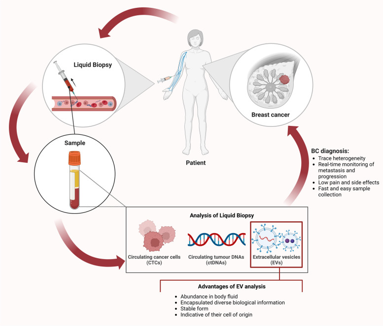
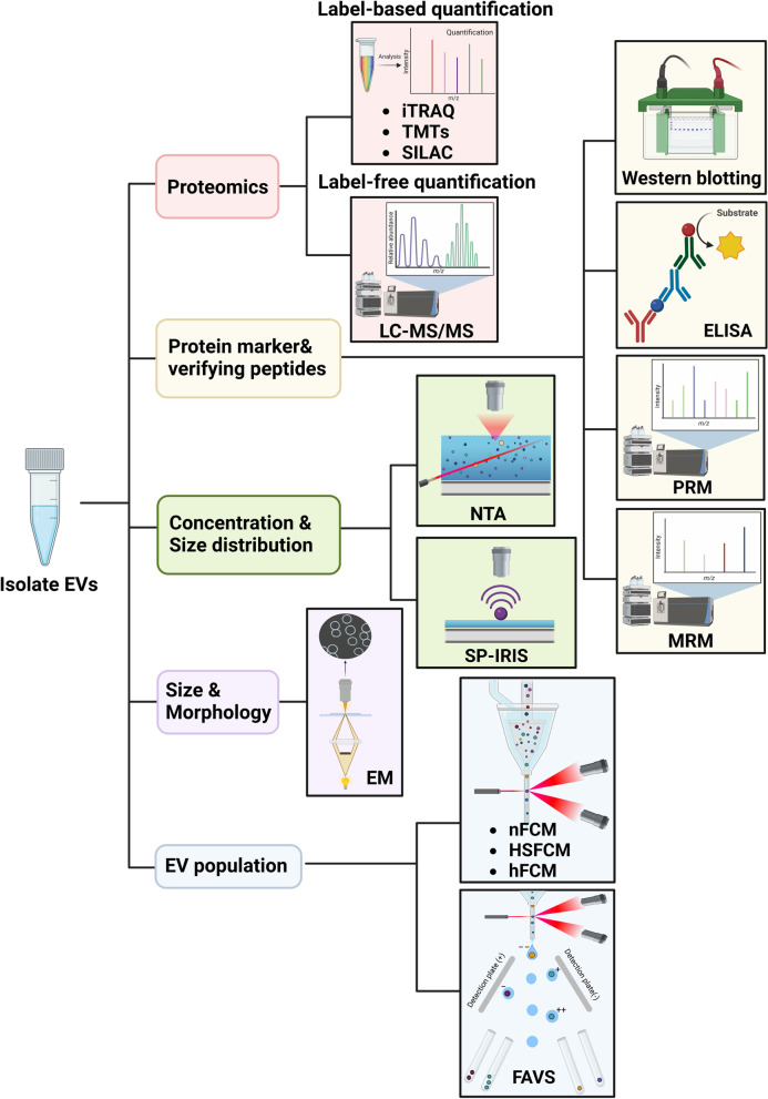
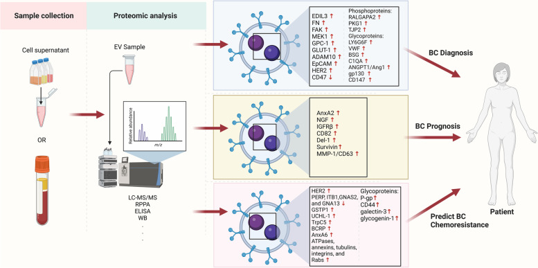
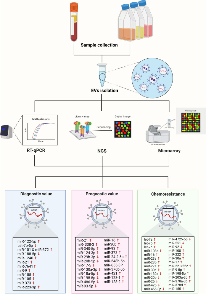
Current clinical tools for breast cancer (BC) diagnosis are insufficient but liquid biopsy of different bodily fluids has recently emerged as a minimally invasive strategy that provides a real-time snapshot of tumour biomarkers for early diagnosis, active surveillance of progression, and post-treatment recurrence. Extracellular vesicles (EVs) are nano-sized membranous structures 50-1000 nm in diameter that are released by cells into biological fluids. EVs contain proteins, nucleic acids, and lipids which play pivotal roles in tumourigenesis and metastasis through cell-to-cell communication. Proteins and miRNAs from small EVs (sEV), which range in size from 50-150 nm, are being investigated as a potential source for novel BC biomarkers using mass spectrometry-based proteomics and next-generation sequencing. This review covers recent developments in sEV isolation and single sEV analysis technologies and summarises the sEV protein and miRNA biomarkers identified for BC diagnosis, prognosis, and chemoresistance. The limitations of current sEV biomarker research are discussed along with future perspective applications.

Keywords: Biomarker; Breast cancer; Diagnosis; Extracellular vesicles; Liquid biopsy; Prognosis.


Conflict of interest statement

The authors declare no competing interests.

Figures

Fig. 1
Fig. 1
Liquid biopsy for breast cancer (BC) diagnosis. Body fluids are collected from BC patients for liquid biopsy analysis. Circulating tumour cells, circulating tumour DNA, and extracellular vesicles can be detected via liquid biopsy. This approach has several advantages in BC diagnosis such as ability to trace heterogeneity, monitoring of tumour metastasis and progression in a real-time, reduced pain and side effects, and it is a faster and easier process to obtain samples compared to tissue biopsy. Among the analytes detected by liquid biopsy, extracellular vesicles have superior characteristics, for example, abundance in various bodily fluids, stable form with encapsulated bioactive molecules, and reflective of their cell of origin. This figure was created with BioRender.com
Fig. 2
Fig. 2
Small extracellular vesicle (sEV) detection and characterisation. sEVs isolated from breast cancer (BC) blood samples and cell lines can be characterised by size, morphology, concentration, and sEV markers. sEV size distribution and concentration can be analysed by nanoparticle tracking analysis (NTA) and single particle interferometric reflectance imaging sensor (SP-IRIS). Various types of electron microscopy (EM) can help classify sEVs by size and morphology. Nano flow cytometry (nFCM), high-sensitivity flow cytometry (HSFCM), high-resolution flow cytometry (hFCM), and fluorescence-activated vesicle sorting (FAVS) can sort sEV subpopulations in the isolated samples using various sEV markers. sEV proteins can be profiled by mass spectrometry-based analysis including label-free and label-based quantification. For verifying sEV protein and peptide markers, multiple reaction monitoring (MRM), parallel reaction monitoring (PRM), western blotting (WB), and enzyme-linked immunosorbent assay (ELISA) can be used. This figure was created with BioRender.com
Fig. 3
Fig. 3
Small extracellular vesicle (sEV) protein biomarkers, including post-translational modifications (PTMs), in breast cancer (BC) diagnosis, prognosis, and chemoresistance. sEVs are collected from cell supernatants and blood samples of BC patients. Isolated sEVs are profiled by proteomic strategies such as liquid chromatography with tandem mass spectrometry (LC–MS/MS) and reverse phase protein microarray (RPPA) and validated by sEV protein detection methods including enzyme-linked immunosorbent assay (ELISA) and western blotting (WB). Various up- or down-regulated proteins in BC sEV samples were identified and can be used in BC diagnosis, prognosis, and prediction of BC chemoresistance. This figure was created with BioRender.com
Fig. 4
Fig. 4
Small extracellular vesicle (sEV) miRNA biomarkers in breast cancer (BC) diagnosis, prognosis, chemoresistance. BC plasma and cell supernatant samples are collected, and the isolated sEVs containing miRNAs are analysed by reverse transcriptase-quantitative polymerase chain reaction (RT-qPCR), next-generation sequencing (NGS), and microarray analysis. The unique sEV miRNA expression profiles of BC samples have value for diagnosis, prognosis, and predicting chemoresistance of BC. This figure was created with BioRender.com

References

    1. Siegel RL, Miller KD, Jemal A. Cancer statistics, 2019. CA Cancer J Clin. 2019;69(1):7–34. doi: 10.3322/caac.21551. - DOI - PubMed
    1. Siegel RL, Miller KD, Fuchs HE, Jemal A. Cancer Statistics, 2021. CA Cancer J Clin. 2021;71(1):7–33. doi: 10.3322/caac.21654. - DOI - PubMed
    1. Group USCSW. U.S. Cancer Statistics Data Visualizations Tool, Based on 2019 Submission Data (1999–2017). US Department of Health and Human Services. 2020.
    1. Indicators CAsNCC. Relative Survival by Stage at Diagnosis (Female Breast Cancer) 2019. https://ncci.canceraustralia.gov.au/relative-survival-stage-diagnosis-fe.... Accessed 20 Sept 2021.
    1. Clarke R, Tyson JJ, Dixon JM. Endocrine resistance in breast cancer–An overview and update. Mol Cell Endocrinol. 2015;418 Pt 3(0 3):220–34. doi: 10.1016/j.mce.2015.09.035. - DOI - PMC - PubMed

Publication types