. 2023 Feb 16;16(1):72.
doi: 10.1186/s13071-023-05660-0.

The therapeutic effect of larval saliva and hemolymph of Lucilia sericata on the treatment of Leishmania major lesion in BALB/c mice946

Affiliations

The therapeutic effect of larval saliva and hemolymph of Lucilia sericata on the treatment of Leishmania major lesion in BALB/c mice946

Sara Rahimi et al. Parasit Vectors. .

Abstract

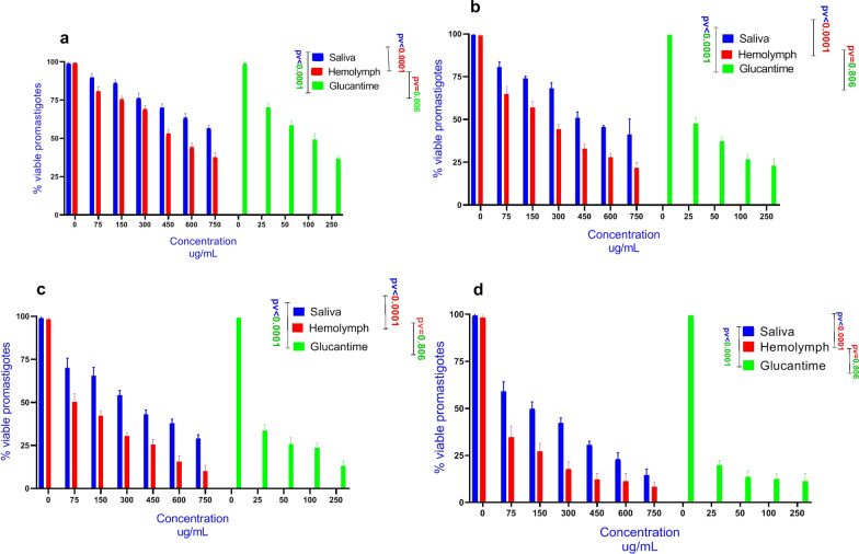
Background: Treatment of cutaneous leishmaniasis (CL) remains a major challenge for the public health and medical community. It has been claimed that natural compounds derived from fly larvae have anti-leishmania properties against some species of Leishmania. The present study aimed at assessing the in vitro effects of larval products of Lucilia sericata against the promastigote and intracellular amastigote forms of Leishmania major. Also, the therapeutic effect of larval products on lesions induced by L. major infection was evaluated in BALB/c mice models.

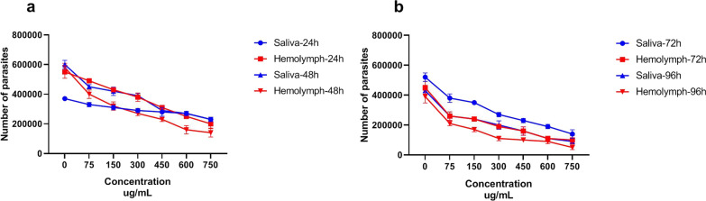
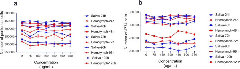
Methods: Parasite specimens and macrophage cells were exposed to varying concentrations of larval products for 24-120 h. Lesion progression and parasite load were investigated in the models to assess the therapeutic effects of the products.

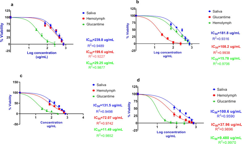
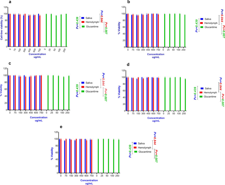
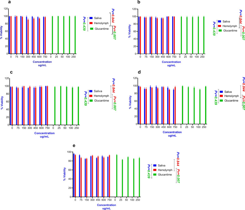
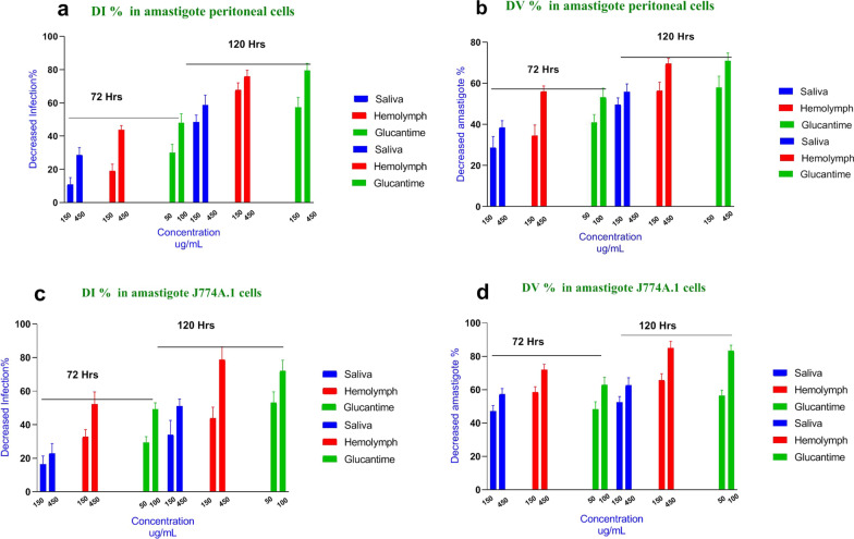
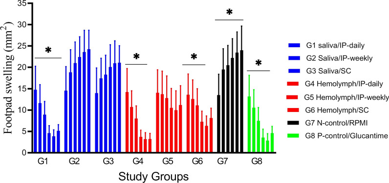
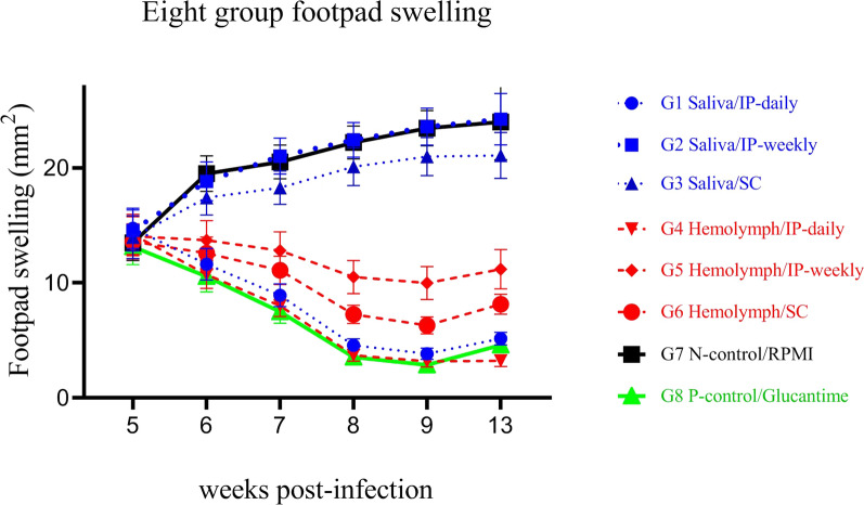
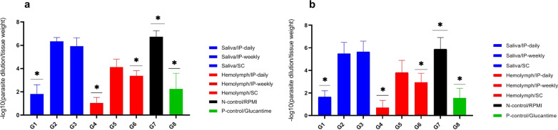
Results: The larval products displayed more potent cytotoxicity against L. major promastigotes. The IC50 values for larval saliva and hemolymph were 100.6 and 37.96 ug/ml, respectively. The IC50 of glucantime was 9.480 ug/ml. Also, the saliva and hemolymph of L. sericata exhibited higher cytotoxicity against the promastigotes of L. major but were less toxic to the macrophage cells. Treatment with leishmanicidal agents derived from larvae of L. sericata decreased the infection rate and the number of amastigotes per infected host cell at all concentrations. Lesion size was significantly (F (7, 38) = 8.54, P < 0.0001) smaller in the treated mice compared with the untreated control group. The average parasite burden in the treated mice groups (1.81 ± 0.74, 1.03 ± 0.45 and 3.37 ± 0.41) was similar to the group treated with a daily injection of glucantime (1.77 ± 0.99) and significantly lower (F (7, 16) = 66.39, P < 0.0001) than in the untreated control group (6.72 ± 2.37).

Conclusions: The results suggest that the larval products of L. sericata were effective against L. major parasites both in vivo and in vitro. However, more clinical trial studies are recommended to evaluate the effects of these larval products on human subjects.

Keywords: Cutaneous leishmaniasis; Glucantime; Leishmania major; Leishmanial activity; Lucilia sericata; Natural compound.


Conflict of interest statement

The authors have stated explicitly that there are no conflicts of interest in connection with this article.

Figures

Fig. 1
Fig. 1
Viability percentage of Leishmania major promastigotes treated with different concentrations of Lucilia sericata larval saliva and hemolymph compared with glucantime. a %Viability of L. major promastigotes treated with different concentrations of larval products at 24 h. b %Viability of L. major promastigotes treated with larval products at 48 h. c %Viability of L. major promastigotes treated with larval products at 72 h. d %Viability of L. major promastigotes treated with larval products at 96 h
Fig. 2
Fig. 2
Number of viable Leishmania major promastigotes treated with Lucilia sericata larval products in different concentrations at different time points and counted in a Neubauer chamber. a Number of viable L. major at 24 and 48 h. b Number of viable L. major at 72 and 96 h
Fig. 3
Fig. 3
Dose-response curves regarding the effect of Lucilia sericata larval-derived products on Leishmania major promastigotes (IC50) using MTT assay. a Larval saliva IC50 vs. larval hemolymph IC50 at 24 h. b Larval saliva IC50 vs. larval hemolymph IC50 at 48 h. c Larval saliva IC50 vs. larval hemolymph IC50 at 72 h. d Larval saliva IC50 vs. larval hemolymph IC50 at 96 h and compared with standard treatment (glucantime)
Fig. 4
Fig. 4
Number of viable cells treated with larval products by trypan blue in different concentrations at different time points and counted in a Neubauer chamber. a Number of peritoneal viable cells treated with larval products. b The number of J774A.1 viable cells treated with larval products
Fig. 5
Fig. 5
Cytotoxicity test of different concentrations of Lucilia sericata larval products on J774A.1 murine macrophage cell line at different time points compared with glucantime. a Cell viability in cells treated with larval products at 24 h. b Cell viability in cells treated with larval products at 48 h. c Cell viability in cells treated with larval products at 72 h. d Cell viability in cells treated with larval products at 96 h. e Cell viability in cells treated with larval products at 120 h
Fig. 6
Fig. 6
Cytotoxicity test of the effects of different concentrations of Lucilia sericata larval products on peritoneal macrophage cell line at different time points compared with glucantime. a Cell viability in cells treated with larval products at 24 h. b Cell viability in cells treated with larval products at 48 h. c Cell viability in cells treated with larval products at 72 h. d Cell viability in cells treated with larval products at 96 h. e Cell viability treated with larval products at 120 h
Fig. 7
Fig. 7
In vitro larval products’ effect on both macrophage types infected with Leishmania major at 72 and 120 h compared with glucantime. a Decrease in infection percentage (DI%) in L. major amastigote peritoneal cells. b Decrease in amastigote viability (DV%) in L. major amastigote peritoneal cells. c Decrease in infection percentage (DI%) in L. major amastigote J774A.1 cells. d Decrease in amastigote viability (DV%) in L. major amastigote J774A.1 cells
Fig. 8
Fig. 8
Progress of lesion size in inoculated BALB/c mice by Leishmania major in study groups at 6 weeks. The bar represents the means ± standard errors of means. There was a significant difference (P < 0.05) between the larval product-treated groups (G1, G4, and G6) and the untreated group of mice (G7)
Fig. 9
Fig. 9
Footpad swelling in BALB/c mice treated with larval products and glucantime (G8) with different routes of administration at 6 weeks post-infection compared to the negative control group (G7)
Fig. 10
Fig. 10
Footpad and lymph node parasite burden in Leishmania major-infected BALB/c mice treated with larval saliva and hemolymph. The number of viable parasites per tissue was determined by the following formula: —log10 (parasite dilution/tissue weight). The bar represents the means ± standard errors of the means. There was a significant difference (P < 0.05) between the larval product-treated groups (G1, G4 and G6) and an untreated group of mice (G7). a Parasite burden in the foot; b parasite burden in lymph nodes

Similar articles

References

    1. Centers for Disease Control and Prevention 2020 [Internet]. [cited 2020 Feb 18]. https://www.cdc.gov/parasites/.
    1. World Health Organization 2021 [Internet]. [cited 2021 May 20]. https://www.who.int/news-room/fact-sheets/detail/leishmaniasis.
    1. McGwire BS, Satoskar A. Leishmaniasis: clinical syndromes and treatment. Int J Med. 2014;107:7–14. - PMC - PubMed
    1. De Menezes JPB, Guedes CES, Petersen ALdOA, Fraga DBM, Veras PST. Advances in the development of new treatments for leishmaniasis. Biomed Res Int. 2015;2015:1–11. - PMC - PubMed
    1. Kassebaum NJ, Arora M, Barber RM, Bhutta ZA, Brown J, Carter A, et al. Global, regional, and national disability-adjusted life-years (DALYs) for 315 diseases and injuries and healthy life expectancy (HALE), 1990–2015: a systematic analysis for the Global burden of disease study 2015. Lancet. 2016;388:1603–1658. doi: 10.1016/S0140-6736(16)31460-X. - DOI - PMC - PubMed

LinkOut - more resources