. 2023 Apr;601(7):1247-1264.
doi: 10.1113/JP284059. Epub 2023 Mar 3.

Cholinergic collaterals arising from noradrenergic sympathetic neurons in mice

Affiliations

Cholinergic collaterals arising from noradrenergic sympathetic neurons in mice

Courtney Clyburn et al. J Physiol. 2023 Apr.

Abstract

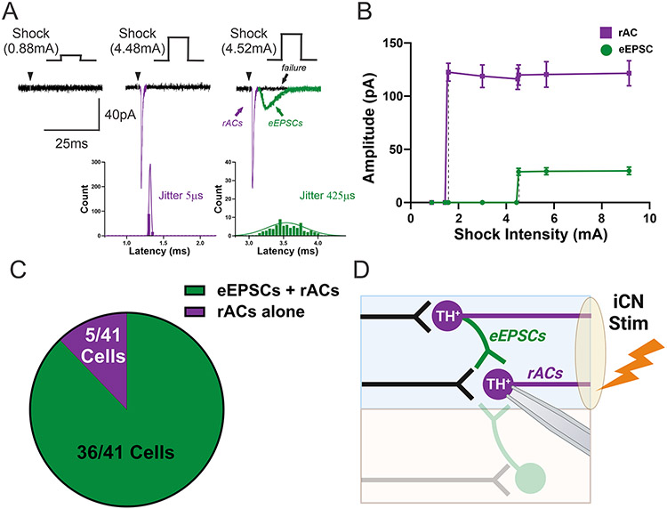
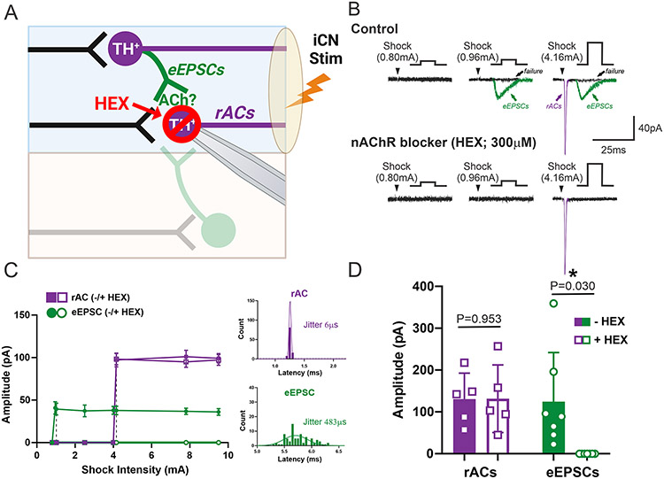
The sympathetic nervous system vitally regulates autonomic functions, including cardiac activity. Postganglionic neurons of the sympathetic chain ganglia relay signals from the central nervous system to autonomic peripheral targets. Disrupting this flow of information often dysregulates organ function and leads to poor health outcomes. Despite the importance of these sympathetic neurons, fundamental aspects of the neurocircuitry within peripheral ganglia remain poorly understood. Conventionally, simple monosynaptic cholinergic pathways from preganglionic neurons are thought to activate postganglionic sympathetic neurons. However, early studies suggested more complex neurocircuits may be present within sympathetic ganglia. The present study recorded synaptic responses in sympathetic stellate ganglia neurons following electrical activation of the pre- and postganglionic nerve trunks and used genetic strategies to assess the presence of collateral projections between postganglionic neurons of the stellate ganglia. Orthograde activation of the preganglionic nerve trunk, T-2, uncovered high jitter synaptic latencies consistent with polysynaptic connections. Pharmacological inhibition of nicotinic acetylcholine receptors with hexamethonium blocked all synaptic events. To confirm that high jitter, polysynaptic events were due to the presence of cholinergic collaterals from postganglionic neurons within the stellate ganglion, we knocked out choline acetyltransferase in adult noradrenergic neurons. This genetic knockout eliminated orthograde high jitter synaptic events and EPSCs evoked by retrograde activation. These findings suggest that cholinergic collateral projections arise from noradrenergic neurons within sympathetic ganglia. Identifying the contributions of collateral excitation to normal physiology and pathophysiology is an important area of future study and may offer novel therapeutic targets for the treatment of autonomic imbalance. KEY POINTS: Electrical stimulation of a preganglionic nerve trunk evoked fast synaptic transmission in stellate ganglion neurons with low and high jitter latencies. Retrograde stimulation of a postganglionic nerve trunk evoked direct, all-or-none action currents and delayed nicotinic EPSCs indistinguishable from orthogradely-evoked EPSCs in stellate neurons. Nicotinic acetylcholine receptor blockade prevented all spontaneous and evoked synaptic activity. Knockout of acetylcholine production in noradrenergic neurons eliminated all retrogradely-evoked EPSCs but did not change retrograde action currents, indicating that noradrenergic neurons have cholinergic collaterals connecting neurons within the stellate ganglion.

Keywords: acetylcholine; co-transmission; collaterals; neurocircuits; sympathetic ganglia; synaptic inputs.


Conflict of interest statement

Author Conflict: No competing interests declared

Figures

Figure 1:
Figure 1:. Genetic strategy for knockout of choline acetyltransferase in noradrenergic neurons.
We used genetics to selectively eliminate ACh synthesis in noradrenergic neurons while leaving preganglionic neurons unaffected. Mice with the genotype Ai9:THCreERT2;ChATfl/fl were treated with tamoxifen to induce Cre recombinase expression in tyrosine hydroxylase (TH) expressing cells. This led to Cre expression and deletion of ChAT/induction of TdTomato (Ai9 transgene) in most, but not all, TH+ neurons (i.e. THChAT KO). Left scale bar = 200μm. Right scale bar = 50μm.
Figure 2:
Figure 2:. T-2 stimulation evokes consistent-latency EPSCs in Control SG neurons
A. Five overlapping consecutive traces illustrates the low variability in the eEPSC waveform, amplitude, and latency in a representative Control SG neuron voltage-clamped at −60mV. Testing relied on stimulation of the T-2 nerve trunk (100 iterations, 200 μs duration, 5 sec interval, 0.1-10.0 mA stimulus range). At a low shock intensity (2.85 mA; black), eEPSCs arrived at nearly invariable times but increases in intensity above a critical level (5.40 mA; grey) abruptly evoked a compound event, eEPSC 1+2. B. This recruitment relationship indicates all-or-none eEPSC amplitudes with sharp transitions consistent with intensity-dependent recruitment of two different postganglionic neurons within the T-2 bundle. Note that the arrival of the second EPSC was very close to eEPSC 1 such that the two events could not be separately distinguished at the highest shock intensities. At the lowest intensity stimuli, the latencies of the isolated eEPSC 1 were distributed normally with a mean jitter of 121 μs (100 iterated eEPSCs) in the inset histogram. Black dotted lines highlight these sharp threshold transitions in response. C. Overall, SG neurons more commonly received multiple synaptic inputs (grey, 19/25 cells) compared to SG neurons receiving only a single input (black; 6/25 cells) from a total of 25 cells from 14 Control mice. D. Such results resemble the conventional view of the pathway of multiple preganglionic sympathetic axons converging to excite (eEPSCs) single postganglionic neurons.
Figure 3:
Figure 3:. Stimulation of the preganglionic nerve trunk (T-2) evokes high and low jitter EPSCs in SG neurons from Control mice
A. B. Ten overlapping consecutive traces illustrating low (A.) and high (B.) jitter EPSCs evoked from orthograde activation of the preganglionic nerve trunk, T-2, from a Control SG neuron voltage clamped at −60 mV using the minimum threshold required to evoke a current. The latency from the shock stimulus to the onset of the current (i.e. jitter) is magnified in the lower right. The distribution of latencies across 100 sweeps is represented in the top graph and demonstrates the jitter of these inputs. The bottom graph illustrates the amplitude of the eEPSC at increasing stimulation intensities and highlights the discrete threshold of these unitary inputs. The black dotted line highlights the small change in stimulation intensity that uncovers this input (bottom graph). C. There is a significant correlation between jitter and latency in the high jitter group (green: r(19) = 0.2059, p = 0.0227, 95% C.I. = 0.071, 0.720 via Pearson’s correlation). The threshold between high and low jitter groups is highlighted in grey. (*p<0.05 via Pearson’s correlation) D. The failure rate is significantly higher in the high jitter group compared to low jitter responses (green and black, respectively: 16.4±17.63% vs. 5.0±4.38%, p = 0.0168, t(22.62) = 2.582, 95% C.I. = −20.58, −2.263, via two-tailed unpaired Welch’s t test, N=19 cells from 14 mice and 6 cells from 5 mice, respectively, data represented as mean ± SD) E. The proportion of THWT SG neurons in the low jitter (black; 6 cells from 5 mice) or high jitter (green; 19 cells from 14 mice) following T-2 stimulation (N = 25 total cells from 18 mice). F. Such results are consistent with a polysynaptic pathway. Electrical stimulation of T-2 may excite a SG neuron with putative collateral projections to the primary recorded cell (Poly 1). The alternative hypothesis is that T-2 stimulation excites an interneuron in the SG that projects to the recorded neuron.
Figure 4:
Figure 4:. Retrograde activation of a postganglionic nerve trunk (iCN) evokes retrograde action currents (rACs) and eEPSCs in SG neurons from Control mice
A. Five overlapping traces illustrating currents evoked from retrograde activation of a postganglionic nerve trunk, the iCN, in a Control SG neuron voltage clamped at −60 mV at three stimulation intensities. At a low stimulation intensity (left, 0.88 mA shock) no observable current is evoked. At an intermediate stimulation intensity, the threshold for a retrograde action current (rAC; i.e. action potential recorded in voltage clamp, middle, purple) is reached. A distribution of latencies across 100 sweeps illustrates the extremely low jitter of rACs (bottom, purple). As the stimulation intensity is increased, the threshold for an eEPSC is reached (right, green). A distribution of latency (bottom, green) illustrates the high jitter of this unitary synaptic input. B. This amplitude at increasing stimulation intensities highlights the discrete threshold of the rAC (purple) and eEPSC (green) unitary event. The black dotted line highlights the small change in stimulation intensity that uncovers each input. C. Graphical representation of the proportion of THWT SG neurons in which eEPSCs and rACs were observed (36/41 (87.8%) of cells from 25 mice, green) or rACs alone (5/41 cells (12.2%) of cells from 25 mice, purple) D. These data are consistent with collateral transmission and rule out a cholinergic interneuron (faded). Electrical stimulation of the iCN could excite a nearby postganglionic neuron with a collateral projection to the primary recorded cell. However, stimulation of the iCN would not affect interneuron activity, suggesting that collateral projections are responsible for retrogradely-activated EPSCs in the SG.
Figure 5:
Figure 5:. Hexamethonium blocks eEPSCs after iCN stimulation but has no effect on rACs in SG neurons from Control mice
A. The aim of this experiment was to determine the neurochemical phenotype of the putative collateral projections in the SG. The nicotinic acetylcholine receptor antagonist, hexamethonium (HEX, 300μM, red) was used to block collateral synaptic transmission after electrical stimulation of a postganglionic nerve trunk, the iCN. B. Five overlapping traces illustrating currents evoked from stimulation of the iCN in a Control SG neuron voltage clamped at −60mV at a range of stimulation intensities before (top) and after (bottom) bath application of HEX. At low stimulation intensities (left, 0.80mA shock). At an intermediate stimulation intensity (middle) the threshold for an eEPSC (green) is reached. At higher stimulation intensities (right), the threshold for a rAC is reached (purple). After bath application of HEX (bottom), eEPSCs were completely blocked and there was no significant effect on rACs. C. HEX (open symbol) had no effect on rAC (purple) amplitude, but completely blocked eEPSCs (green). The distribution of latencies across 100 sweeps illustrates the low jitter of rACs and the high jitter of eEPSCs after iCN stimulation. D. HEX (open) had no effect on rAC amplitude (purple: 130.5±61.80 pA vs. 131.4±80.78 pA, p = 0.9531, t(4) = 0.0626, 95% C.I. = −35.20, 36.82 via two-tailed paired Student’s t test, N = 5 cells from 3 mice) but completely blocked eEPSCs (green: 124.6±117.30 pA vs. 0.0±0.00 pA, p = 0.0308, t(6) = 2.810, 95% C.I. = −233.1,−16.10, via two-tailed paired Welch’s t test, N = 7 cells from 5 mice, data represented as mean±SD).
Figure 6:
Figure 6:. Inhibition of collateral cholinergic transmission reduces spontaneous EPSC frequency
A. The aim of this experiment was to determine the neurochemical phenotype of the collateral projections in the SG. The nicotinic acetylcholine receptor antagonist, hexamethonium (HEX, 300μM, red) was used to block collateral synaptic transmission and measure the effects on spontaneous EPSCs (sEPSCs). B. Representative voltage clamp recordings (−60mV) of sEPSCs before (black) and after (red) bath application of HEX (300μM). Note that HEX completely blocked all sEPSC activity. C. HEX (red) completely blocked all sEPSC activity in THWT SG neurons (0.367±0.2781 events/sec vs. 0.002±0.0044 events/sec, p = 0.0427, t(4) = 2.933, 95% C.I. = −0.7111, −0.0195, via two-tailed paired Welch’s t test, N = 5 cells from 3 mice data represented as mean±SD). D. A genetic strategy was used to conditionally knockout choline acetyltransferase (ChAT) and, subsequently, cholinergic transmission from tyrosine hydroxylase+ (TH+; i.e. noradrenergic) neurons (THChAT KO). This will block cholinergic transmission from collateral projections while leaving preganglionic cholinergic inputs and putative cholinergic interneuron inputs intact. E. F. A representative voltage clamp recording (−60 mV) of sEPSCs in THWT neurons (E.), and THChAT KO (F.). Note the significant decrease in sEPSC frequency compared to THWT controls. G. There is a significant decrease in sEPSC frequency in THChAT KO neurons (0.087±0.0612 events/s vs. 0.307±0.3560 events/s, p = 0.0054, t(25.76) = 3.305, 95% C.I. = −0.3691, −0.0709, via two tailed unpaired Welch’s t test, N = 20 cells from 11 mice and 25 cells from 12 mice, respectively, data represented as mean±SD). H. There is no significant difference in the sEPSC amplitude in THChAT KO neurons (22.9±13.33 pA vs. 26.2±11.69 pA, p = 0.3808, t(38.20) = 0.8866, 95% C.I. = −11.11, 4.342, via two tailed unpaired Student’s t test, N = 20 cells from 11 mice and 25 cells from 12 mice, respectively, data represented as mean±SD). I. Representative sEPSCs from control (black) and THChAT KO (blue) SG neurons illustrating the current waveform.
Figure 7:
Figure 7:. Genetic knockdown of cholinergic signaling from noradrenergic neurons significantly reduces the proportion of high jitter cells and cells that receive multiple inputs
A. Electrical stimulation of T-2 was used to evoke orthograde eEPSCs and to assess the differences in eEPSC jitter between THWT and THChAT KO cells. B. Ten overlapping traces illustrating low jitter EPSCs evoked from T-2 stimulation in a THChAT KO SG neuron voltage clamped at −60mV using the minimum shock threshold. The jitter is magnified in the lower right. The distribution of latencies across 100 sweeps (top graph) illustrates the low jitter of these events. The bottom graph illustrates the amplitude of eEPSCs at increasing stimulation intensities and illustrates the discrete threshold of unitary input unitary. The black dotted line highlights the small change in stimulation intensity that uncovers this input. C. There is a significant decrease in the proportion of high jitter (open) cells in THChAT KO (blue) cells compared to THWT (black) cells (0/15 neurons (0%) from 9 mice vs. 19/25 neurons (24%) from 18 mice, p<0.0001, via Fisher’s exact test, Figure 7C, THWT Control data also represented in Figure 3E). D. There is a significant decrease in the proportion of cells that received multiple inputs (open) in THChAT KO (blue) neurons compared to THWT (black) Controls (5/15 neurons (33.3%) from 9 mice vs. 19/25 (76%) from 18 mice, (p = 0.0180, via Fisher’s exact test, Figure 7D, THWT data also represented in Figure 2C).
Figure 8:
Figure 8:. Genetic knockdown of cholinergic signaling from noradrenergic neurons significantly reduces the proportion of cells that receive eEPSCs after retrograde activation of the iCN
A. Electrical stimulation of the iCN was used to evoke rACs and eEPSCs and to assess differences in eEPSCs between Control THWT and THChAT KO cells. The effects of HEX on rACs were also assessed. B. Five overlapping traces illustrating currents evoked from retrograde activation of the iCN in a THChAT KO SG neuron voltage clamped at −60mV at a range of stimulation intensities before (top) and after (bottom) HEX. At low stimulation intensities (left, 2.20mA shock) no current is evoked. At an intermediate stimulation intensity the threshold for a rAC (middle, purple) is uncovered. However, eEPSCs were not uncovered through the range of stimulation intensities assessed. Bath application of HEX (bottom) had no effect on the rACs. The distribution of the latencies across 100 sweeps is represented in the top graph and illustrates very low jitter of rACs. The bottom graph illustrates the amplitude of the rACs at increasing stimulation intensities and highlights the sharp threshold of the unitary event. D. HEX (open) had no significant effect on rAC (blue) amplitude (222.0±170.72 pA vs. 202.3±136.18 pA, p = 0.3117, t(5) = 1.125, 95% C.I. = −64.86, 25.37, via two tailed paired Student’s t test, data represented as mean±SD). E. There is a significant decrease in the proportion of cells that exhibited eEPSCs and rACs (open) in THChAT KO (blue) mice compared to THWT (black) controls (3/21, 7.6% of cells from 12 THChAT KO mice vs. 36/41, 87.8% of cells from 25 control mice, p<0.0001, via Fisher’s exact test, THWT data also represented in Figure 4C).

Comment in

Similar articles

Cited by

References

    1. Bao J, Ma HY, Schuster A, Lin YM & Yan W. (2013). Incomplete cre-mediated excision leads to phenotypic differences between Stra8-iCre; Mov10l1(lox/lox) and Stra8-iCre; Mov10l1(lox/Delta) mice. Genesis 51, 481–490. - PMC - PubMed
    1. Bardsley EN & Paterson DJ. (2020). Neurocardiac regulation: from cardiac mechanisms to novel therapeutic approaches. J Physiol 598, 2957–2976. - PMC - PubMed
    1. Barrett MS, Hegarty DM, Habecker BA & Aicher SA. (2022). Distinct morphology of cardiac- and brown adipose tissue-projecting neurons in the stellate ganglia of mice. Physiol Rep 10, e15334. - PMC - PubMed
    1. Bosnjak ZJ & Kampine JP. (1989). Cardiac sympathetic afferent cell bodies are located in the peripheral nervous system of the cat. Circ Res 64, 554–562. - PubMed
    1. Bosnjak ZJ, Seagard JL & Kampine JP. (1982). Peripheral neural input to neurons of the stellate ganglion in dog. Am J Physiol 242, R237–243. - PubMed

Publication types

Substances