[Preprint]. 2023 Feb 9:2023.02.09.527862.
doi: 10.1101/2023.02.09.527862.

PNMA2 forms non-enveloped virus-like capsids that trigger paraneoplastic neurological syndrome

Affiliations

PNMA2 forms non-enveloped virus-like capsids that trigger paraneoplastic neurological syndrome

Junjie Xu et al. bioRxiv. .

Update in

Abstract

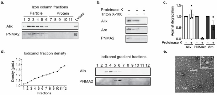
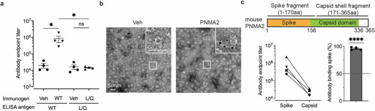
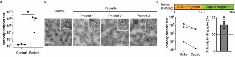
The paraneoplastic Ma antigen (PNMA) genes are associated with cancer-induced paraneoplastic syndromes that present with neurological symptoms and autoantibody production. How PNMA proteins trigger a severe autoimmune disease is unclear. PNMA genes are predominately expressed in the central nervous system with little known functions but are ectopically expressed in some tumors. Here, we show that PNMA2 is derived from a Ty3 retrotransposon that encodes a protein which forms virus-like capsids released from cells as non-enveloped particles. Recombinant PNMA2 capsids injected into mice induce a robust autoimmune reaction with significant generation of autoantibodies that preferentially bind external "spike" PNMA2 capsid epitopes, while capsid-assembly-defective PNMA2 protein is not immunogenic. PNMA2 autoantibodies present in cerebrospinal fluid of patients with anti-Ma2 paraneoplastic neurologic disease show similar preferential binding to PNMA2 "spike" capsid epitopes. These observations suggest that PNMA2 capsids released from tumors trigger an autoimmune response that underlies Ma2 paraneoplastic neurological syndrome.


Conflict of interest statement

Competing interests C.F. is a consultant for Tessera Therapeutics, Inc. and HAYA Therapeutics, Inc. J.D.S is a co-founder of VNV, LLC and a consultant for Aera Therapeutics, Inc.

Figures

Figure 1.
Figure 1.. Ty3/mdg4 retrotransposon-derived PNMA2 encodes proteins self-assembling into virus-like capsids.
a. Structure and evolution of PNMA2. The figure is derived from the Vertebrate Multiz Alignment and Conservation track at the UCSC Genome Browser (hg38) and shows the phylogenetic relationship (left) and level of sequence conservation (right) for a subset of vertebrate genomes across the mRNA sequence and shared promoter region of PNMA2 (red) and DPYSL2 (green) gene models. There are three transposable elements annotated by RepeatMasker within the PNMA2 mRNA: a Mam-Gyp-int element (purple box) that gave rise to the Gag-like coding sequence, and two Alu elements (grey boxes) embedded within the 3’ UTR. The arrows depict the predicted transcription start sites for PNMA2 and DPYSL2. The conservation track shows that PNMA2 mRNA sequence is deeply conserved across placental mammals (except for the two Alu elements which are primate-specific insertions), but not other vertebrates, while DPYSL2 and promoter region are conserved more deeply across vertebrate evolution pointing at their earlier origins. b. Single-cell expression of PNMA2 mRNA in the human cortex (right panel) and mouse cortex and hippocampus (left panel). c. PNMA2 single molecule fluorescent in situ hybridization probe (RNAscope) and negative control probe were used to detect PNMA2 mRNA in wild-type mouse (2 months old) hippocampal slice. Scale bar: 400μm. d. Representative negative-stained EM images of purified recombinant mouse PNMA2 (mPNMA2) capsids and human PNMA2 (hPNMA2) capsids. Scale bar: 50nm. e. Surface representation of mPNMA2 as resolved from cryo-EM, viewed down the two-fold axis. The spike densities are not resolved. f. Atomic model of the T=1 mPNMA2 capsid. CANTD is depicted in purple and the CACTD in cyan. g. External view of the isolated five-fold capsomere. h. One of 60 mPNMA2 monomers required to form the T=1 capsids.
Figure 2.
Figure 2.. Endogenous mPNMA2 is released by neurons as non-enveloped capsids.
a. Media was collected from primary cultured cortical neurons (DIV15-16) after 24h incubation and fractionated using size exclusion chromatography (SEC). Fractions were run on a gel and blotted for mPNMA2 protein and ALIX, a canonical EV marker. mPNMA2 protein is released in early fractions that contain EV proteins. b. The early fractions (–4) from SEC were pooled and blotted for Arc, mPNMA2, and ALIX. Fractions were incubated with Proteinase K (200μg/mL) with or without detergent present (1% Triton-X) for 10mins. Representative Western blots show that mPNMA2 protein was sensitive to Proteinase K degradation without detergent present. c. Quantification of Western blot in b (n=3 experiments). Error bars indicate mean ± s.e.m. d. The early fractions (–4) from SEC were fractionated using ultracentrifugation. An iodixanol gradient was used to separate proteins by density and size. mPNMA2 protein was enriched in fraction 6, while ALIX was enriched in fractions 3 and 4. e. Representative negative-stained EM image of non-enveloped mPNMA2 capsids isolated from iodixanol gradient fraction 6 in d.
Figure 3.
Figure 3.. mPNMA2 capsids injected into mice induce PNMA2 autoantibody production.
a. Mice were injected intraperitoneally with vehicle (n=4), 5μg purified mPNMA2 capsids (n=4) or 5μg mPNMA2 L/Q protein (n=4) and blood sera collected 3 weeks after injections. Sera were analyzed for antibody production using ELISA, using 2μg/mL mPNMA2 capsids or mPNMA2 L/Q protein as the antigen coated on the plates. mPNMA2 capsid-injected mice produced robust PNMA2 autoantibodies, whereas vehicle and mPNMA2 L/Q injections did not elicit autoantibody production. (*One-way ANOVA with post-hoc pairwise comparisons by Tukey’s test, P=0.0146. Vehicle vs. mPNMA2 WT (using mPNMA2 capsids as antigen): P=0.0308; Vehicle vs. mPNMA2 L/Q (using mPNMA2 L/Q as antigen): P>0.9999); mPNMA2 WT vs. mPNMA2 L/Q: P=0.0296.). b. Representative negative-stained EM images of purified mPNMA2 capsids immunogold labelled with mouse PNMA2 autoantibodies in serum collected from PNMA2 capsid-injected mice. c. Purified mPNMA2 spike fragments and capsid shell fragments (see schematic) were used as ELISA antigens to map the epitopes of PNMA2 autoantibodies from PNMA2 capsid-injected mice. The schematic shows PNMA2 protein regions located on the spike and the capsid domains of capsids, as determined by the cryo-EM structure. PNMA2 autoantibodies preferentially bind to the spike fragments. (****One sample t-test, P<0.0001, null hypothesis of 50% binding). Error bars indicate mean ± s.e.m. WT: wild type; L/Q: L270QL325Q; Veh: vehicle.
Figure 4.
Figure 4.. PNMA2 autoantibodies in CSF from human patients with paraneoplastic neurological syndrome preferentially bind to hPNMA2 capsids.
a. An ELISA, using purified hPNMA2 capsids as the antigen, was used to quantify PNMA2 autoantibodies in CSF from patients diagnosed with or without PNMA2-related paraneoplastic neurological syndrome.(n=3 for each group, *Mann-Whitney test, P=0.05). b. Representative negative-stained EM images of hPNMA2 capsids labeled with immunogold using patient CSF from control or paraneoplastic patients. c. ELISAs using hPNMA2 spike and capsid shell fragments as antigens show patient CSF PNMA2 autoantibodies preferentially bind to the spike fragment (*One sample t-test, P=0.0356, null hypothesis of 50% binding). Error bars indicate mean ± s.e.m.

References

    1. Rosenfeld M. R., Eichen J. G., Wade D. F., Posner J. B., Dalmau J., Molecular and clinical diversity in paraneoplastic immunity to Ma proteins. Annals of Neurology: Official Journal of the American Neurological Association and the Child Neurology Society 50, 339–348 (2001). - PubMed
    1. Dalmau J. et al., Ma1, a novel neuron-and testis-specific protein, is recognized by the serum of patients with paraneoplastic neurological disorders. Brain 122, 27–39 (1999). - PubMed
    1. Voltz R. et al., A Serologic Marker of Paraneoplastic Limbic and Brain-Stem Encephalitis in Patients with Testicular Cancer. New England Journal of Medicine 340, 1788–1795 (1999). - PubMed
    1. Takaji M., Komatsu Y., Watakabe A., Hashikawa T., Yamamori T., Paraneoplastic antigen-like 5 gene (PNMA5) is preferentially expressed in the association areas in a primate specific manner. Cerebral Cortex 19, 2865–2879 (2009). - PMC - PubMed
    1. Schüller M., Jenne D., Voltz R., The human PNMA family: novel neuronal proteins implicated in paraneoplastic neurological disease. Journal of neuroimmunology 169, 172–176 (2005). - PubMed

Publication types