. 2023 Jan 17;6(2):320-333.
doi: 10.1021/acsptsci.2c00232. eCollection 2023 Feb 10.

Development of Brain-Derived Bioscaffolds for Neural Progenitor Cell Culture

Affiliations

Development of Brain-Derived Bioscaffolds for Neural Progenitor Cell Culture

Julia C Terek et al. ACS Pharmacol Transl Sci. .

Abstract

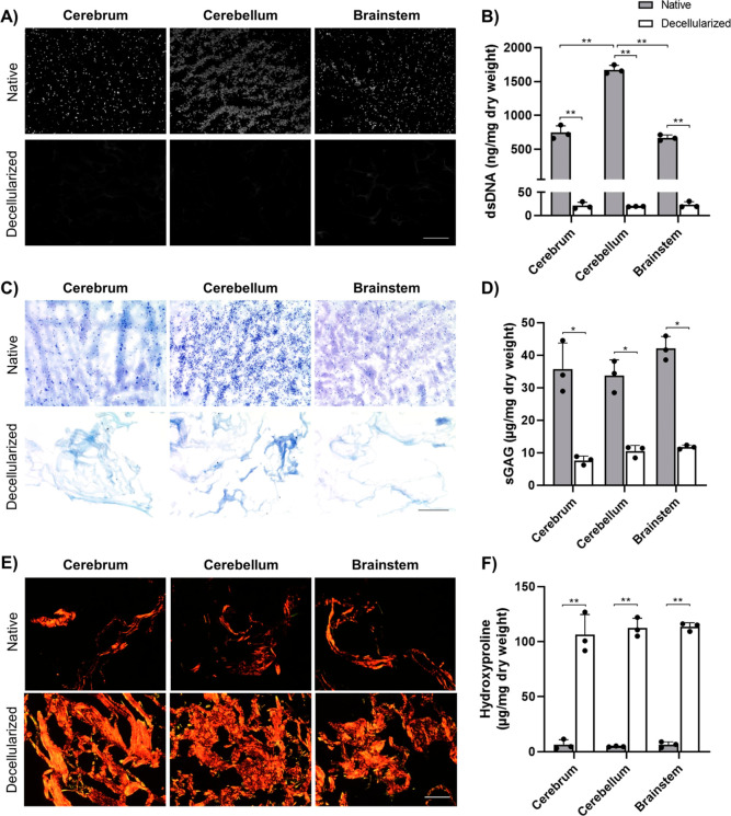
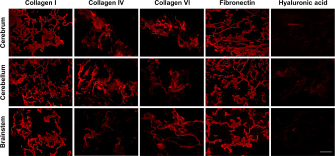
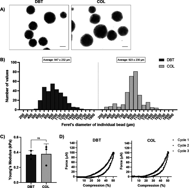
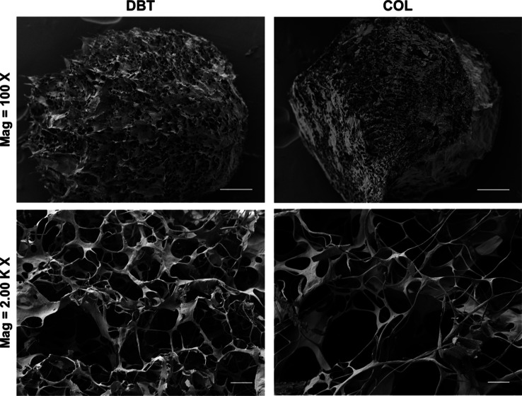
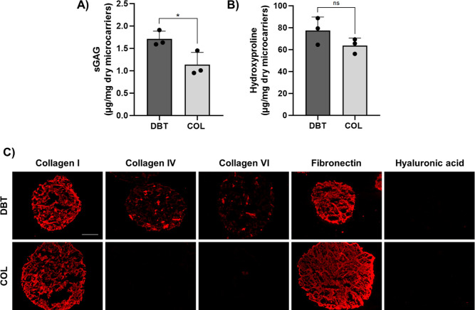
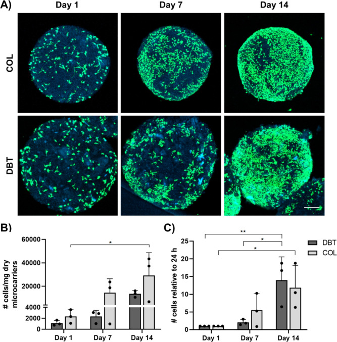
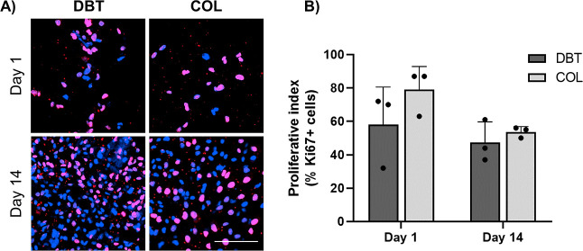
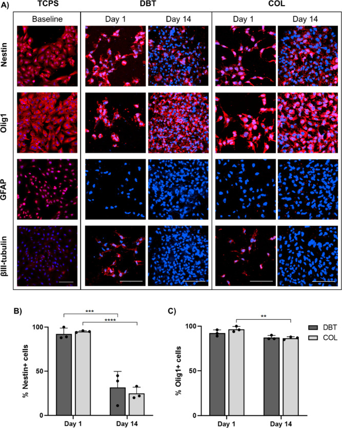
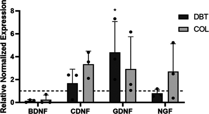
Biomaterials derived from brain extracellular matrix (ECM) have the potential to promote neural tissue regeneration by providing instructive cues that can direct cell survival, proliferation, and differentiation. This study focused on the development and characterization of microcarriers derived from decellularized brain tissue (DBT) as a platform for neural progenitor cell culture. First, a novel detergent-free decellularization protocol was established that effectively reduced the cellular content of porcine and rat brains, with a >97% decrease in the dsDNA content, while preserving collagens (COLs) and glycosaminoglycans (GAGs). Next, electrospraying methods were applied to generate ECM-derived microcarriers incorporating the porcine DBT that were stable without chemical cross-linking, along with control microcarriers fabricated from commercially sourced bovine tendon COL. The DBT microcarriers were structurally and biomechanically similar to the COL microcarriers, but compositionally distinct, containing a broader range of COL types and higher sulfated GAG content. Finally, we compared the growth, phenotype, and neurotrophic factor gene expression levels of rat brain-derived progenitor cells (BDPCs) cultured on the DBT or COL microcarriers within spinner flask bioreactors over 2 weeks. Both microcarrier types supported BDPC attachment and expansion, with immunofluorescence staining results suggesting that the culture conditions promoted BDPC differentiation toward the oligodendrocyte lineage, which may be favorable for cell therapies targeting remyelination. Overall, our findings support the further investigation of the ECM-derived microcarriers as a platform for neural cell derivation for applications in regenerative medicine.


Conflict of interest statement

The authors declare no competing financial interest.

Figures

Figure 1
Figure 1
New detergent-free protocol effectively decellularized porcine cortex, cerebellum, and brainstem. (A) Representative DAPI nuclear staining (gray) of native and decellularized pig brain samples showing no visible cell nuclei following decellularization. Scale bar = 200 μm. (B) Quantitative analysis of the dsDNA content by PicoGreen assay showing a significant reduction in dsDNA in the decellularized samples (n = 3 and N = 3; **p < 0.0001). (C) Representative toluidine blue staining of GAG (purple) showing retention of GAGs following decellularization. Scale bar = 200 μm. (D) Quantitative analysis of the sGAG content by DMMB assay showing 20–30% GAG retention in the decellularized samples (n = 3 and N = 3; *p < 0.001). (E) Representative picrosirius red staining of COL fibers (red/yellow) showing a dense network of thick and thin COL fibers in the decellularized tissues. Scale bar = 200 μm. (F) Quantitative analysis of the COL content by hydroxyproline assay showing a significant relative enrichment in the COL content following decellularization (n = 3 and N = 3; **p < 0.0001).
Figure 2
Figure 2
Immunohistochemical analyses confirmed the presence of various types of COL and fibronectin following decellularization. Representative staining showing the retention of multiple types of COL and fibronectin, with similar staining patterns in the decellularized porcine cerebrum, cerebellum, and brainstem samples (n = 3). Minimal amounts of hyaluronic acid were detected in the cerebrum and brainstem samples, and no laminin, keratan sulfate, or brevican were detected in any region. Scale bar = 200 μm.
Figure 3
Figure 3
DBT and COL microcarriers have similar shape, size, and stiffness. (A) Representative brightfield images of DBT and COL microcarriers showing their spherical shape. Scale bars = 400 μm. (B) Size distribution plots showing Feret’s diameter of the microcarriers following rehydration in PBS. Average diameter was not significantly different between the two groups when analyzed with an unpaired t test (n = 100 and N = 3). (C) Young’s moduli of the DBT and COL microcarriers, with no significant difference measured between the two groups (n = 6 and N = 3). (D) Representative force versus deformation curves for DBT and COL microcarriers. Data from three cycles are shown with compression at a strain rate of 0.01 s–1.
Figure 4
Figure 4
Representative SEM images visualizing the ECM ultrastructure of the DBT and COL microcarriers. Both microcarrier types had a porous and complex fibrous ultrastructure. Scale bars = 100 μm (top row), 5 μm (bottom row).
Figure 5
Figure 5
DBT microcarriers have a more complex composition than COL microcarriers. (A) Quantitative analysis of the sGAG content by DMMB assay showing that the DBT microcarriers contained significantly more sGAG (n = 3–6 and N = 3; *p < 0.05). (B) Quantitative analysis of the COL content, showing no significant difference in the hydroxyproline content between the DBT and COL microcarriers (n = 3 and N = 3). (C) Representative IF staining showing that the DBT microcarriers contained multiple types of COL and fibronectin, while only type I COL and fibronectin were detected in the COL microcarriers (n = 3 and N = 2). No hyaluronic acid was detected in either group. Scale bar = 200 μm.
Figure 6
Figure 6
ECM-derived microcarriers support the attachment and growth of BDPCs under dynamic culture. (A) Representative LIVE/DEAD staining visualized through confocal microscopy showing the presence of calcein+ live cells (green) on the surface of the microcarriers (blue), with a qualitative increase in the density observed over time. Scale bar = 200 μm. (B) dsDNA content was assessed by PicoGreen assay to quantify the cell content on the microcarriers at various timepoints. Cell density significantly increased between days 1 and 14 on the COL microcarriers (n = 3 and N = 3; *p < 0.05). (C) Cell density data normalized to day 1 showing a significant increase on the DBT and COL microcarriers between days 1 and 14, confirming that both platforms supported BDPC expansion (n = 3 and N = 3; *p < 0.05).
Figure 7
Figure 7
BDPCs retain a high proliferative index over 2 weeks of culture under dynamic conditions on the DBT and COL microcarriers. (A) Representative Ki67 IF staining in rat BDPCs cultured dynamically on the DBT and COL microcarriers for 1 or 14 days within the spinner flask bioreactors, visualized through confocal microscopy. Scale bar = 100 μm. (B) Quantitative assessment of the proliferative index showing that a high percentage of the cells expressed Ki67 at both timepoints (n = 4–8 and N = 3).
Figure 8
Figure 8
Dynamic culture of BDPCs on DBT and COL microcarriers modifies their expression of neural lineage markers at the protein level. (A) Representative IF staining visualized through confocal microscopy showing the presence and distribution of the neural progenitor marker nestin, oligodendrocyte marker Olig1, astrocyte marker GFAP, and neuronal marker βIII-tubulin in the rat BDPCs cultured over 2 weeks within the spinner flask bioreactors on the DBT and COL microcarriers (n = 3–5 and N = 3) relative to BDPCs cultured on TCPS as a baseline control. Prior to seeding, the BDPCs displayed robust expression of nestin, Olig1, GFAP, and βIII-tubulin. Following culture on both DBT and COL microcarriers, expression of GFAP and βIII-tubulin was not detected, while robust expression of Olig1 was observed, suggestive of differentiation toward an oligodendrocyte lineage. Scale bars = 100 μm. (B) Manual counting of nestin+ cells showing a significant decrease between days 1 and 14 on both DBT and COL microcarriers (n = 3–10 and N = 3; ***p < 0.001 and ****p < 0.0001). (C) Manual count of Olig1+ cells showing a slight decrease between days 1 and 14 on COL microcarriers, but no change was observed on DBT microcarriers (n = 3–7 and N = 3; **p < 0.005).
Figure 9
Figure 9
Gene expression of GDNF in the BDPCs was significantly enhanced on DBT microcarriers compared to culture on TCPS. Relative gene expression of the NTFs, BDNF, CDNF, GDNF, and NGF, in BDPCs cultured on DBT and COL microcarriers was analyzed. The hatched line indicates baseline levels in BDPCs cultured on TCPS. GDNF expression in BDPCs was significantly enhanced following culture on the DBT microcarriers relative to the baseline samples (n = 3 and N = 3; *p < 0.05).

Similar articles

Cited by

References

    1. Gaskin J.; Gomes J.; Darshan S.; Krewski D. Burden of neurological conditions in Canada. Neurotoxicology 2017, 61, 2–10. 10.1016/j.neuro.2016.05.001. - DOI - PubMed
    1. Horner P. J.; Gage F. H. Regenerating the damaged central nervous system. Nature 2000, 407, 963–970. 10.1038/35039559. - DOI - PubMed
    1. Mirahmadi M.; Rezanejadbardaji H.; Irfan-Maqsood M.; Mokhtari M. J.; Naderi-Meshkin H. Stem Cell Therapy for Neurodegenerative Diseases: Strategies for Regeneration against Degeneration. J. Genes Cells 2017, 3, 22.10.15562/gnc.54. - DOI
    1. Chamberlain K. A.; Nanescu S. E.; Psachoulia K.; Huang J. K. Oligodendrocyte regeneration: Its significance in myelin replacement and neuroprotection in multiple sclerosis. Neuropharmacology 2016, 110, 633–643. 10.1016/j.neuropharm.2015.10.010. - DOI - PMC - PubMed
    1. Quan L.; Uyeda A.; Muramatsu R. Central nervous system regeneration: the roles of glial cells in the potential molecular mechanism underlying remyelination. Inflammation Regener. 2022, 42, 7.10.1186/s41232-022-00193-y. - DOI - PMC - PubMed

LinkOut - more resources