. 2022 Dec 19;28(4):10.7196/AJTCCM.2022.v28i4.248.
doi: 10.7196/AJTCCM.2022.v28i4.248. eCollection 2022.

Role of chest radiograph in predicting the need for ventilator support in COVID-19 patients

Affiliations

Role of chest radiograph in predicting the need for ventilator support in COVID-19 patients

G Patnayak et al. Afr J Thorac Crit Care Med. .

Abstract

Background: COVID-19 disease, a pandemic for more than two years, has major morbidity and mortality related to pulmonary involvement. Chest radiography is the main imaging tool for critically ill patients. As the availability of arterial blood gas analysis is limited in the Level I and II healthcare centres, which are major partners in providing healthcare in resource-limited times, we planned the present study.

Objectives: To assess the role of chest radiography in predicting the need for oxygen/ventilator support in critically ill COVID-19 patients.

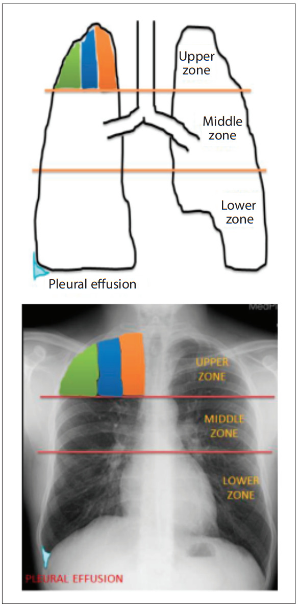
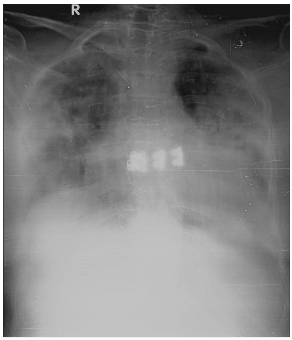
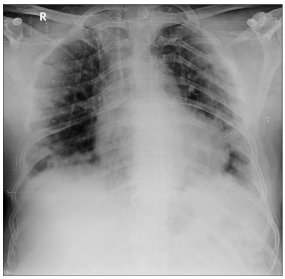
Methods: This hospital-based, retrospective study included 135 patients who needed oxygen/ventilator support and had optimal-quality chest radiographs at admission. All the chest X-rays were evaluated and a severity score was calculated on a predesigned pro forma. Statistical evaluation of the data obtained was done using appropriate tools and methods.

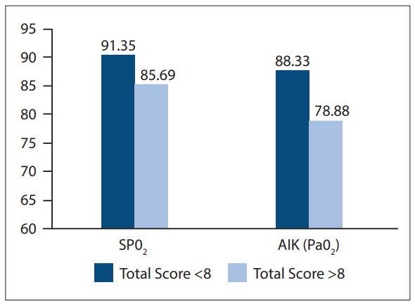
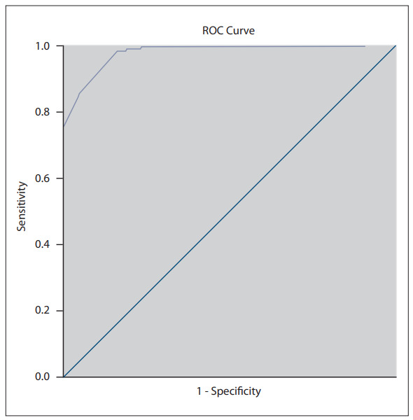
Results: Males outnumbered females, with a mean age of 54.35 ± 14.49 years. More than 72% of patients included in our study needed ventilator support while the rest needed oxygen support. There was a significant statistical correlation between the chest radiograph severity score and SPO2 /PaO2 levels in our study. Using a cut-off value >8 for the chest radiograph severity score in predicting the need for ventilator support in a Covid-19 patient, the sensitivity, specificity and accuracy was 85.7%, 92.5% and 89.5%, respectively.

Conclusion: Chest radiography remains the mainstay of imaging in critically ill COVID-19 patients when they are on multiple life-support systems. Though arterial blood gas analysis is the gold standard tool for assessing the need for oxygen/ventilator support in these patients, the severity score obtained from the initial chest radiograph at the time of admission may also be used as a screening tool. Chest radiography may predict the need for oxygen/ventilator support, allowing time for patients to be moved to an appropriate-level healthcare centre, thus limiting morbidity and mortality.

Keywords: COVID-19; Chest radiography; ventilator.


Conflict of interest statement

Conflicts of interest: None.

Figures

Fig. 1
Fig. 1
Schematic diagram and corresponding chest radiograph showing arbitrary division of three lung zones and percentage of involvement by consolidation or ground-glass opacity of that respective zone, i.e. score 1 for one-third (33%) involvement (shown by green colour), 2 for one-third to two-thirds (34 - 66%) involvement (blue colour) and 3 for two-thirds (66%) involvement (shown by orange colour). A score of point one is given for presence of pleural effusion for each side (shown by sky-blue colour).
Fig. 2
Fig. 2
Chest radiograph showing opacity involving two-thirds (67%) area of bilateral middle and lower lung zones with obscuration of bilateral costophrenic angles, representing chest radiograph severity score of 14. This patient required ventilatory support.
Fig. 3
Fig. 3
Chest radiograph showing opacity involving more than 33% but less than 66% area of the bilateral middle lung zones and more than two-thirds (67%) area of bilateral lower lung zones without obscuration of bilateral costophrenic angles, representing chest radiograph severity score of 10. This patient required ventilatory support.
Fig. 4
Fig. 4
Chest radiograph showing opacity involving less than 33% area of the right upper lung zone and more than 33% but less than 66% area of right lower lung zone with right-sided pleural effusion, representing chest radiograph severity score of 4. This patient did not require ventilatory support.
Fig. 5
Fig. 5
Chest radiograph showing opacity involving more than 33% but less than 66% area of the right upper lung zone, less than 33% area of the left upper lung zone and more than two-thirds (67%) area of the bilateral middle and lower lung zones with obscuration of bilateral costophrenic angles, representing chest radiograph severity score of 17. This patient required ventilatory support.
Fig. 6
Fig. 6
Chest radiograph showing opacity involving less than 33% area of the bilateral upper and middle lung zones, more than 33% but less than 66% area of the right lower lung zone and more than two-thirds (67%) area of left lower lung zone with obscuration of bilateral costophrenic angles, representing chest radiograph severity score of 11. This patient required ventilatory support.
Fig. 7
Fig. 7
Graphical representation of comparison of SPO2 and PaO2 with total CXR severity score
Fig. 8
Fig. 8
The ROC curve

Similar articles

Cited by

References

    1. Xiang C, Huang L, Xia L. Mobile chest X-ray manifestations of 54 deceased patients with coronavirus disease 2019: Retrospective study. Medicine. 2020;13(99):46. - PMC - PubMed
    1. Salehi S, Abedi A, Balakrishnan S, Gholamrezanezhad A. Coronavirus disease 2019 (COVID-19): A systematic review of imaging findings in 919 patients. Am J Roentgenol. 2020;1:1–7. - PubMed
    1. Toussie D, Voutsinas N, Finkelstein M, et al. Clinical and chest radiography features determine patient outcomes in young and middle age adults with COVID-19. Radiology. 2020;14:201754. - PMC - PubMed
    1. Balbi M, Caroli A, Corsi A, et al. Chest X-ray for predicting mortality and the need for ventilatory support in COVID-19 patients presenting to the emergency department. Eur Radiology. 2020;8:1–4. - PMC - PubMed
    1. Xiao N, Cooper JG, Godbe JM, et al. Chest radiograph at admission predicts early intubation among inpatient COVID-19 patients. Eur Radiology. 2020;13:1–8. - PMC - PubMed

LinkOut - more resources