. 2023 Jan 18:28:100593.
doi: 10.1016/j.bbih.2023.100593. eCollection 2023 Mar.

pKr-2 induces neurodegeneration via upregulation of microglial TLR4 in the hippocampus of AD brain

Affiliations

pKr-2 induces neurodegeneration via upregulation of microglial TLR4 in the hippocampus of AD brain

Sehwan Kim et al. Brain Behav Immun Health. .

Abstract

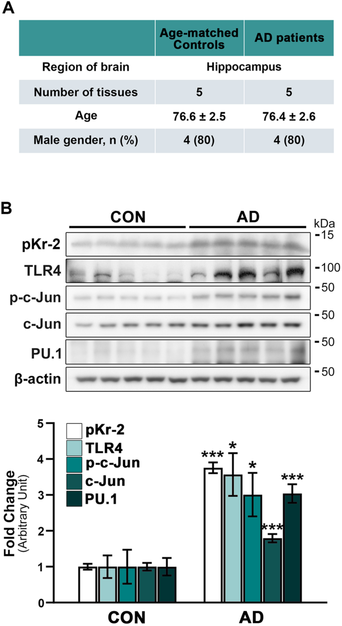
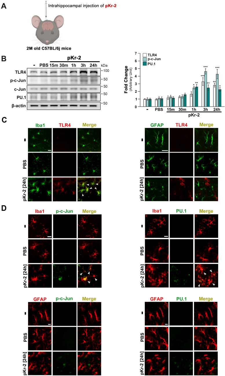
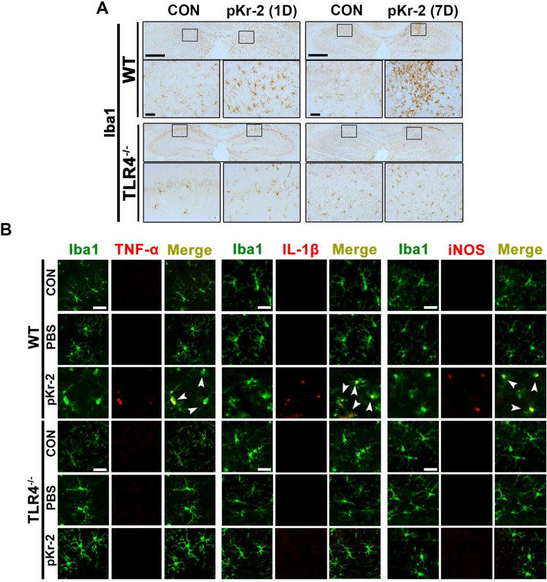
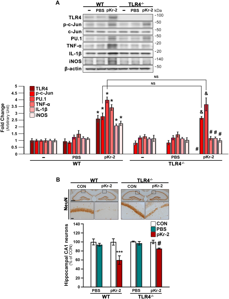
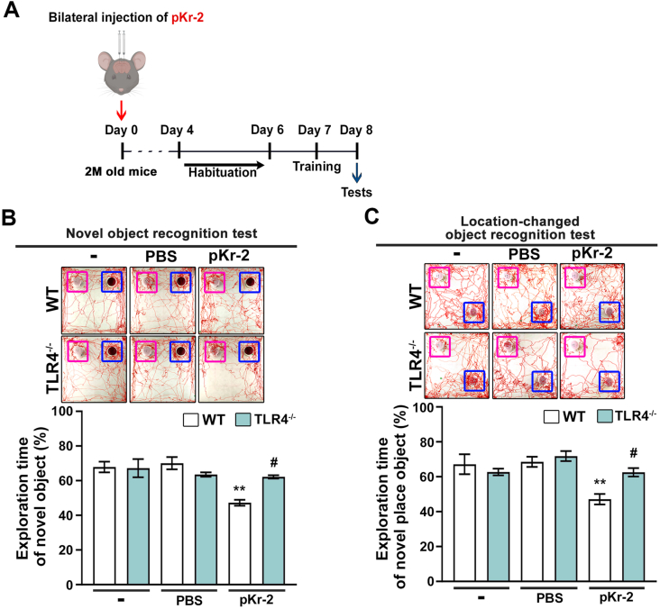
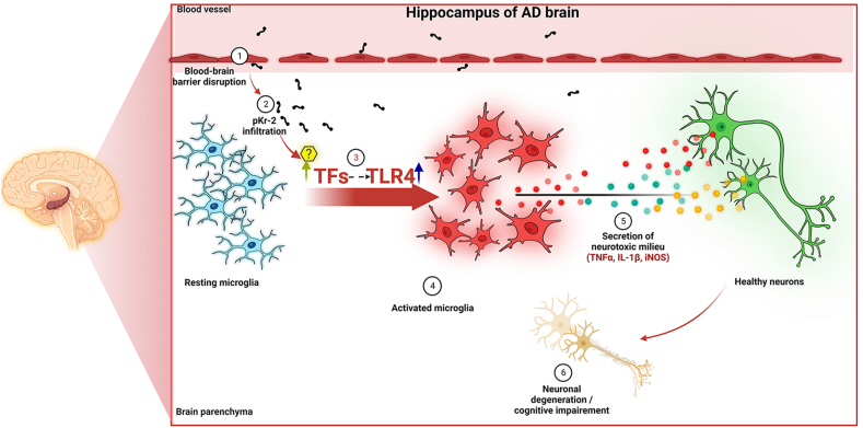
We recently demonstrated that prothrombin kringle-2 (pKr-2) derived from blood-brain barrier (BBB) disruption could induce hippocampal neurodegeneration and object recognition impairment through neurotoxic inflammatory responses in the five familial Alzheimer's disease mutation (5XFAD) mice. In the present study, we aimed to determine whether pKr-2 induces microglial activation by stimulating toll-like receptor 4 (TLR4) upregulation and examine whether this response contributes to pKr-2-induced neuroinflammatory damage in the hippocampi of mice models. We observed that inflammatory responses induced by pKr-2 administration in the hippocampi of wild-type mice were significantly abrogated in TLR4-deficient mice (TLR4-/-), and caffeine supply or rivaroxaban treatment that inhibits the overexpression of hippocampal pKr-2 reduced TLR4 upregulation in 5XFAD mice, resulting in the inhibition of neuroinflammatory responses. Similar to the expression patterns of pKr-2, TLR4, and the TLR4 transcription factors, PU.1 and p-c-Jun, seen in the postmortem hippocampal tissues of Alzheimer's disease (AD) patients, our results additionally showed the influence of transcriptional regulation on TLR4 expression following pKr-2 expression in triggering the production of neurotoxic inflammatory mediators. Therefore, we conclude that pKr-2 may play a role in initiating upregulation of microglial TLR4, consequently inducing hippocampal neurodegeneration. Furthermore, the control of pKr-2-induced microglial TLR4 could be a useful therapeutic strategy against hippocampal neurodegeneration in AD.

Keywords: Alzheimer's disease; Hippocampus; Microglia; Neurodegeneration; Neuroinflammation; Prothrombin kringle-2; Toll-like receptor 4.


Conflict of interest statement

The authors declare that they have no competing interests.

Figures

Fig. 1
Fig. 1
Upregulation of pKr-2, TLR4, and TLR4 transcription factors in postmortem hippocampal tissues of patients with AD. (A) Details of the human postmortem hippocampal tissues are obtained from the Victorian Brain Bank Network. (B) Western blot analysis shows significantly increased protein levels of pKr-2, TLR4, p-c-Jun, c-Jun, and PU.1 in the postmortem hippocampal tissues of patients with AD compared to age-matched controls (CON). *p < 0.05 and ***p < 0.001 vs. CON (t-test; n = 5 for each group).
Fig. 2
Fig. 2
Upregulation of TLR4 and its transcription factors in the hippocampi of pKr-2-treated mice. (A) Schematic representing the intrahippocampal injection in the mouse brain. Created with http://biorender.com. To investigate whether there are changes in the protein levels of TLR4 and its transcription factors at early time points after pKr-2 overexpression, we examined the protein levels of p-c-Jun and PU.1 as transcriptional factors for TLR4 expression following an intrahippocampal unilateral (right side) injection of pKr-2 in the male mouse brain in a time-dependent manner (15 m, 30 m, 1 h, 3 h and 24 h after pKr-2 injection). (B) Western blotting for TLR4, p-c-Jun, c-Jun, and PU.1 expression in the hippocampi at 15 min, 30 min, 1 h, 3 h, and 24 h following intrahippocampal injection of pKr-2. *p < 0.05, **p < 0.01, and ***p < 0.001 vs. intact controls (one-way ANOVA with Tukey's post-hoc analysis; n = 5 for each group). (C) Immunofluorescence double staining for Iba1 (green) and TLR4 (red) or GFAP (green) and TLR4 (red) in the hippocampal CA1 region on day 1 following PBS or pKr-2 injection. White arrowheads indicate an increase in TLR4 expression co-localized within the microglia. Scale bar, 10 μm. (D) Immunofluorescence double staining for Iba1 (red) and p-c-Jun (green), or Iba1 and PU.1 (green), GFAP (red) and p-c-Jun (green), or GFAP (red) and PU.1 (green) in the hippocampal CA1 regions at 24 h after PBS or pKr-2 injection. Scale bar, 10 μm. (For interpretation of the references to color in this figure legend, the reader is referred to the Web version of this article.)
Fig. 3
Fig. 3
Inhibition of pKr-2-induced microglial activation resulting in neuroinflammation in the hippocampi of TLR4−/−mice (A) Hippocampal sections obtained from male WT and TLR4−/− mice are immunostained with anti-Iba1 antibody (brown color) on 1 and 7 days following pKr-2 administration. The image in each rectangular box is magnified in the bottom panel. Scale bars, 500 and 50 μm, respectively. (B) Immunofluorescence double staining for Iba1 (green) and TNF-α (red), Iba1 (green) and IL-1β (red), and Iba1 (green) and iNOS (red) in the hippocampi of WT and TLR4−/− mice at 1 day following pKr-2 administration. White arrowheads indicate microglial cells co-localized with each inflammatory molecule. Scale bar, 20 μm. (For interpretation of the references to color in this figure legend, the reader is referred to the Web version of this article.)
Fig. 4
Fig. 4
pKr-2 administration leads to neurotoxicity mediated by TLR4 induction in the hippocampus in vivo. To examine the protein levels of neuroinflammatory molecules at the early time point after pKr-2 overexpression, we checked 1 day after pKr-2 injection in the hippocampi of male WT and TLR4−/− mice. Moreover, to investigate the neurotoxicity caused by the pKr-2 injection, we checked the hippocampi of male WT and TLR4−/− mice, 7 days after the injection. (A) Western blot analysis of TLR4, p-c-Jun, PU.1, TNF-α, IL-1β, and iNOS protein levels in the hippocampus at day 1 following pKr-2 administration. *p < 0.001 vs. non-treated WT mice; &p < 0.001 vs. non-treated TLR4−/− mice; #p < 0.001 vs. pKr-2-treated WT mice; NS, no significance (two-way ANOVA with Tukey's post-hoc analysis; n = 5 for each group). (B) Representative images of immunohistochemical staining for anti-NeuN at day 7 following pKr-2 administration. Each image within a rectangular box is magnified in the bottom panel. Scale bars, 500 and 50 μm, respectively. The histogram quantitatively demonstrates NeuN-positive neurons in the counting area of the ipsilateral injection side compared to those of the contralateral control side (CON). ***p < 0.001 vs. CON; #p < 0.05 vs. pKr-2-treated WT mice (two-way ANOVA with Tukey's post hoc analysis; n = 5 for each group).
Fig. 5
Fig. 5
Intrahippocampal administration of pKr-2 induces cognitive impairment in WT mice. (A) Behavioral tests (Novel object recognition test, Location-changed object recognition test) for assessing object recognition are conducted on day 8 following bilateral injections of pKr-2 into the hippocampal CA1 regions of male WT and TLR4−/− mice. (B) Novel object recognition tests (familiar object: purple square; novel object: blue square). Results are represented as the time ratio exploring the novel object (novel object/total object exploring time). **p < 0.01 vs. non-treated WT mice; #p < 0.05 vs. pKr-2-treated WT mice (two-way ANOVA with Tukey's post-hoc analysis; n = 8 for each group). (C) Object location recognition tests (familiar object: purple square; location-changed object: blue square). Results are represented as the time ratio exploring the location-changed object (location-changed object/total object exploring time). **p < 0.01 vs. non-treated WT mice; #p < 0.05 vs. pKr-2-treated WT mice (two-way ANOVA with Tukey's post-hoc analysis; n = 8 for each group). (For interpretation of the references to color in this figure legend, the reader is referred to the Web version of this article.)
Fig. 6
Fig. 6
Inhibition of pKr-2 expression by caffeine supply or rivaroxaban treatment decreases TLR4 transcription factors in the hippocampi of 5XFAD mice. (A) Experimental schematic demonstrating caffeine (0.6 mg/mL) supply. (B) Western blot analysis of pKr-2, TLR4, p-c-Jun, PU.1 protein levels in the hippocampi of male and female WT and 5XFAD mice aged seven months following caffeine supply. *p < 0.001 vs. WT mice; #p < 0.001 vs. unsupplied 5XFAD mice; (one-way ANOVA with Tukey's post-hoc analysis; n = 5 for each group). (C) Experimental schematic demonstrating rivaroxaban (2 mg/kg/day) oral treatment. (D) Western blot analysis of pKr-2, TLR4, p-c-Jun, PU.1 protein levels in the hippocampi of male and female WT and 5XFAD mice aged three months following rivaroxaban oral administration. *p < 0.001 vs. WT mice; #p < 0.001 vs. non-treated 5XFAD mice; (one-way ANOVA with Tukey's post hoc analysis; n = 5 for each group).
Fig. 7
Fig. 7
Schematic representation of pKr-2-induced neurodegeneration in the hippocampus of the adult brain. Increased pKr-2 due to blood-brain barrier breakdown can lead to translocation of pKr-2 to the microglia, resulting in microglial activation via induction of microglial toll-like receptor 4 (TLR4) and its associated transcription factors (TFs). Furthermore, the activated microglia may induce hippocampal neurodegeneration by producing neuroinflammatory molecules. These observations suggest that pKr-2 upregulation is one of the major neuroinflammatory mechanisms associated with microglial activation leading to neurotoxic events in the hippocampus of the AD brain. This figure was created with http://biorender.com.

Similar articles

Cited by

References

    1. Arai T., Miklossy J., Klegeris A., Guo J.P., McGeer P.L. Thrombin and prothrombin are expressed by neurons and glial cells and accumulate in neurofibrillary tangles in Alzheimer disease brain. J. Neuropathol. Exp. Neurol. 2006;65:19–25. - PubMed
    1. Bachiller S., Jimenez-Ferrer I., Paulus A., Yang Y., Swanberg M., Deierborg T., Boza-Serrano A. Microglia in neurological diseases: a road map to brain-disease dependent-inflammatory response. Front. Cell. Neurosci. 2018;12:488. - PMC - PubMed
    1. Baker S.K., Chen Z.L., Norris E.H., Revenko A.S., MacLeod A.R., Strickland S. Blood-derived plasminogen drives brain inflammation and plaque deposition in a mouse model of Alzheimer's disease. Proc. Natl. Acad. Sci. U.S.A. 2018;115:E9687–E9696. - PMC - PubMed
    1. Behre G., Whitmarsh A.J., Coghlan M.P., Hoang T., Carpenter C.L., Zhang D.E., Davis R.J., Tenen D.G. c-Jun is a JNK-independent coactivator of the PU.1 transcription factor. J. Biol. Chem. 1999;274:4939–4946. - PubMed
    1. Bevins R.A., Besheer J. Object recognition in rats and mice: a one-trial non-matching-to-sample learning task to study 'recognition memory. Nat. Protoc. 2006;1:1306–1311. - PubMed

LinkOut - more resources