

An Inexpensive 3D Printed Mouse Model of Successful, Complication-free Long Bone Distraction Osteogenesis
- PMID: 36798717
- PMCID: PMC9925097
- DOI: 10.1097/GOX.0000000000004674
An Inexpensive 3D Printed Mouse Model of Successful, Complication-free Long Bone Distraction Osteogenesis
Abstract
Distraction osteogenesis (DO) is used for skeletal defects; however, up to 50% of cases exhibit complications. Previous mouse models of long bone DO have been anecdotally hampered by postoperative complications, expense, and availability. To improve clinical techniques, cost-effective, reliable animal models are needed. Our focus was to develop a new mouse tibial distractor, hypothesized to result in successful, complication-free DO.
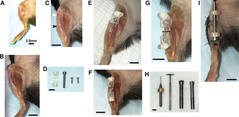
Methods: A lightweight tibial distractor was developed using CAD and 3D printing. The device was fixed to the tibia of C57Bl/6J mice prior to osteotomy. Postoperatively, mice underwent 5 days latency, 10 days distraction (0.15 mm every 12 hours), and 28 days consolidation. Bone regeneration was examined on postoperative day 43 using micro-computed tomography (μCT) and Movat's modified pentachrome staining on histology (mineralized volume fraction and pixels, respectively). Costs were recorded. We compared cohorts of 11 mice undergoing sham, DO, or acute lengthening (distractor acutely lengthened 3.0 mm).
Results: The histological bone regenerate was significantly increased in DO (1,879,257 ± 155,415 pixels) compared to acute lengthening (32847 ± 1589 pixels) (P < 0.0001). The mineralized volume fraction (bone/total tissue volume) of the regenerate was significantly increased in DO (0.9 ± 0.1) compared to acute lengthening (0.7 ± 0.1) (P < 0.001). There was no significant difference in bone regenerate between DO and sham. The distractor was relatively low cost ($11), with no complications.
Conclusions: Histology and µCT analysis confirmed that the proposed tibial DO model resulted in successful bone formation. Our model is cost-effective and reproducible, enabling implementation in genetically dissectible transgenic mice.
Copyright © 2023 The Authors. Published by Wolters Kluwer Health, Inc. on behalf of The American Society of Plastic Surgeons.
Conflict of interest statement
Disclosure: The authors have no financial interest to declare in relation to the content of this article.
Figures


Similar articles
-
Administration of allogeneic mesenchymal stem cells in lengthening phase accelerates early bone consolidation in rat distraction osteogenesis model.Stem Cell Res Ther. 2020 Mar 20;11(1):129. doi: 10.1186/s13287-020-01635-5. Stem Cell Res Ther. 2020. PMID: 32197646 Free PMC article.
-
A Mouse Distraction Osteogenesis Model.J Vis Exp. 2018 Nov 14;(141). doi: 10.3791/57925. J Vis Exp. 2018. PMID: 30507900
-
Histomorphometry of distraction osteogenesis in a caprine tibial lengthening model.J Bone Miner Res. 1998 Jan;13(1):1-9. doi: 10.1359/jbmr.1998.13.1.1. J Bone Miner Res. 1998. PMID: 9443783
-
Demineralized Bone Matrix Injection in Consolidation Phase Enhances Bone Regeneration in Distraction Osteogenesis via Endochondral Bone Formation.Clin Orthop Surg. 2015 Sep;7(3):383-91. doi: 10.4055/cios.2015.7.3.383. Epub 2015 Aug 13. Clin Orthop Surg. 2015. PMID: 26330963 Free PMC article.
-
Management of Osseous Defects in the Tibia: Utilization of External Fixation, Distraction Osteogenesis, and Bone Transport.Clin Podiatr Med Surg. 2021 Jan;38(1):111-116. doi: 10.1016/j.cpm.2020.09.006. Clin Podiatr Med Surg. 2021. PMID: 33220740 Review.
Cited by
-
A 3D-Printed Dummy for Training Distal Phalanx Amputation in Mice.Animals (Basel). 2024 Apr 22;14(8):1253. doi: 10.3390/ani14081253. Animals (Basel). 2024. PMID: 38672401 Free PMC article.
References
-
- Keating JF, Simpson AH, Robinson CM. The management of fractures with bone loss. J Bone Joint Surg Br. 2005;87:142–150. - PubMed
-
- Houben RH, Kotsougiani D, Friedrich PF, et al. . Outcomes of vascularized bone allotransplantation with surgically induced autogenous angiogenesis in a large animal model: bone healing, remodeling, and material properties. J Reconstr Microsurg. 2020;36:82–92. - PubMed
-
- Ilizarov GA. The tension-stress effect on the genesis and growth of tissues: part II. The influence of the rate and frequency of distraction. Clin Orthop Relat Res. 1989;(239):263–285. - PubMed
LinkOut - more resources
Full Text Sources
Miscellaneous