Review
. 2022 Oct 6;8(2):86-109.
doi: 10.4244/AIJ-D-22-00022. eCollection 2022 Oct.

Coronary physiology in the catheterisation laboratory: an A to Z practical guide

Affiliations
Review

Coronary physiology in the catheterisation laboratory: an A to Z practical guide

Simone Fezzi et al. AsiaIntervention. .

Abstract

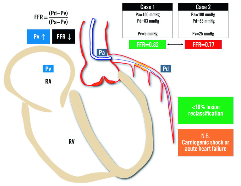
Coronary revascularisation, either percutaneous or surgical, aims to improve coronary flow and relieve myocardial ischaemia. The decision-making process in patients with coronary artery disease (CAD) remains largely based on invasive coronary angiography (ICA), even though until recently ICA could not assess the functional significance of coronary artery stenoses. Invasive wire-based approaches for physiological evaluations were developed to properly assess the ischaemic relevance of epicardial CAD. Fractional flow reserve (FFR) and later, instantaneous wave-free ratio (iFR), were shown to improve clinical outcomes in several patient subsets when used for coronary revascularisation guidance or deferral and for procedural optimisation of percutaneous coronary intervention (PCI) results. Despite accumulating evidence and positive guideline recommendations, the adoption of invasive physiology has remained quite low, mainly due to technical and economic issues as well as to operator-resistance to change. Coronary image-based computational physiology has been recently developed, with promising results in terms of accuracy and a reduction in computational time, costs, radiation exposure and risks for the patient. Lastly, the integration of intracoronary imaging and physiology allows for individualised PCI treatment, aiming at complete relief of ischaemia through optimised morpho-functional immediate procedural results. Instead of a conventional state-of-the-art review, this A to Z dictionary attempts to provide a practical guide for the application of coronary physiology in the catheterisation laboratory, exploring several methods, their pitfalls, and useful tips and tricks.


Conflict of interest statement

W. Wijns received a research grant and honoraria from MicroPort (TARGET AC trial); is the co-founder of Argonauts, an innovation facilitator; and is the senior medical advisor at Rede Optimus Research and Corrib Core Laboratory. S. Tu is a consultant for and received research grants from Pulse Medical. The other authors have no conflicts of interest to declare.

Figures

Figure 1
Figure 1. Coronary physiology and non-culprit lesions of acute coronary syndromes: potential pitfalls.
Potential scenarios affecting the reliability of invasive physiological assessment of non-culprit lesions in ACS: A. residual non-flow-limiting high-risk vulnerable plaques; B. microvascular impairment and blunted vasodilatory ability leading to reduced hyperaemic coronary flow. ACS: acute coronary syndrome; CCS: chronic coronary syndrome; CFR: coronary flow reserve; DS: diameter stenosis; FCT: fibrous cap thickness; FFR: fractional flow reserve; IMR: index of microcirculatory resistance; LCBI: lipid-core burden index; MACE: major adverse cardiac events; MLA: minimal lumen area; MVO: microvascular obstruction; NC: non-culprit; NHPR: non-hyperaemic pressure ratios; NPV: negative predictive value; PB: plaque burden; PPV: positive predictive value; TCFA: thin-cap fibroatheroma
Figure 2
Figure 2. Provisional bifurcation stenting: jailed side branch functional assessment.
Following SB provisional stenting the functional evaluation of the jailed side branch should be considered in case of ostial SB “pinching”. In case of FFR>0.80/NHPR>0.89, further SB treatment is not recommended, while in case of abnormal values, ostial SB balloon inflation, POT and eventual SB stenting are suggested. FFR: fractional flow reserve; MV: main vessel; NHPR: non-hyperaemic pressure ratios; POT: proximal optimisation technique; SB: side branch
Figure 3
Figure 3. Representative examples of μQFR computation.
Upper panels show right (A) and left anterior descending (B) coronary artery disease. The lumen contours and side branches of the target vessels are automatically delineated and superimposed on the angiographic images. A step-down reference diameter function is reconstructed based on Murray’s bifurcation fractal law and used for μQFR computation. The lower panels provide a virtual point-by-point physiologic mapping of the target vessel. DN: distal normal; LAD: left anterior descending artery; LM: left main; PN: proximal normal; QFR: quantitative flow ratio; μQFR: Murray’s law-based QFR; RCA: right coronary artery
Figure 4
Figure 4. Computed tomography-derived quantitative flow ratio.
The framework of CT-QFR computation includes: A) lumen contour and coronary tree automatic reconstruction and CT-QFR computation; B) patient-specific virtual hyperaemic flow estimation; C) QFR computational fluid dynamics equations. CT: computed tomography; HFV: hyperaemic flow velocity; LM: left main; QFR: quantitative flow ratio; RCA: right coronary artery; RFV: resting flow velocity. Adapted with permission from Li et al.
Figure 5
Figure 5. Physiological patterns of epicardial atherosclerosis: pitfalls and clinical relevance.
PW-based and angio-derived indices allow the reconstruction of a point-by-point physiological map along the diseased coronary artery. The physiological pattern is relevant for the appropriate CAD management. CABG: coronary artery bypass grafting; CFR: coronary flow reserve; FFR: fractional flow reserve; iFR: instantaneous wave-free ratio; OMT: optimal medical therapy; PCI: percutaneous coronary intervention; PW: pressure wire; QFR: quantitative flow ratio; ΔP: change in pressure
Figure 6
Figure 6. Impact of the amount of supplied myocardial mass on invasive physiology assessment.
The larger the amount of “viable” myocardial mass perfused, the higher the pressure gradient for any given stenosis: A) in case of normal myocardium, an epicardial stenosis on the proximal LAD has a different haemodynamic relevance compared to the second marginal branch, despite superimposable angiographic DS and OCT derived MLA; B) after a myocardial infarction with necrosis the amount of viable myocardial mass is reduced and the same stenosis yields a lower haemodynamic relevance; C) conversely, in the case of a collateral-donor vessel, the amount of subtended myocardial mass is larger and the pressure gradient higher. DS: diameter stenosis; FFR: fractional flow reserve; iPa: instantaneous aortic pressure; iPd: instantaneous distal pressure; LAD: left anterior descending; MLA: minimal lumen area; NHPR: non-hyperaemic pressure ratios; OCT: optical coherence tomography; Pa: aortic pressure; Pd: distal pressure; PCI: percutaneous coronary intervention; QCA: quantitative coronary angiography
Figure 7
Figure 7. Influence of central venous pressure on fractional flow reserve measurement.
An increase of central venous pressure is related to a progressive reduction of the FFR value: the same Pd and Pa values measured across an epicardial stenosis provide a negative FFR value in cases with normal Pv (Case 1) or a positive FFR value in cases with elevated Pv (Case 2). Even though normal- to moderately-increased Pv does not affect FFR in a clinically relevant way, FFR calculated without accounting for severely increased Pv can be significantly overestimated in case of acute heart failure or cardiogenic shock. FFR: fractional flow reserve; Pa: aortic pressure; Pd: distal pressure; Pv: venous pressure; RA: right atrium; RV: right ventricle
Figure 8
Figure 8. Hyperaemic agents compendium: mechanism of action and user manual.
Posology (grey panels), advantages (green panels) and pitfalls/side effects (red panels). A1-A2: adenosine receptors; AC: adenylyl cyclase; AMP: adenosine monophosphate; ATP: adenosine triphosphate; AV: atrioventricular; cAMP: cyclic adenosine monophosphate; IC: intracoronary; IV: intravenous; LCA: left coronary artery; PDE-3: phosphodiesterase-3; RCA: right coronary artery; SA: sinoatrial
Figure 9
Figure 9. Non-hyperaemic pressure ratios. iFR is the average Pd/Pa measured in the wave-free period. dPR is the average Pd/Pa along the whole diastole.
DFR is defined as average Pd/Pa measured in the phase with Pa lower than mean Pa. RFR is the lowest mean Pd/Pa detected inside the whole cardiac cycle. DFR: diastolic hyperaemia-free ratio; dPR: diastolic pressure ratio; iFR: instantaneous wave-free ratio; Pa: aortic pressure; Pd: distal pressure; RFR: resting full-cycle ratio. Adapted with permissions from Kogame et al.
Figure 10
Figure 10. Tips and tricks for invasive physiology in left main bifurcation.
Three scenarios are presented: isolated LM bifurcation disease (A); concomitant LM bifurcation and LAD disease (B); concomitant LM bifurcation, LAD and LCx disease (C). FFR: fractional flow reserve; LAD: left anterior descending; LCx: left circumflex; LM: left main; MV: main vessel; NHPR: non-hyperaemic pressure ratio; PCI: percutaneous coronary intervention; PW: pressure wire; SB: side branch
Figure 11
Figure 11. Invasive physiology for the guidance of multivessel disease revascularisation.
The use of invasive physiology in case of multivessel disease allows a reclassification of lesion severity, reducing the number of haemodynamically significant lesions. A patient with angiographic 3-vessel disease actually presents a haemodynamically significant single-vessel disease (LAD) at the FFR evaluation, reducing the SYNTAX score from 28 to 14. FFR: fractional flow reserve; iPa: instantaneous aortic pressure; iPd: instantaneous distal pressure; LAD: left anterior descending; MACE: major adverse cardiac events; MI: myocardial infarction; Pa: aortic pressure; Pd: distal pressure
Figure 12
Figure 12. Coronary physiology in the catheterisation laboratory, the complete “arsenal”: pros and cons.
The different physiology indices are classified according to a traffic light colour-code, from strength (green) to weakness (red) or intermediate grading (orange). μQFR: Murray's law-based quantitative flow ratio; CCTA: coronary computed tomography angiography; cFFR: contrast FFR; DFR: diastolic hyperaemia-free ratio; DPR: diastolic pressure ratio; FFR: fractional flow reserve; FFRangio: fractional flow reserve angio; FFRCT: computed tomography–derived FFR; iFR: instantaneous wave-free ratio; OFR: optical flow ratio; PCI: percutaneous coronary intervention; Pd/Pa: cresting distal coronary pressure to aortic pressure ratio; QFR: quantitative flow ratio; QFRCT: computed tomography-derived QFR; RCT: randomised clinical trials; RFR: resting full-cycle ratio; UFR: ultrasonic flow ratio; vFFR: vessel FFR
Figure 13
Figure 13. Computation of simulated residual OFR and post-PCI OFR.
A1) ICA and FFR value (*) of LAD before PCI. A2) Cross-sectional proximal reference (I), minimum lumen area (II-III), distal reference (IV) and 3D reconstructed artery. A3) The computed OFR along the vessel is presented by a virtual pressure pullback. Vessel pre-PCI OFR is 0.63, with a drop across the lesion of 0.30 and a simulated residual OFR of 0.93. B1) ICA and FFR (*) after PCI. B2) Cross-sectional OCT showing incomplete stent apposition and underexpansion. B3) Post-PCI OFR of 0.85 and virtual OFR pullback showing a diffuse in-stent pressure drop of 0.08. FFR: fractional flow reserve; ICA: invasive coronary angiography; LAD: left anterior descending artery; MEI: minimum expansion index; OCT: optical coherence tomography, OFR: optical flow ratio; PCI: percutaneous coronary intervention. Adapted with permission from Ding et al.
Figure 14
Figure 14. Post-PCI physiology: state-of-the-art.
Post-PCI physiological assessment detects suboptimal functional improvement in a large proportion of cases with apparently optimal angiographic results and could have a prognostic relevance in terms of vessel-oriented adverse events. FFR: fractional flow reserve; MACE: major adverse cardiac events; NHPR: non-hyperaemic pressure ratios; OMT: optimal medical therapy; PCI: percutaneous coronary intervention; QFR: quantitative flow ratio; TVR: target vessel revascularisation; VOCE: vessel-oriented composite endpoint
Figure 15
Figure 15. Serial stenoses: the cross-talk phenomenon.
Coronary flow physiology is affected by the complex interplay between serial stenoses. The proximal lesion (1) reduces flow and pressure leading to an increase gradient across the distal lesion and FFR reduction (severity overestimation). Conversely the distal lesion might increase the inter-lesion pressure (S2) reducing the pressure gradient across the proximal one, thus leading to FFR increase (severity underestimation). FFR: fractional flow reserve; Pa: aortic pressure; Pd: distal pressure; Pv: venous pressure; Qn: hypothetical maximal myocardial flow without stenosis; Qs: maximal myocardial flow in the presence of stenosis. Adapted with permission from Pijls et al.
Figure 16
Figure 16. Invasive physiology assessment and aortic stenosis.
Interstitial fibrosis and microvascular dysfunction due to aortic stenosis affect both hyperaemic and resting functional indices. A) The increase of LVEDP leads to a rise in resting coronary flow, falsely overestimating NHPR values, while the blunted vasodilatory ability reduces peak hyperaemic coronary flow, underestimating FFR. B) After valve replacement, the reduction of afterload leads to an increase of NHPR values, with negligible impact on FFR values. AVA: aortic valve area; FFR: fractional flow reserve; LV: left ventricular; LVEDP: left ventricular end-diastolic pressure; MG: mean gradient; NHPR: non-hyperaemic pressure ratios; Pa: aortic pressure; Pd: distal pressure; TAVI: transcatheter aortic valve implantation
Figure 17
Figure 17. Invasive physiology assessment in the catheterisation laboratory: a practical guide.
FFR: fractional flow reserve; IC: intracoronary; IV: intravenous; LCA: left coronary artery; LM: left main; NHPR: non-hyperaemic pressure ratios; Pa: aortic pressure; Pd: distal pressure; PW: pressure wire; RCA: right coronary artery; RVD: reference vessel diameter
Figure 18
Figure 18. Physiology-imaging angiography coregistration: a step forward towards precision medicine.
M1 to M6 and N1 to N6 are the side branches detected on angiography and OCT images, respectively. All side branches are automatically and accurately matched by the software (AngioPlus, version 3.0, Pulse Medical Imaging Technology), where M1 to M6 correspond to N1 to N6, respectively. OCT: optical coherence tomography; QFR: quantitative flow ratio
Figure 19
Figure 19. Virtual PCI: a case example.
A) The combined FFR, iFR and QFR assessment of a mid-LAD stenosis identifying a significant reduction of coronary flow (FFR 0.78; iFR 0.84; QFR 0.73) and a mixed pattern at the iFR/QFR-pullback (a focal drop in the mid segment with diffuse disease proximally and distally). The expected iFR and QFR after the focal lesion treatment were 0.93 and 0.90, respectively. B) Repeated physiology assessment post-PCI providing acceptable results (FFR 0.91, iFR 0.96 and QFR 0.90). The residual diffuse disease proximally and distally to the stented segment are confirmed at the PW- and QFR-pullback. FFR: fractional flow reserve; iFR: instantaneous wave-free ratio; LAD: left anterior descending; PCI: percutaneous coronary intervention; PW: pressure wire; QFR: quantitative flow ratio

Similar articles

Cited by

References

    1. Brueren BR, ten Berg, Suttorp MJ, Bal ET, Ernst JM, Mast EG, Plokker HW. How good are experienced cardiologists at predicting the hemodynamic severity of coronary stenoses when taking fractional flow reserve as the gold standard. Int J Cardiovasc Imaging. 2002;18:73–6. - PubMed
    1. Tonino PA, Fearon WF, De Bruyne, Oldroyd KG, Leesar MA, Ver Lee, Maccarthy PA, Van’t Veer, Pijls NH. Angiographic versus functional severity of coronary artery stenoses in the FAME study fractional flow reserve versus angiography in multivessel evaluation. J Am Coll Cardiol. 2010;55:2816–21. - PubMed
    1. Adjedj J, Westra J, Ding D, Yang J, Wijns W, Tu S. Computational non-invasive physiological assessment of coronary disease. In The PCR - EAPCI Textbook Percutaneous Interventional Cardiovascular Medicine. 2021.
    1. Jüni P. PCI for Nonculprit Lesions in Patients with STEMI — No Role for FFR. N Engl J Med. 2021;385:370–1. - PubMed
    1. De Maria, Alkhalil M, Borlotti A, Wolfrum M, Gaughran L, Dall’Armellina E, Langrish JP, Lucking AJ, Choudhury RP, Kharbanda RK, Channon KM, Banning AP. Index of microcirculatory resistance-guided therapy with pressure-controlled intermittent coronary sinus occlusion improves coronary microvascular function and reduces infarct size in patients with ST-elevation myocardial infarction: the Oxford Acute Myocardial Infarction - Pressure-controlled Intermittent Coronary Sinus Occlusion study (OxAMI-PICSO study). EuroIntervention. 2018;14:e352–9. - PubMed