. 2023 Mar 4;40(3):msad037.
doi: 10.1093/molbev/msad037.

Dual Functions of labial Resolve the Hox Logic of Chelicerate Head Segments

Affiliations

Dual Functions of labial Resolve the Hox Logic of Chelicerate Head Segments

Guilherme Gainett et al. Mol Biol Evol. .

Abstract

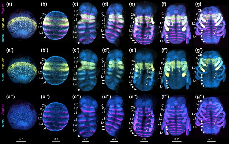
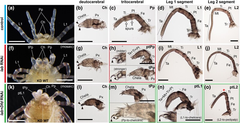
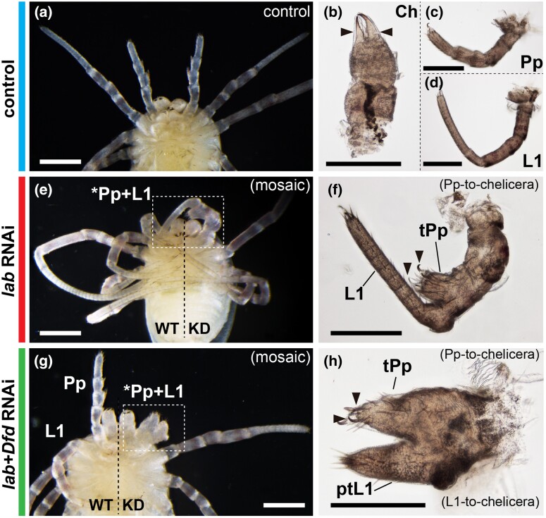
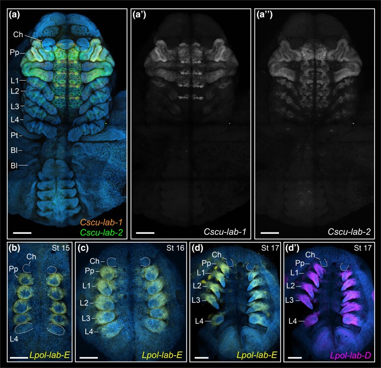
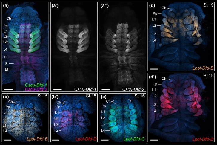
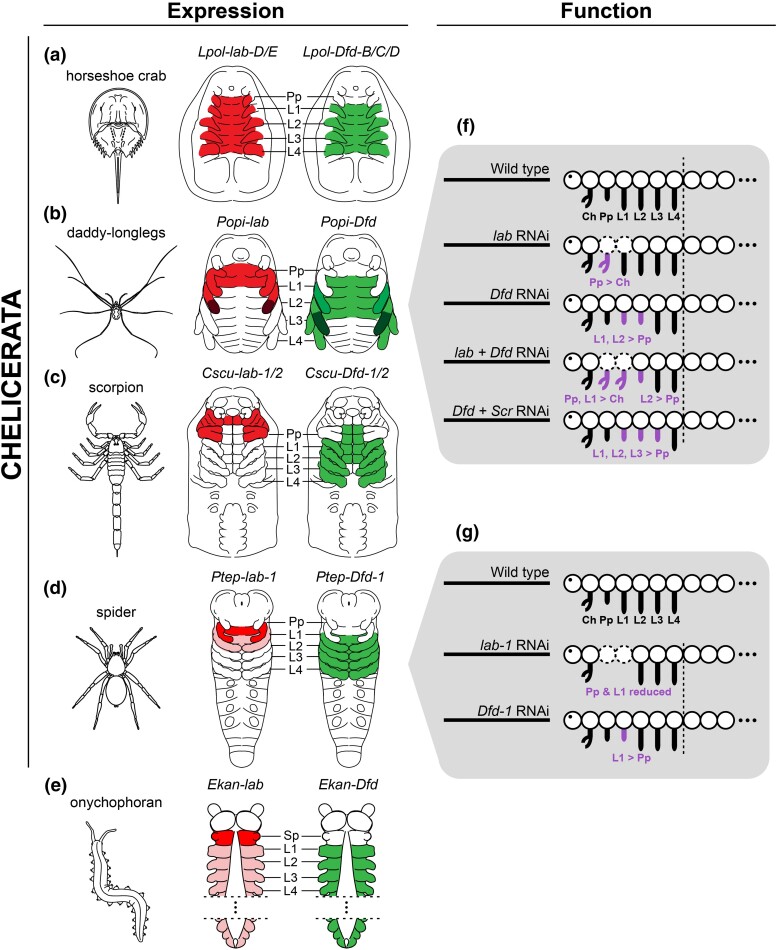
Despite an abundance of gene expression surveys, comparatively little is known about Hox gene function in Chelicerata. Previous investigations of paralogs of labial (lab) and Deformed (Dfd) in a spider have shown that these play a role in tissue maintenance of the pedipalp segment (lab-1) and in patterning the first walking leg identity (Dfd-1), respectively. However, extrapolations of these data across chelicerates are hindered by the existence of duplicated Hox genes in arachnopulmonates (e.g., spiders and scorpions), which have resulted from an ancient whole genome duplication (WGD) event. Here, we investigated the function of the single-copy ortholog of lab in the harvestman Phalangium opilio, an exemplar of a lineage that was not subject to this WGD. Embryonic RNA interference against lab resulted in two classes of phenotypes: homeotic transformations of pedipalps to chelicerae, as well as reduction and fusion of the pedipalp and leg 1 segments. To test for combinatorial function, we performed a double knockdown of lab and Dfd, which resulted in a homeotic transformation of both pedipalps and the first walking legs into cheliceral identity, whereas the second walking leg is transformed into a pedipalpal identity. Taken together, these results elucidate a model for the Hox logic of head segments in Chelicerata. To substantiate the validity of this model, we performed expression surveys for lab and Dfd paralogs in scorpions and horseshoe crabs. We show that repetition of morphologically similar appendages is correlated with uniform expression levels of the Hox genes lab and Dfd, irrespective of the number of gene copies.

Keywords: Hox1; Arthropoda; Opiliones; Xiphosura; serial homology; tritocerebrum.


Figures

Fig. 1.
Fig. 1.
Wild type expression of Popi-lab and Popi-en across embryonic stages. Anterior at top. (a) Wholemount, ventrolateral view of the germ disc. (b) Wholemount, ventral view. (cg): Flatmount, ventral view. Nuclei in blue (Hoechst). Popi-en: magenta. Popi-lab: yellow. (ag): Hoechst, Popi-lab and Popi-en multiplexed. (a′–g′): Hoechst and Popi-lab, same embryos as previous row. (a″g″): Hoechst and Popi-en, same embryos as previous row. Arrowheads: Popi-en stripes marking the posterior of each segment. Arrows: dots of Popi-lab expression adjacent to the ventral midline. Ch, chelicera; Pp, pedipalp; L1–L4, L1–L4 legs; st, stage number. Scale bars: 200 µm.
Fig. 2.
Fig. 2.
Popi-lab knockdown results in homeotic transformations. Brightfield images of hatchlings (postembryos) of control (dH2O-injected) (ae), Popi-lab RNAi (fj), and Popi-lab + Popi-Dfd RNAi (ko) experiments. (a), (f) and (k) are ventral views of the anterior prosoma. Other panels are dissected appendages in lateral view with proximal at right. (b, g, and l): Deutocerebral appendage (chelicera). (c, h, and m): Tritocerebral appendage. (d, i, and n): Appendage of the L1 segment. (e, j, and o): Appendage of the L2 segment. Note that because the first instar's cuticle is already secreted and visible inside the postembryo cuticle, two sets of claws are discernible in some panels. Black arrowhead, claw; red asterisk, missing metatarsus (Mt); white arrows, pedipalpal spurs; Ch, chelicera; Pp, pedipalp; tPp, transformed pedipalp; ptPp, partially transformed pedipalp; L1–L2, L1–L2 appendages; ptL1–ptL2, partially transformed L1–L2 appendages; Ta, tarsus; Mt, metatarsus; Ti, tibia; Pt, patella; Fe, femur; Tr, trochanter; Px, proximal cheliceral segment; WT, wild type side of individual; KD, knockdown side of individual. Scale bars: 200 µm.
Fig. 3.
Fig. 3.
Popi-lab knockdown results in fusion of the pedipalp and L1 leg. Brightfield images of hatchlings (postembryos) of control (dH2O-injected) (ad), Popi-lab RNAi (ef), and Popi-lab + Popi-Dfd RNAi (gh) experiments. Phenotypes in (gh) present both homeosis and fusion. (a, e, and g) are ventral views of the anterior prosoma. Other panels are dissected appendages in lateral view. Mosaic individuals in (e) and (g) are affected on the right side of the picture. Black arrowhead, claw; Ch, chelicera; Pp, pedipalp; tPp, transformed pedipalp; L1, L1 appendage; ptL1, partially transformed L1 leg; WT, wildtype side of individual; KD, knockdown side of individual. Scale bars: 200 µm.
Fig. 4.
Fig. 4.
HCR in situ hybridization of labial paralogs in the scorpion C. sculpturatus (aa″) and the horseshoe crab L. polyphemus (bd′). Maximum intensity projections of flat mounted embryos in ventral view. Hoechst counter staining in blue. Same letters indicate same embryo (multiplexed). (a) Merged projections of Cscu-lab-1 (orange), Cscu-lab-2 (green). Overlap of lab paralogs appears in yellow. Note that apparent labial expression in the posterior chelicera and labrum is in the tritocerebral tissue; overlapping of these regions is incurring by the maximum intensity projection. (a′) Single channel projection of Cscu-lab-1. (a″) Single channel projection of Cscu-lab-2. (bd) Lpol-lab-E expression (yellow). (d″) Lpol-lab-D expression (magenta). Ch, chelicera; Pp, pedipalp; L1–L4, L1–L4 legs. Scale bars: 200 µm.
Fig. 5.
Fig. 5.
HCR in situ hybridization of Deformed paralogs in the scorpion C. sculpturatus (aa″) and the horseshoe crab L. polyphemus (bd′). Maximum intensity projections of flat mounted embryos in ventral view. Hoechst counter staining in blue. Same letters indicate same embryo (multiplexed). (a) Merged projections of Cscu-Dfd-1 (green), Cscu-Dfd-2 (magenta). (b) Lpol-Dfd-B expression (orange). (b′) Lpol-Dfd-D expression (red). (c) Lpol-Dfd-C expression (green). (d) Lpol-Dfd-B expression (orange). (d′) Lpol-Dfd-D expression (red). Ch, chelicera; Pp, pedipalp; L1–L4, L1–L4 legs. Scale bars: 200 µm.
Fig. 6.
Fig. 6.
Summary of labial (red; left schematics) and Deformed (green; right schematics) expression and functional data in Chelicerata and Onychophora (outgroup). Darker shades reflect concentration of expression. (a) Horseshoe crab L. polyphemus. (b) Daddy-longlegs P. opilio, after Sharma et al. (2012a), and Gainett et al. (2021). (c) Scorpion C. sculpturatus. (d) Spider P. tepidariorum, after Schwager et al. ([2017]). (e) Onychophoran E. kanangrensis, after Eriksson et al (2010) and Janssen et al. (2014). (f) Summary of phenotypic outcomes of Hox RNAi experiments in P. opilio. Dotted body segments indicate segment reduction. Data for the single knockdown of Popi-Dfd and Popi-Scr knockdown after Gainett et al. (2021). (g) Summary of phenotypic outcomes of Ptep-lab-1 and Ptep-Dfd-1 RNAi experiments in P. tepidariorum, after Pechmann et al. (2015). Ch, chelicera; Pp, pedipalp; L1–L4, L1–L4 legs; Sp, slime papilla.

Similar articles

Cited by

References

    1. Abzhanov A, Kaufman TC. 1999. Homeotic genes and the arthropod head: expression patterns of the labial, proboscipedia, and Deformed genes in crustaceans and insects. Proc Natl Acad Sci U S A. 96:10224–10229. - PMC - PubMed
    1. Angelini DR, Liu PZ, Hughes CL, Kaufman TC. 2005. Hox gene function and interaction in the milkweed bug Oncopeltus fasciatus (Hemiptera). Dev Biol. 287:440–455. - PubMed
    1. Arnaud F, Bamber RN. 1988. The Biology of Pycnogonida. Adv Mar Biol 24:1–96.
    1. Auman T, Chipman AD. 2018. Growth zone segmentation in the milkweed bug Oncopeltus fasciatus sheds light on the evolution of insect segmentation. BMC Evol Biol. 18:178. - PMC - PubMed
    1. Ballesteros JA, Santibáñez-López CE, Baker CM, Benavides LR, Cunha TJ, Gainett G, Ontano AZ, Setton EVW, Arango CP, Gavish-Regev E, et al. . 2022. Comprehensive species sampling and sophisticated algorithmic approaches refute the monophyly of Arachnida. Mol Biol Evol. 39:msac021. - PMC - PubMed

Publication types

Substances