Review
. 2023 Mar;16(3):e012019.
doi: 10.1161/CIRCINTERVENTIONS.122.012019. Epub 2023 Feb 17.

Transcatheter Electrosurgery: A Narrative Review

Affiliations
Review

Transcatheter Electrosurgery: A Narrative Review

Christopher G Bruce et al. Circ Cardiovasc Interv. 2023 Mar.

Abstract

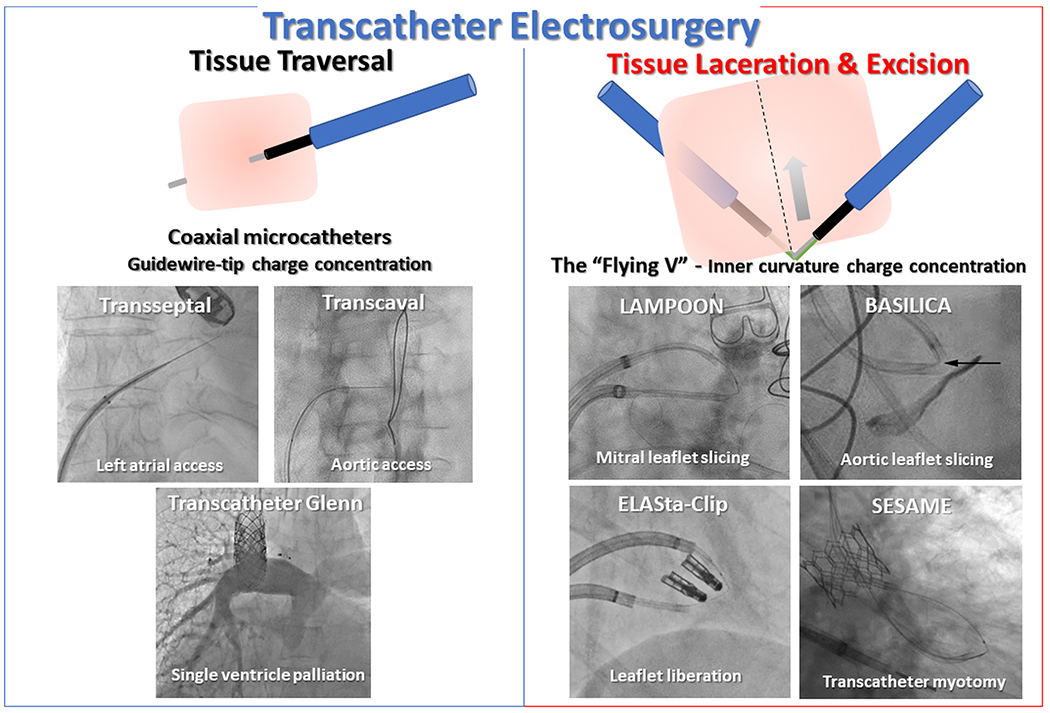
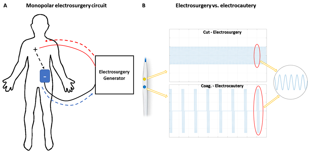
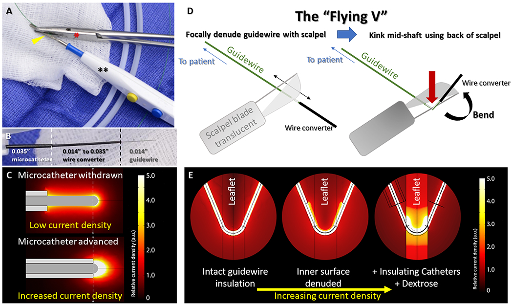
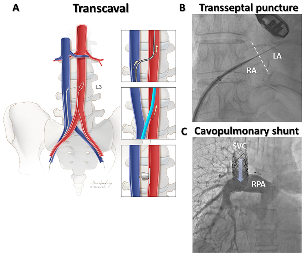
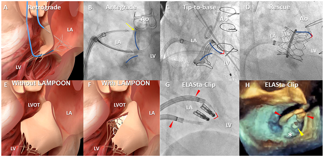
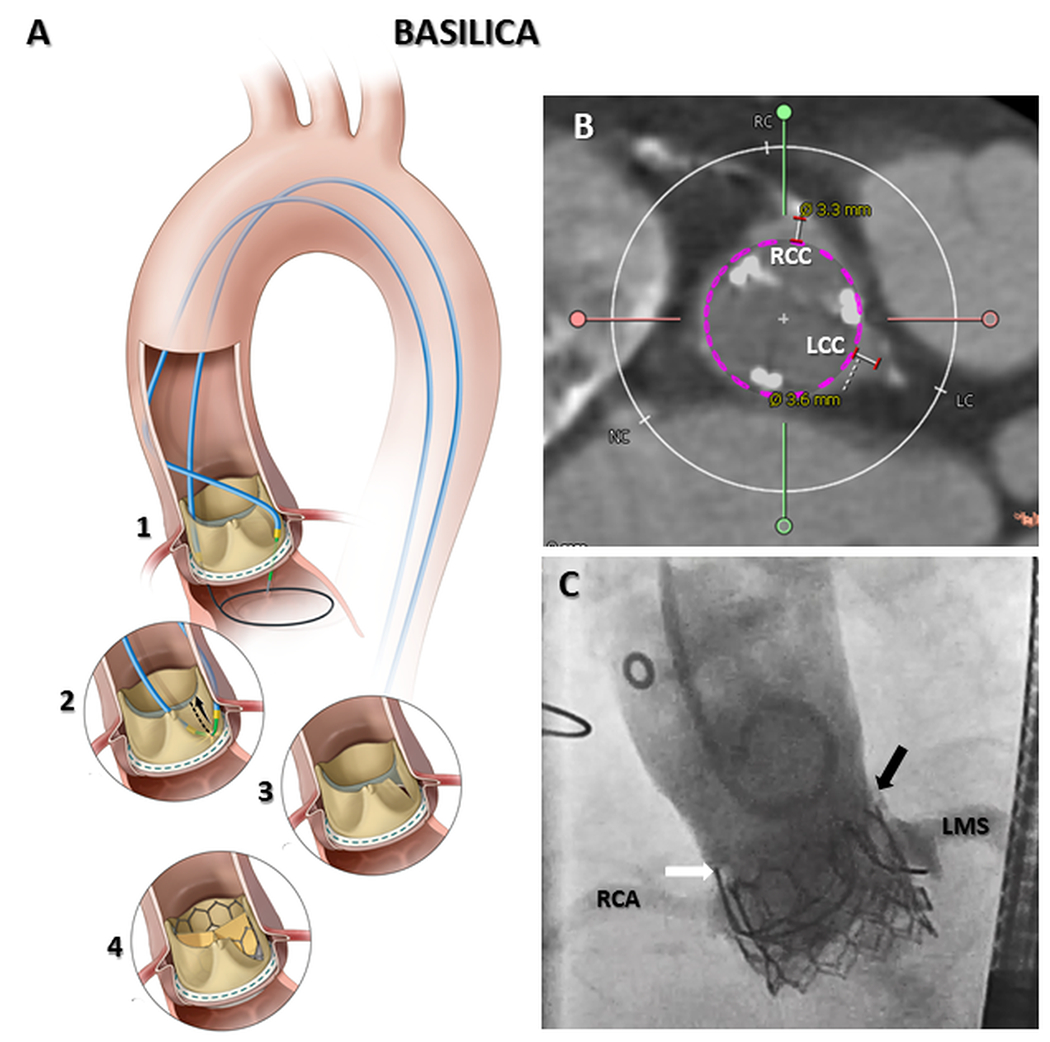
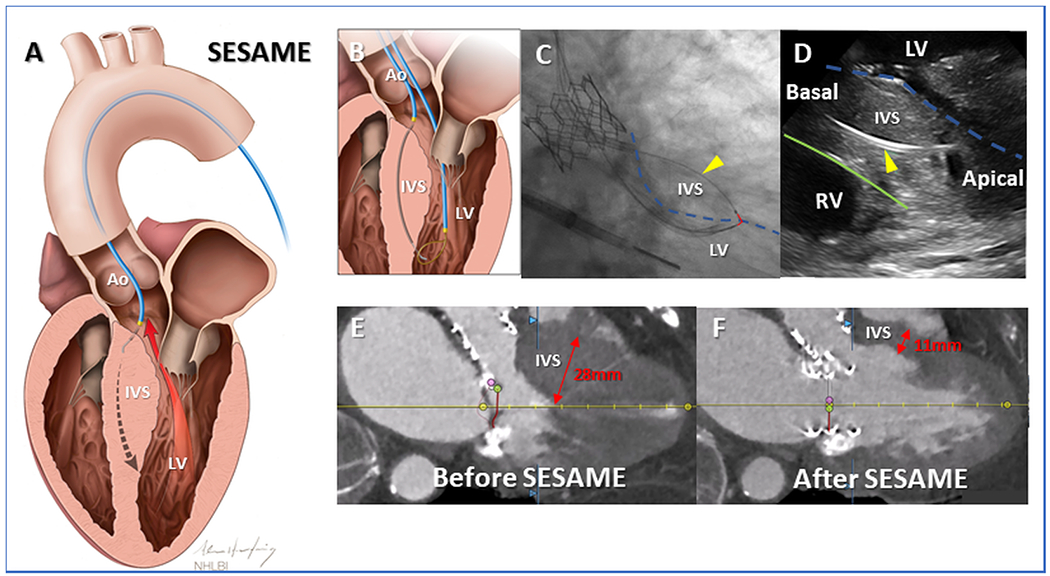
Transcatheter electrosurgery describes the ability to cut and traverse tissue, at a distance, without an open surgical field and is possible using either purpose-built or off-the-shelf devices. Tissue traversal requires focused delivery of radiofrequency energy to a guidewire tip. Initially employed to cross atretic pulmonary valves, tissue traversal has enabled transcaval aortic access, recanalization of arterial and venous occlusions, transseptal access, and many other techniques. To cut tissue, the selectively denuded inner curvature of a kinked guidewire (the Flying-V) or a single-loop snare is energized during traction. Adjunctive techniques may complement or enable contemporary transcatheter procedures, whereas myocardial slicing or excision of ectopic masses may offer definitive therapy. In this contemporary review we discuss the principles of transcatheter electrosurgery, and through exemplary clinical applications highlight the range of therapeutic options offered by this versatile family of procedures.

Keywords: catheter; catheterization; electrode; electrosurgery; transcatheter aortic valve replacement.


Figures

Fig 1.
Fig 1.
Clinical applications of transcatheter electrosurgery.
Fig 2.
Fig 2.. Monopolar electrosurgery circuit and radiofrequncy waveforms.
Monopolar electrosurgical circuits (A) consist of an active electrode(+) that receives current from an electrosurgery generator (red interrupted-arrow) and conducts through the body (black interrupted-arrow) to a dispersive electrode(blue patch), placed on the patient’s skin, and thereafter back to the generator (blue interrupted-arrow). (B) Continuous versus intermittent radiofrequency application (“duty-cycle”) creates different electrosurgical effects. Continuous (100% ‘on’) radiofrequency energy (top panel) vaporizes cells and cuts tissue at the point of maximum current density adjacent to the active electrode. In (“low duty cycle”) electrocautery(bottom panel), interrupted radiofrequency energy causes tissue heating, protein denaturation, and blood coagulation. Reprinted from Khan et al. JACC, 2020.
Fig 3.
Fig 3.. Guidewire modifications that enable transcatheter electrosurgery.
(A) The denuded back-end of a guidewire(yellow arrowhead) is clamped to an electrosurgery pencil(**) by hemostatic forceps(red*). (B) A ‘crossing-system’ for electrosurgical tissue traversal consists of an 0.014” guidewire inside a hubless-locking wire-converter, inside a 0.035” microcatheter. (C) Microcatheters increase current density at the guidewire tip increasing electrosurgical efficiency. Focal denudation and kinking the mid-shaft of the guidewire (D) create the “Flying V”. When placed at the target tissue, inner curvature denudation focuses charge and increases current density (E). Microcatheter insulation and dextrose infusion further enhance charge concentration. Reprinted from Khan et al. JACC, 2020.
Fig 4.
Fig 4.. Clinical applications – Tissue Traversal.
In transcaval aortic access (A), the electrified guidewire is advanced through the walls of inferior vena cava and abdominal aorta where it is ensnared. Exchange for a stiff guidewire permits large bore access to the aorta in patients with unsuitable femoral arteries. The transcaval tract is closed with a nitinol vascular occluder. (B) Electrosurgical transseptal puncture using the dilator of a deflectable sheath for insulation. (C) A transcatheter superior cavopulmonary shunt was following electrosurgical traversal from superior vena cava to right pulmonary artery. RA=Right atrium; LA=Left atrium; SVC=Superior vena cava; RPA=Right pulmonary artery. Reprinted from Greenbaum et al. JACC, 2017.
Fig 5.
Fig 5.. Clinical applications – Tissue laceration; Mitral leaflet modification.
The original, retrograde LAMPOON (A) crossed the base of the A2 mitral scallop across the aortic valve. Technical refinements include an antegrade approach (B) across the interventricular septum. One catheter is positioned on the atrial surface at the base (yellow arrow) of the A2 mitral leaflet (blue overlay) from where the guidewire is electrified towards a snare in the LVOT. Tip-to-base LAMPOON (C) lacerates the mitral leaflet (blue overlay) in reverse, without leaflet traversal, in patients with a suitable backstop such as a surgical prosthesis. The flying V (red-outline) is positioned on the tip and withdrawn until it meets a hard-stop. Rescue LAMPOON (D) similarly lacerates from tip backwards to slice long, overhanging leaflets causing dynamic LVOT obstruction after transcatheter mitral valve replacement. LAMPOON techniques uncover cells otherwise draped with anterior mitral valve leaflet tissue (E&F). (G) A pair of deflectable sheaths(red arrowheads) for ELASta-Clip to liberate MitraClips from the anterior mitral leaflet to enable TMVR. (H) Positioning of sheaths(red arrowheads) anterior to the MitraClip(yellow arrowhead) allows TMVR to pin the posterior leaflet(white asterisk) harmlessly. Ao=Aorta; LA=Left atrium; LV=Left ventricle; LVOT=Left ventricular outflow tract.
Fig 6.
Fig 6.. Clinical applications – Tissue laceration; BASILICA.
Tissue traversal at the base of the offending aortic leaflet (A1), followed by tissue laceration (A2) creates a midline slice in target aortic leaflet (A3). BASILICA enables leaflet splay following transcatheter heart valve implantation, preventing coronary artery obstruction (A4). In cases where both right and left coronary ostia are at risk (B) BASILICA can be performed on both leaflets in a ‘doppio’ procedure. In (C) brisk coronary flow remains following TAVR despite surgical valve leaflets being visible above left (black arrow) and right (white arrow) aortic sinuses. RCA=Right coronary artery; RCC=Right coronary cusp; LCC=Left coronary cusp; LMS=Left main stem. Reprinted from Khan et al. JACC: Cardiovascular Interventions, 2018.
Fig 7.
Fig 7.. Clinical applications – Myocardial slicing.
(A) In SESAME transcatheter septal myotomy, a guidewire traverses the interventricular septum, defining length and depth of subsequent myotomy. Once ensnared (B) the flying V (C, red overlay) is positioned on the left ventricular endocardium(broken blue-outline). Fluoroscopic (C) and intracardiac echocardiographic (D) appearance of catheters, one wholly within septal myocardium(yellow arrowhead), safely deep to right ventricular endocardium (green outline), positioned ready to cut muscle during guidewire electrification. SESAME creates space in the left ventricular outflow tract to enable TMVR (E&F). Ao=Aorta; IVS=Interventricular septum; LV=Left ventricle; RV=Right ventricle. Reprinted from Khan et al. Circulation: Cardiovascular Interventions, 2022.

References

    1. Khan JM, Lederman RJ. Transcatheter electrosurgery in bipolar or monopolar modes. Catheter Cardiovasc Interv. 2018;91:1052–1053. doi: 10.1002/ccd.27620 - DOI - PMC - PubMed
    1. Khan JM, Rogers T, Greenbaum AB, Babaliaros VC, Yildirim DK, Bruce CG, Herzka DA, Schenke WH, Ratnayaka K, Lederman RJ. Transcatheter Electrosurgery: JACC State-of-the-Art Review. J Am Coll Cardiol. 2020;75:1455–1470. doi: 10.1016/j.jacc.2020.01.035 - DOI - PMC - PubMed
    1. Rosenthal E, Qureshi SA, Chan KC, Martin RP, Skehan DJ, Jordan SC, Tynan M. Radiofrequency-assisted balloon dilatation in patients with pulmonary valve atresia and an intact ventricular septum. Br Heart J. 1993;69:347–351. doi: 10.1136/hrt.69.4.347 - DOI - PMC - PubMed
    1. Akagi T, Hashino K, Maeno Y, Ishii M, Sugimura T, Kawano T, Kato H. Balloon dilatation of the pulmonary valve in a patient with pulmonary atresia and intact ventricular septum using a commercially available radiofrequency catheter. Pediatr Cardiol. 1997;18:61–63. doi: 10.1007/s002469900112 - DOI - PubMed
    1. Alwi M, Geetha K, Bilkis AA, Lim MK, Hasri S, Haifa AL, Sallehudin A, Zambahari R. Pulmonary atresia with intact ventricular septum percutaneous radiofrequency-assisted valvotomy and balloon dilation versus surgical valvotomy and Blalock Taussig shunt. J Am Coll Cardiol. 2000;35:468–476. doi: 10.1016/s0735-1097(99)00549-5 - DOI - PubMed

Publication types