. 2023 Nov;17(11):2257-2274.
doi: 10.1002/1878-0261.13397. Epub 2023 Jul 14.

MET exon 14 skipping mutation is a hepatocyte growth factor (HGF)-dependent oncogenic driver in vitro and in humanised HGF knock-in mice

Affiliations

MET exon 14 skipping mutation is a hepatocyte growth factor (HGF)-dependent oncogenic driver in vitro and in humanised HGF knock-in mice

Marie Fernandes et al. Mol Oncol. 2023 Nov.

Abstract

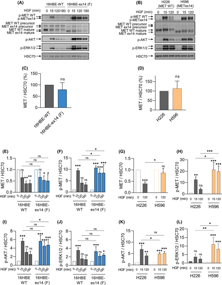
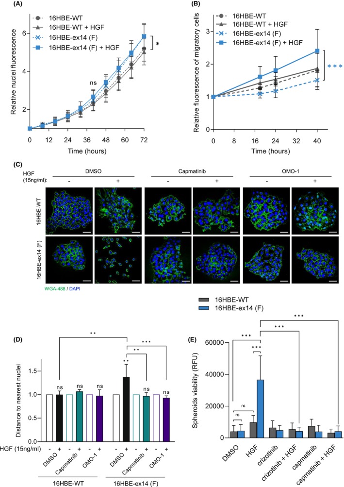
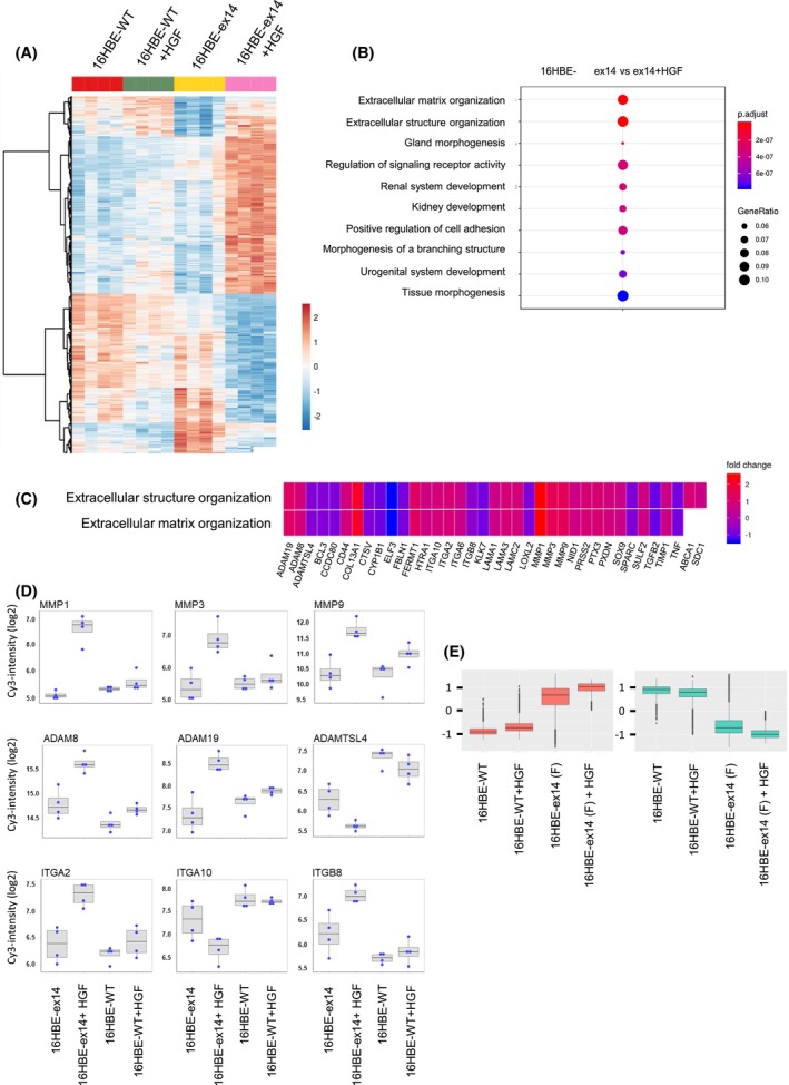
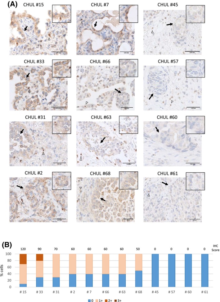
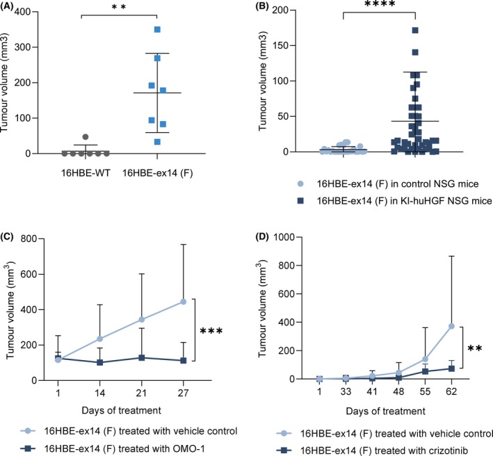
Exon skipping mutations of the MET receptor tyrosine kinase (METex14), increasingly reported in cancers, occur in 3-4% of non-small-cell lung cancer (NSCLC). Only 50% of patients have a beneficial response to treatment with MET-tyrosine kinase inhibitors (TKIs), underlying the need to understand the mechanism of METex14 oncogenicity and sensitivity to TKIs. Whether METex14 is a driver mutation and whether it requires hepatocyte growth factor (HGF) for its oncogenicity in a range of in vitro functions and in vivo has not been fully elucidated from previous preclinical models. Using CRISPR/Cas9, we developed a METex14/WT isogenic model in nontransformed human lung cells and report that the METex14 single alteration was sufficient to drive MET-dependent in vitro anchorage-independent survival and motility and in vivo tumorigenesis, sensitising tumours to MET-TKIs. However, we also show that human HGF (hHGF) is required, as demonstrated in vivo using a humanised HGF knock-in strain of mice and further detected in tumour cells of METex14 NSCLC patient samples. Our results also suggest that METex14 oncogenicity is not a consequence of an escape from degradation in our cell model. Thus, we developed a valuable model for preclinical studies and present results that have potential clinical implication.

Keywords: hepatocyte growth factor; lung cancer; preclinical models; targeted therapies; transcriptomic; tyrosine kinase receptor.


Conflict of interest statement

PJ reports personal fees and non‐financial support from Novartis and Boehringer Ingelheim FRANCE, non‐financial support from Pierre Fabre, Chugai Pharma and MYLAN MEDICAL SAS, outside the submitted work. SH participates in advisory board for Brystol‐Myers Squibb and Boehringer Ingelheim. CD reports personal fees and non‐financial support from AstraZeneca, Novartis pharma SAS, Roche SAS, Boehringer Ingelheim France, Pfizer, outside the submitted work. MCC participates in advisory boards for Pfizer and Roche. ABC participated in advisory boards or received honoraria from Abbvie, Amgen, Astra‐Zeneca, Bristol‐Myers Squibb, Merck & Co, Pfizer, Roche, Novartis, Takeda, Janssen, Sanofi and received grants payed to ABC's institution from Novartis, Merck, Roche. TP headed OCTIMET Oncology NV and DeuterOncology NV. MFe, BH, SP, VG, AV, BDL, AM, GW, PG, JPM, SS, EC, JS, TS, PF, LG, MFi, DT, SK and ZK declare no competing financial interests.

Figures

Fig. 1
Fig. 1
Activation of exon 14 spliced MET is dependent on HGF, and results in sustained downstream signalling. (A) 16HBE cells expressing either WT MET or METex14, following CRISPR/Cas9 editing and (B) H226, expressing MET WT and H596 cells expressing endogenous MET ex14, were grown for 48 h (A) and 24 h (B) and serum‐starved 1 h before treatment with 50 ng·mL−1 HGF for the times indicated. For each condition, whole cell lysates were resolved by SDS/PAGE and analysed by western blotting with the indicated antibodies. Arrows indicate MET WT or ex14 precursor or beta‐chain mature forms. Quantification by densitometry, normalised to a loading control (HSC70) is represented as fold change of MET expression in unstimulated cells (C, D), and of MET expression (E, G) or MET, AKT and ERK1/2 phosphorylation (F, H–L) levels upon HGF activation in each cell line. Data are means of three experiments, −/+ SD, Two‐way ANOVA test, *P < 0.05, **P < 0.01, ***P < 0.001, ns, not significant.
Fig. 2
Fig. 2
Transforming capacities of 16HBE‐ex14 cells are dependent on HGF stimulation in vitro. (A) Real‐time cell numbers of adherent 16HBE cell lines, stimulated or not by 20 ng·mL−1 of HGF, was determined through viable nuclei labelling and live imaging in an IncuCyte ZOOM over 72 h. (B) Migration of DilC12‐labelled 16HBE cells was determined in a transwell assay, with or without HGF (20 ng·mL−1) in the medium of the lower chamber. Fluorescence of migrating cells was measured over time and expressed as relative mean migration. (C, D) 16HBE cells were seeded at a low density on coverslips in full media and stimulated or not by 15 ng·mL−1 of HGF in the presence of DMSO, capmatinib or OMO‐1 (1 μm). (C) Representative confocal sections of cells fixed at day 6 are shown (scale bar: 50 μm). (D) Distances from the centroid of each nucleus to the second nearest nucleus represented as fold change with HGF over without HGF. (E) The viability of non‐adherent spheroids of 16HBE cells seeded in ultra‐low attachment plates with low‐serum media containing HGF (20 ng·mL−1), crizotinib (1 μm) or capmatinib (1 μm), was evaluated after 14 days by reduction of resazurin (Alamar Blue reagent). All data are means of three experiments with at least three wells per condition, Two‐way ANOVA test −/+ SD, *P < 0.05, **P < 0.01, ***P < 0.001, ns, not significant.
Fig. 3
Fig. 3
Heat map and gene ontology (GO) enrichment of genes differentially expressed in 16HBE WT or 16HBE‐ex14 cells in response to HGF. 16HBE cells expressing either MET WT or METex14 were grown 24 h, serum‐starved 2 h and treated for 24 h with HGF (20 ng·mL−1). mRNA was extracted and gene expression determined by DNA microarray. (A) Heat map of genes significantly differentially expressed in response to HGF (P‐value (adj) < 0.05 and absolute fold change > 1.5) between indicated conditions (n = 4 for each condition). Scale bar represents relative gene expression changes scaling. (B) Dot plot of GO enrichment of genes significantly differentially expressed in 16HBE cells stimulated or not with HGF according to the ‘biological process’ annotations of Gene Ontology. Dot size represents ratio of significantly differentiated genes. Colour scale represents P‐value (adjusted). (C) Heatmap‐like functional classification of gene list for the subgroups of ‘extracellular structure and matrix organization’ GO enrichment, with colour indicating the fold change of each gene comparing 16HBE‐ex14 cells activated with HGF to basal conditions. (D) Box‐and‐whisker plot of selection from the significantly differentiated GO ‘extracellular matrix organization’ analysis (intensity in log2) of MMP, ADAM and integrin families. All genes were significantly differentially regulated in the transcriptomic analysis of 16HBE‐WT cells and 16HBE‐ex14 cells activated by HGF. The boxplot shows the 25th, 50th and 75th percentiles while the blue points show the normalised expression values for each sample (n = 4 per condition). The median is indicated by the line across the box. (E) Box‐and‐whisker plot of the normalised expression of the top 100 most deregulated genes in the 16HBE‐ex14 cells activated with HGF to basal conditions. The comparison, performed for the four experimental conditions, show upregulated genes in red and downregulated genes in green.
Fig. 4
Fig. 4
HGF expression in METex14 NSCLC patient tumours. (A) HGF immunostaining (brown) and nuclear staining with haematoxylin (blue) on FFPE tumour samples from NSCLC patients harbouring METex14 mutations. Empty arrowheads indicate examples of stromal cells known to express HGF and arrows indicate examples of tumour cells. Scale bar = 0.05 mm. (n = 1) (B) Samples were semi‐quantified by pathologist visual scoring of staining on a scale of 0–3+. A semiquantitative scoring was performed by multiplying the percentage of positive stained cells by the intensity of labelling.
Fig. 5
Fig. 5
MET exon 14 loss sensitises tumours to MET‐TKIs in the presence of HGF. In vivo tumour xenograft experiments were performed with humanised HGF knock‐in NSG (KI‐huHGF) or control NSG mice. (A) Scatter plots of the tumour volumes (mm3) at end point in KI‐huHGF mice xenografted with 16HBE‐WT or 16HBE‐ex14 cells (n = 7 per group). (B) Scatter plots of the tumour volumes (mm3) 8 weeks after xenografts of 16HBE‐ex14 cells in control NSG (n = 26) or KI‐huHGF (n = 44) mice. (C) Tumour growth curves of 16HBE‐ex14 in KI‐huHGF mice treated, when tumours reached a mean volume of 100 mm3, with 48 mg·kg−1·day−1 of OMO‐1 or vehicle control (n = 8 per group). (D) Tumour growth curves of 16HBE‐ex14 in KI‐huHGF mice treated, 30 days after cell xenografts, with 50 mg·kg−1·day−1 of crizotinib or vehicle control (n = 14 per group). (A, B) Statistical analyses were performed with a nonparametric Mann–Whitney test. (C, D) Data are mean tumour volumes (mm3) of indicated number of tumours, −/+ SD. Statistical analyses were performed with a two‐way ANOVA test to compare the whole curves. **P ≤ 0.01, ***P ≤ 0.001, ****P ≤ 0.0001.

References

    1. Bedard PL, Hyman DM, Davids MS, Siu LL. Small molecules, big impact: 20 years of targeted therapy in oncology. Lancet. 2020;395:1078–88. - PubMed
    1. Lynch TJ, Bell DW, Sordella R, Gurubhagavatula S, Okimoto RA, Brannigan BW, et al. Activating mutations in the epidermal growth factor receptor underlying responsiveness of non‐small‐cell lung cancer to gefitinib. N Engl J Med. 2004;350:2129–39. - PubMed
    1. Liu X, Jia Y, Stoopler MB, Shen Y, Cheng H, Chen J, et al. Next‐generation sequencing of pulmonary Sarcomatoid carcinoma reveals high frequency of actionable MET gene mutations. J Clin Oncol. 2016;34:794–802. - PubMed
    1. Kawakami H, Okamoto I, Okamoto W, Tanizaki J, Nakagawa K, Nishio K. Targeting MET amplification as a new oncogenic driver. Cancers (Basel). 2014;6:1540–52. - PMC - PubMed
    1. Tovar EA, Graveel CR. MET in human cancer: germline and somatic mutations. Ann Transl Med. 2017;5:205. - PMC - PubMed