. 2023 Apr;145(4):479-496.
doi: 10.1007/s00401-023-02551-7. Epub 2023 Feb 17.

Variants in DTNA cause a mild, dominantly inherited muscular dystrophy

Affiliations

Variants in DTNA cause a mild, dominantly inherited muscular dystrophy

Andres Nascimento et al. Acta Neuropathol. 2023 Apr.

Abstract

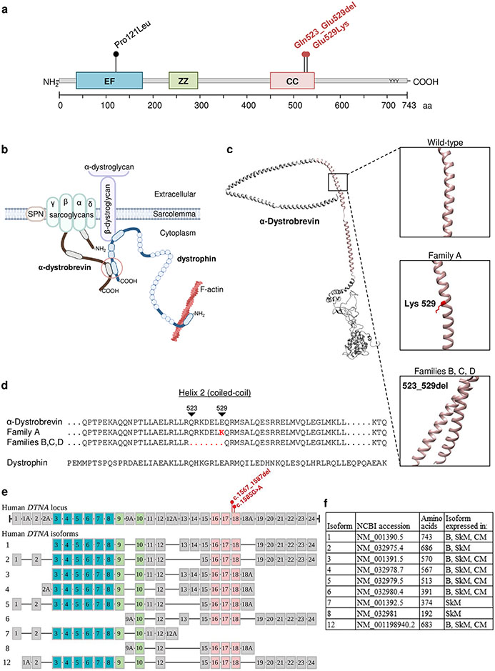
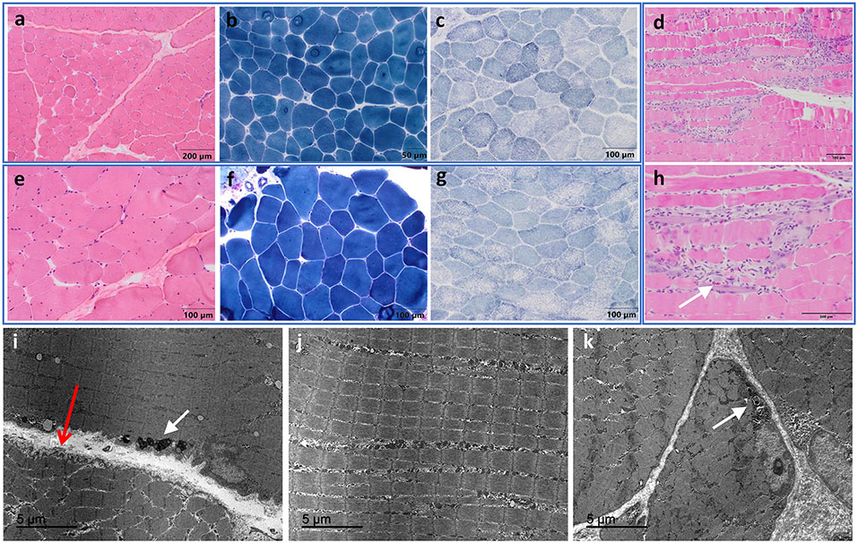
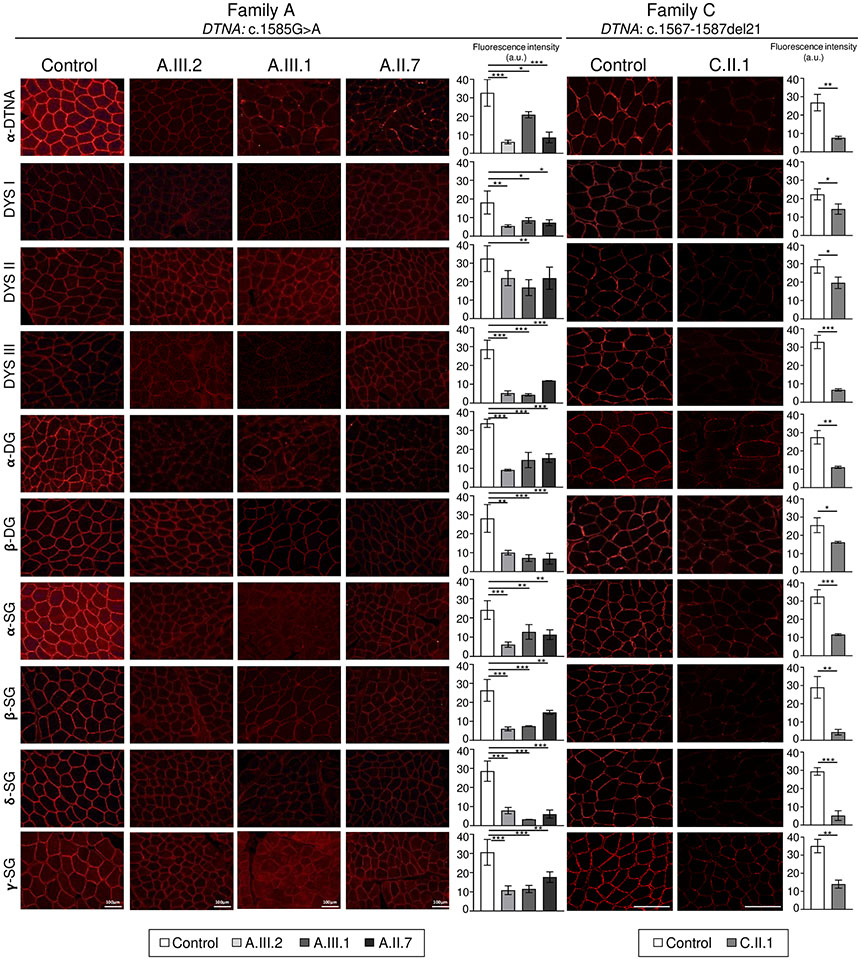
DTNA encodes α-dystrobrevin, a component of the macromolecular dystrophin-glycoprotein complex (DGC) that binds to dystrophin/utrophin and α-syntrophin. Mice lacking α-dystrobrevin have a muscular dystrophy phenotype, but variants in DTNA have not previously been associated with human skeletal muscle disease. We present 12 individuals from four unrelated families with two different monoallelic DTNA variants affecting the coiled-coil domain of α-dystrobrevin. The five affected individuals from family A harbor a c.1585G > A; p.Glu529Lys variant, while the recurrent c.1567_1587del; p.Gln523_Glu529del DTNA variant was identified in the other three families (family B: four affected individuals, family C: one affected individual, and family D: two affected individuals). Myalgia and exercise intolerance, with variable ages of onset, were reported in 10 of 12 affected individuals. Proximal lower limb weakness with onset in the first decade of life was noted in three individuals. Persistent elevations of serum creatine kinase (CK) levels were detected in 11 of 12 affected individuals, 1 of whom had an episode of rhabdomyolysis at 20 years of age. Autism spectrum disorder or learning disabilities were reported in four individuals with the c.1567_1587 deletion. Muscle biopsies in eight affected individuals showed mixed myopathic and dystrophic findings, characterized by fiber size variability, internalized nuclei, and slightly increased extracellular connective tissue and inflammation. Immunofluorescence analysis of biopsies from five affected individuals showed reduced α-dystrobrevin immunoreactivity and variably reduced immunoreactivity of other DGC proteins: dystrophin, α, β, δ and γ-sarcoglycans, and α and β-dystroglycans. The DTNA deletion disrupted an interaction between α-dystrobrevin and syntrophin. Specific variants in the coiled-coil domain of DTNA cause skeletal muscle disease with variable penetrance. Affected individuals show a spectrum of clinical manifestations, with severity ranging from hyperCKemia, myalgias, and exercise intolerance to childhood-onset proximal muscle weakness. Our findings expand the molecular etiologies of both muscular dystrophy and paucisymptomatic hyperCKemia, to now include monoallelic DTNA variants as a novel cause of skeletal muscle disease in humans.

Keywords: Dystrophin; Muscle sarcolemmal proteins; Myalgia; Rhabdomyolysis; hyperCKemia; α-Dystrobrevin.


Conflict of interest statement

Conflicts of interest None of the authors has any conflict of interest to disclose.

Figures

Figure 1.
Figure 1.. Pedigrees of the four families with DTNA variants included in this study.
Arrows indicate probands, circles indicate females, squares indicate males, filled in symbols denote affected individuals (the gray colour indicates a milder phenotype), and a question mark in the box indicates that the phenotype is not known. Asterisks mark individuals from whom DNA samples were obtained.
Figure 2.
Figure 2.. In silico analyses of α-dystrobrevin variants found in the four families included in our cohort.
(a) α-dystrobrevin domain structure (Uniprot: Q9Y4J8) and location of the variants found in families A, B, C, and D (in red), as well as the previously reported variant associated with left ventricular non-compaction (LVNC) cardiomyopathy (in black). (b) Partial schematic representation of the dystrophin glycoprotein complex (DGC). (c) Modeled 3D structure of wild-type α-dystrobrevin with coiled-coil domain indicated in salmon. Wild-type and affected individuals modeled residues indicated in boxes. All residues in all 3D structures were modelled at >90% confidence using Phyre2 software. (d) Sequence alignment of part of the coiled-coil domains of wild-type and mutated α-dystrobrevin and wild-type dystrophin. Abbreviations: EF, EF hand region; ZZ, zinc-binding domain; CC, coiled-coil. (e) The genomic structure of human DTNA indicating the two variants found in the four families. Also shown is a schematic representation of the exons included in 9 isoforms of DTNA that are expressed in skeletal muscle. Exons encoding the calcium-binding EF hand domains are shaded in teal, exons encoding the zinc finger domains are shaded in lime green, and exons encoding the coiled-coil domain are shaded in salmon. (f) The selected DTNA isoforms (Uniprot: Q9Y4J8-1 through Q9Y4J8-8 and Q9Y4J8-13) are listed with National Center for Biotechnology Information (NCBI) transcript accession number, number of amino acids, and tissues in which the isoforms are highly expressed. B, brain; SkM, skeletal muscle; CM, cardiac muscle.
Figure 3.
Figure 3.. Muscle histology findings from biopsies of individuals with pathogenic DTNA variants.
The biopsy from individual A.III.2 was taken at 17 years (a-c) and the biopsy from individual A.III.1 at 23 years (e-g). Variability in fibre size (range: 31-105 μm in individual A.III.2; range: 37-178 μm in individual A.III.1) and fibres with internalized nuclei (10.9% in individual A.III.2 and 26% in individual A.III.1) were observed on hematoxylin and eosin (H&E) and modified Gomori trichrome stainings, as well as slightly increased endomysial or perimysial connective tissue with fibrosis or fatty replacement (a, b, e, f). An irregular intermyofibrillar pattern, with small areas devoid of staining for oxidative enzymes, were identified with SDH (c, g). Biopsy of individual B.III.3 showed necrotic and regenerating muscle fibers, as well as some eosinophils (white arrow in h) (d, h). Electron microscopy performed in individual A.III.1 showed muscle fibres with fibre size variability and normal sarcomeric structure. (i-k) Slight folds into the extracellular space were observed in the sarcolemma, as well as minimal redundancy of the basement membrane into the extracellular space, with no thickening of the basal lamina (long thin red open arrow in i). Note that the orientation of the lower fiber is not optimal. Non-specific lipofuscin granules (white arrow in i) and pseudomyelinic debris (white arrow in k) were observed. Scale bars are indicated in the panels.
Figure 4.
Figure 4.. Immunofluorescence showed significantly reduced signal intensity in several sarcolemmal proteins in individuals from families A and C, who carry the DTNA c.1585G>A; p.Glu529Lys and c.1567_1587del21; p.Gln523_Glu529del7 variants.
Representative images of α-dystrobrevin and different dystrophin-glycoprotein complex proteins in healthy controls and affected individuals (family A: A.III.2, A.III.1, A.II.7; family C: C.II.1) and quantification of fluorescence intensity signal (mean ± SD; n = 3; statistical analyses were performed using Student’s t-test (family C) and one-way ANOVA followed by the Tukey-Kramer post hoc test (family A). *, p<0.05; **, p<0.01; ***, p<0.001. In addition to reduced α-dystrobrevin immunoreactivity at the sarcolemma, variable reductions in immunoreactivity levels of other proteins in the dystrophin-glycoprotein complex were identified, including dystrophin, α, β, δ and γ-sarcoglycans and α and β-dystroglycans. β-DG, β-dystroglycan; DYS1, dystrophin antibody 1 (rod domain); DYS2, dystrophin antibody 2 (C-terminal domain); DYS3, dystrophin antibody 3 (N-terminal domain); α-DG, α-dystroglycan; α-SG, α-sarcoglycan; β-SG, β-sarcoglycan; δ-SG, δ-sarcoglycan; γ-SG, γ-sarcoglycan; α-DTNA, α-dystrobrevin. Scale bars, 100 μm; a.u., arbitrary units.
Figure 5.
Figure 5.. Neuromuscular junction imaging.
Immunofluorescence stains for α-dystrobrevin were merged with images of fluorescently-labeled α-bungarotoxin staining acetylcholine receptors to show that the neuromuscular junctions in individual A.III.1 were thinner compared to control neuromuscular junctions (arrows). Scale bars: 50 μm.
Figure 6.
Figure 6.. Immunoprecipitation and GST pulldown assays.
(a) Lysates were prepared from C2C12 cells that were transiently transfected with WT-DTNA (WT) fused to GST, variant-DTNA (Del) fused to GST, and empty vector containing GST as a negative control (EV). We also included a non-transfected cell control (NT). Input and GST pull downs were analyzed by immunoblotting with anti-syntrophin, anti-GST, and anti-GAPDH antibodies (the asterisk indicates the GAPDH band). The anti-syntrophin immunoblot of the GST pull down shows decreased syntrophin for the deletion construct versus WT-DTNA, while the anti-GST immunoblot shows no discernable difference in α-dystrobrevin levels. In contrast, anti-syntrophin immunoblot of the whole cell lysate reveals no discernable differences in syntrophin levels. Anti-GAPDH was used as a loading control. (b) Quantification of syntrophin in GST pulldowns normalized to GST-α-dystrobrevin.

Similar articles

Cited by

References

    1. Amici DR, Pinal-Fernandez I, Mázala DAG, Lloyd TE, Corse AM, Christopher-Stine L, Mammen AL, Chin ER (2017) Calcium dysregulation, functional calpainopathy, and endoplasmic reticulum stress in sporadic inclusion body myositis. Acta neuropathologica communications 5:24. doi: 10.1186/s40478-017-0427-7 - DOI - PMC - PubMed
    1. Belhasan DC, Akaaboune M (2020) The role of the dystrophin glycoprotein complex on the neuromuscular system. Neuroscience Letters 722:134833. doi: 10.1016/j.neulet.2020.134833 - DOI - PMC - PubMed
    1. Böhm S, Jin H, Hughes SM, Roberts RG, Hinits Y (2008) Dystrobrevin and dystrophin family gene expression in zebrafish. Gene Expression Patterns 8:71–78. doi: 10.1016/j.modgep.2007.10.004 - DOI - PMC - PubMed
    1. Bruels CC, Li C, Mendoza T, Khan J, Reddy HM, Estrella EA, Ghosh PS, Darras BT, Lidov HGW, Pacak CA, Kunkel LM, Modave F, Draper I, Kang PB (2019) Identification of a pathogenic mutation in ATP2A1 via in silico analysis of exome data for cryptic aberrant splice sites. Molecular Genetics and Genomic Medicine 7:e552. doi: 10.1002/mgg3.552 - DOI - PMC - PubMed
    1. Cagliani R, Comi GP, Tancredi L, Sironi M, Fortunate F, Giorda R, Bardoni A, Moggio M, Prelle A, Bresolin N, Scarlato G (2001) Primary beta-sarcoglycanopathy manifesting as recurrent exercise-induced myoglobinuria. Neuromuscular Disorders 11:389–394. doi: 10.1016/S0960-8966(00)00207-8 - DOI - PubMed

Publication types