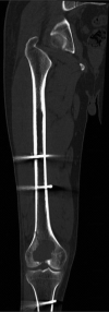
Giant cell tumor of bone with H3F3B mutation: A case report
- PMID: 36800629
- PMCID: PMC9936042
- DOI: 10.1097/MD.0000000000032995
Giant cell tumor of bone with H3F3B mutation: A case report
Abstract
Rationale: Giant cell tumor of bone is a locally aggressive and rarely metastasizing neoplasm that typically affects the ends of long bones or the axial skeleton of young to middle-aged adults. As many as 69% to 100% of giant cell tumors harbor H3F3A gene mutations, while H3F3B gene mutations have rarely been reported.
Patient concerns: A 53-year-old male patient who underwent right distal femoral tumor resection.
Diagnoses: Preoperative CT plain scan indicated giant cell tumor of bone with pathological fracture. Laboratory findings were as follows: serum calcium was 2.23 mmol/L (reference range: 2.1-2.55 mmol/L) and serum phosphorus was 1.35 mmol/L (reference range: 0.81-1.45 mmol/L).
Interventions: The histological morphology showed the typical features of a conventional GCT. The immunoprecipitation analysis results were as follows: H3.3G34W(-), H3.3G34R(-), H3.3G34V(-), and H3K36M(-). Sanger sequencing showed that the H3F3A and H3F3B gene mutations were wild type. The high-throughput gene sequencing results revealed the H3F3B gene mutations H3.3p.Gly35Trp and H3.3p.Val36Leu.
Outcomes: The patient was stable with no recurrence in 12 months follow-up.
Lessons: Giant cell tumor of bone with H3F3B gene mutations is extremely rare. In the pathological diagnosis of bone tumors, we need to analyze clinical presentation, imaging features, histology, immunophenotype, and cytogenetic/molecular alterations, in order to get a correct diagnosis.
Copyright © 2023 the Author(s). Published by Wolters Kluwer Health, Inc.
Conflict of interest statement
The authors have no funding and conflicts of interest to disclose.
Figures


Similar articles
-
Immunohistochemistry for histone H3G34W and H3K36M is highly specific for giant cell tumor of bone and chondroblastoma, respectively, in FNA and core needle biopsy.Cancer Cytopathol. 2018 Aug;126(8):552-566. doi: 10.1002/cncy.22000. Epub 2018 May 14. Cancer Cytopathol. 2018. PMID: 29757500 Free PMC article.
-
The identification of H3F3A mutation in giant cell tumour of the clivus and the histological diagnostic algorithm of other clival lesions permit the differential diagnosis in this location.BMC Cancer. 2018 Apr 2;18(1):358. doi: 10.1186/s12885-018-4291-z. BMC Cancer. 2018. PMID: 29609578 Free PMC article. Review.
-
Mutation Analysis of H3F3A and H3F3B as a Diagnostic Tool for Giant Cell Tumor of Bone and Chondroblastoma.Am J Surg Pathol. 2015 Nov;39(11):1576-83. doi: 10.1097/PAS.0000000000000512. Am J Surg Pathol. 2015. PMID: 26457357
-
Malignant Giant Cell Tumor of Bone: A Clinicopathologic Series of 28 Cases Highlighting Genetic Differences Compared With Conventional, Atypical, and Metastasizing Conventional Tumors.Am J Surg Pathol. 2025 Jun 1;49(6):539-553. doi: 10.1097/PAS.0000000000002387. Epub 2025 Mar 13. Am J Surg Pathol. 2025. PMID: 40077813
-
Histone H3.3 mutation in giant cell tumor of bone: an update in pathology.Med Mol Morphol. 2020 Mar;53(1):1-6. doi: 10.1007/s00795-019-00238-1. Epub 2019 Nov 20. Med Mol Morphol. 2020. PMID: 31748824 Review.
Cited by
-
Giant Cell Tumor With Secondary Aneurysmal Bone Cyst in the Left Distal Humerus: A Case Report.Cureus. 2024 Jul 27;16(7):e65507. doi: 10.7759/cureus.65507. eCollection 2024 Jul. Cureus. 2024. PMID: 39188432 Free PMC article.
References
-
- Amelio JM, Rockberg J, Hernandez RK, et al. . Population-based study of giant cell tumor of bone in Sweden (1983-2011). Cancer Epidemiol. 2016;42:82–9. - PubMed
-
- The 2020 World Health Organization Classification of Soft Tissue and Bone Tumours. 5th Edition - PubMed
-
- Eckardt JJ, Grogan TJ. Giant cell tumor of bone. Clin Orthop Relat Res. 1986:45–58. - PubMed
Publication types
MeSH terms
Substances
LinkOut - more resources
Full Text Sources
Medical
Research Materials

