Review
. 2023 Mar 13;41(3):421-433.
doi: 10.1016/j.ccell.2023.01.009. Epub 2023 Feb 16.

Metabolic programming and immune suppression in the tumor microenvironment

Affiliations
Review

Metabolic programming and immune suppression in the tumor microenvironment

Emily N Arner et al. Cancer Cell. .

Abstract

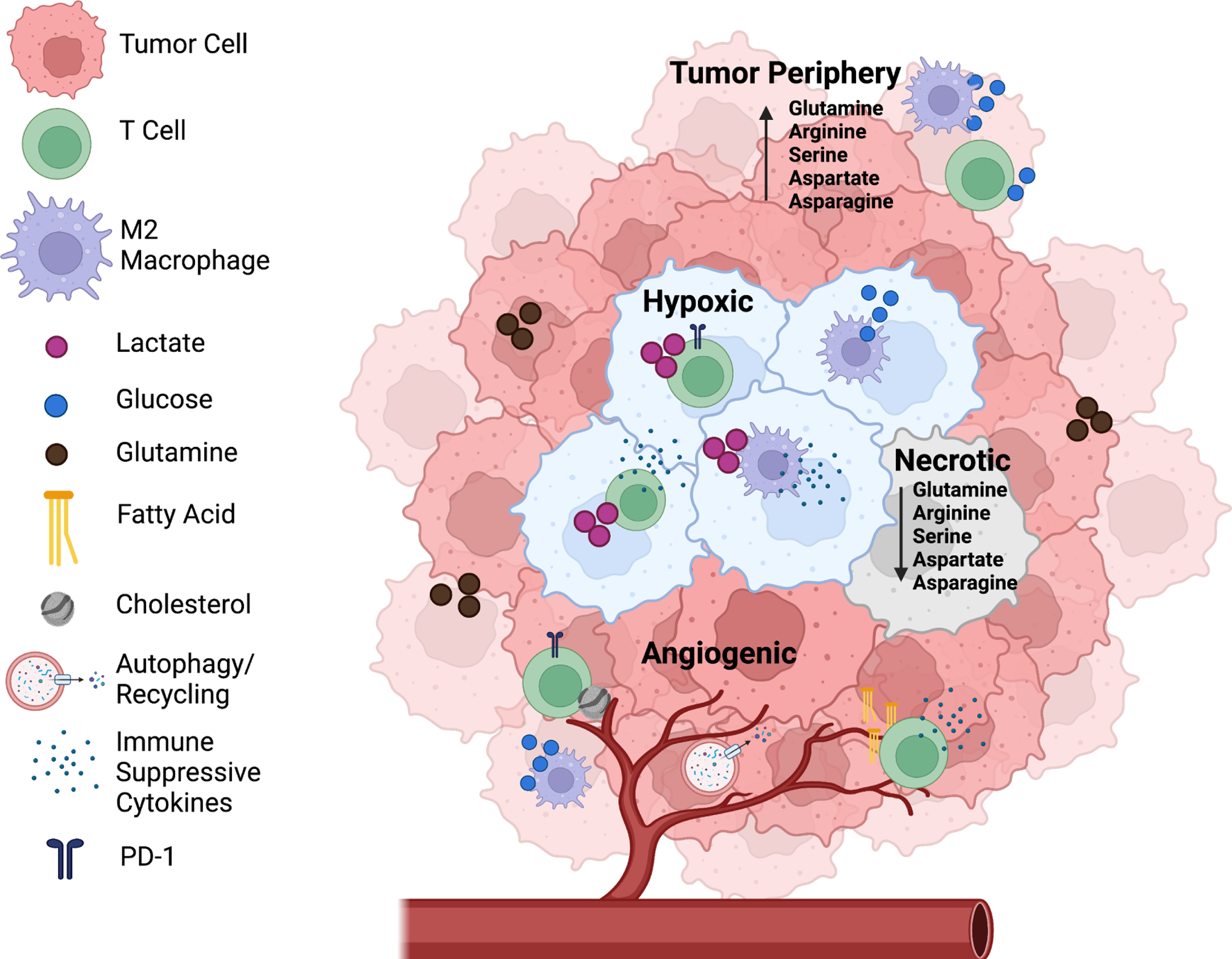
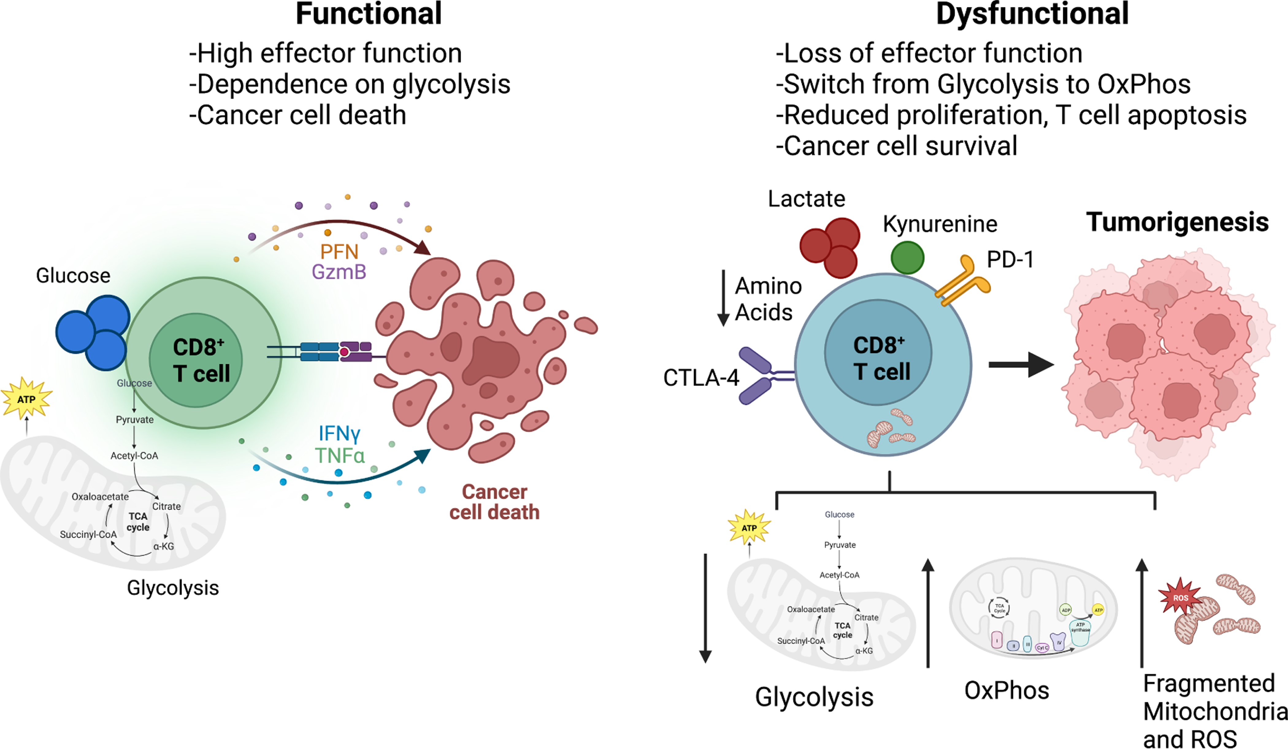
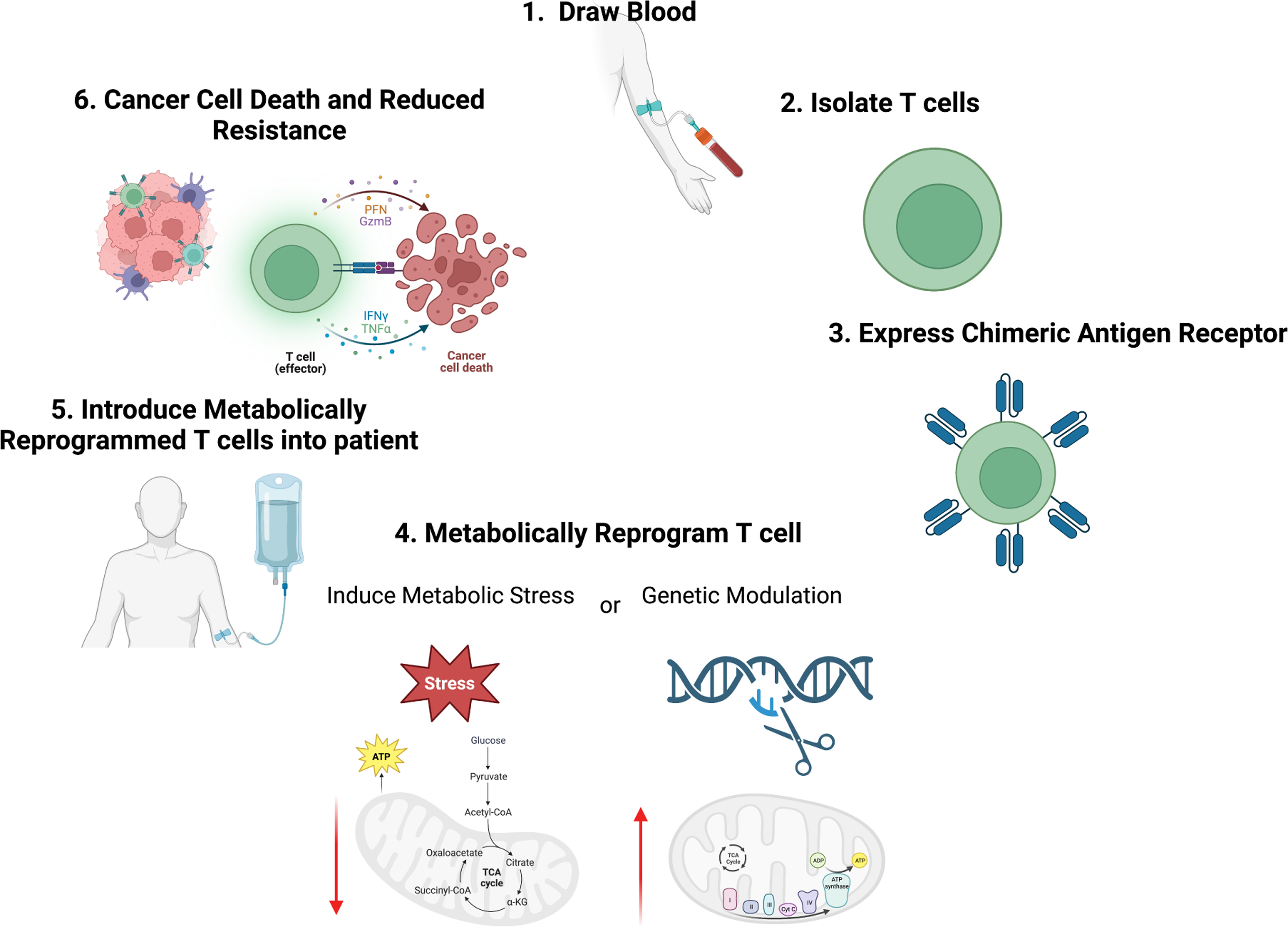
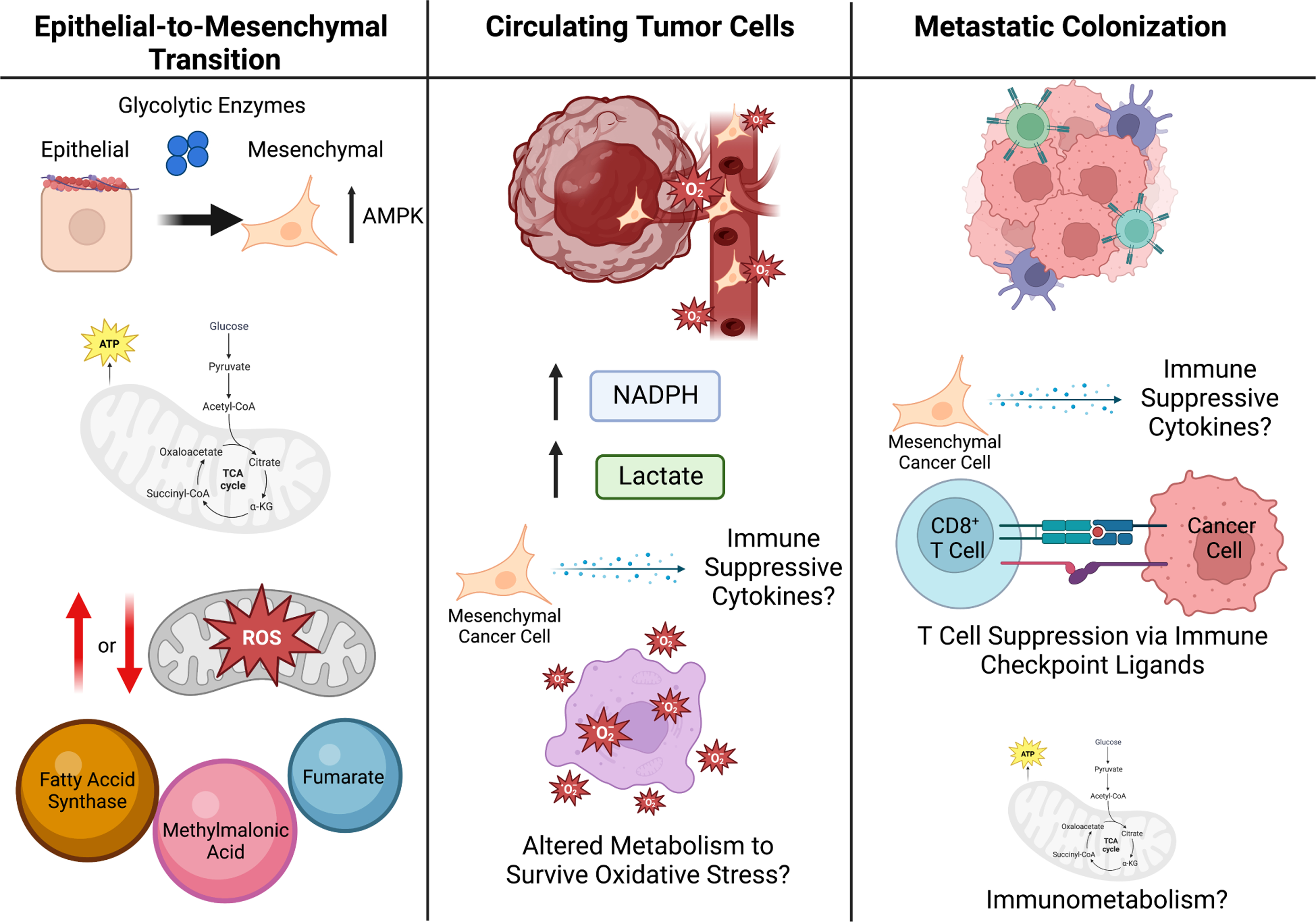
Increased glucose metabolism and uptake are characteristic of many tumors and used clinically to diagnose and monitor cancer progression. In addition to cancer cells, the tumor microenvironment (TME) encompasses a wide range of stromal, innate, and adaptive immune cells. Cooperation and competition between these cell populations supports tumor proliferation, progression, metastasis, and immune evasion. Cellular heterogeneity leads to metabolic heterogeneity because metabolic programs within the tumor are dependent not only on the TME cellular composition but also on cell states, location, and nutrient availability. In addition to driving metabolic plasticity of cancer cells, altered nutrients and signals in the TME can lead to metabolic immune suppression of effector cells and promote regulatory immune cells. Here we discuss how metabolic programming of cells within the TME promotes tumor proliferation, progression, and metastasis. We also discuss how targeting metabolic heterogeneity may offer therapeutic opportunities to overcome immune suppression and augment immunotherapies.

Keywords: immune; metabolism; metastasis; plasticity; tumor microenvironment.


Conflict of interest statement

Declaration of interests J.C.R. holds stock equity in Sitryx and Caribou and within the past 2 years has received unrelated research support, travel, or honoraria from Sitryx, Caribou, Nirogy, Kadmon, Calithera, Tempest, Merck, Mitobridge, and Pfizer.

Figures

Figure 1.
Figure 1.. Metabolic Heterogeneity in the Tumor Microenvironment is Location Dependent.
The tumor microenvironment (TME) is composed of many different cells that support or restrain tumorigenesis, including cancer cells and pro- and anti-tumor immune cells. Just as the TME has a diversity of cells, the TME is also metabolically heterogeneous. In hypoxic regions of the tumor (blue cancer cells), cancer cells often secrete lactate, which has been shown to inhibit effector T cell activation while promoting suppressive Tregs to drive immune suppression. Additionally, lactate promotes differentiation and polarization of TAMs towards a more pro-tumorigenic M2-like phenotype, which secrete immunosuppressive cytokines. In angiogenic tumors, blood vessels branch irregularly which leads to regions with increased autophagy to make up for the lack of delivered nutrients. Less angiogenic regions typically use glucose as their main source of energy. Necrotic tumor regions arise in part because of severe lack of nutrients and have been shown to have decreased levels of amino acids such as glutamine, arginine, asparagine, serine, and aspartate compared to tumor peripheries.
Figure 2.
Figure 2.. Dysregulated Cytotoxic T Cell Metabolism.
Cytotoxic T cells act directly to kill cancer cells by secreting inflammatory cytokines as well as cell lytic molecules such as granzyme. In a highly functioning state, CD8+ effector T cells are dependent on glycolysis, which promotes inflammation. Metabolic immune suppression occurs when T cells from tumors develop a wide range of metabolic adaptations or dysfunctions that prevent anti-tumor activity. These include dysregulated and fragmented mitochondria, increased ROS production, and reduced glycolysis. In the TME, cancer cells secrete metabolites that effect the function of immune cells within the TME and promote T cell exhaustion such as lactate, cholesterol, and kynurenine, a by-product of tryptophan catabolism. Once exhausted, T cells are unable survive and function to secrete cytokines and express inhibitory receptors such as PD-1 and CTLA-4, which reduce the function of effector T cells by inhibiting glycolysis and upregulating oxidative phosphorylation of the cells. Additionally, amino acids such as arginine, tryptophan, and serine may be limited in the TME, thus inhibiting cytotoxic T cells as they rely on these nutrients for proliferation and function.
Figure 3.
Figure 3.. Metabolic Rewiring to Reduce Metabolic Immune Suppression.
Adoptive T cell therapies target cytotoxic T cells by engineering T cells from patients to express chimeric antigen receptors (CAR) specific to tumor antigens. To reduce metabolic immune suppression of T cells in the TME and increase their metabolic capacity, it may be possible to metabolically rewire T cells either by inducing metabolic stress or through genetic modification. These strategies may be able to increase T cell metabolic plasticity and fitness to turn tumors that are typically immune “cold” due to poor nutrient conditions to immune “hot”, thus increasing anti-tumor immunity and reducing resistance to therapies such as immune checkpoint.
Figure 4.
Figure 4.. Metabolic Plasticity in Metastasis.
To escape the primary tumor and form metastases, cells undergo EMT (left panel). Glycolytic enzymes are attached to the cell cytoskeleton, as such it is possible that during cytoskeleton rearrangement in the EMT process, these glycolytic enzymes are released to promote glycolysis during EMT. The production of mitochondrial ROS within the cancer cells has been suggested to both promote and reduce EMT and metastatic potential of cancer cells, highlighting the likelihood that this process is highly dependent on the genetic context and local TME. Additionally, some metabolites act as pro-EMT signaling molecules, such as fumarate, methylmalonic acid, and fatty-acid synthase (FASN). Once in circulation (middle panel), cancer cells undergo oxidative stress and thus cell-death, therefore it is likely the cancer cells that are able to survive oxidative stress have a metabolic advantage resulting in their survival. Additionally, CTCs have been shown to upregulate NADPH production and lactate uptake, which diverts glucose carbon into the oxidate PPP and increases their antioxidant capacity. It is likely that cytokines and metabolites within the blood stream may influence both immune surveillance of CTCs as well as cancer cell immune evasion. Once cancer cells enter the metastatic site (right panel), they must act on the tumor microenvironment within the metastatic site to suppress immune surveillance, which can be done metabolically, such as by presenting immune checkpoint ligands. Additionally, immunometabolism likely plays a significant role in this process and may provide a therapeutic vulnerability for the treatment of metastatic cancer.

References

    1. Li X, Sun X, and Carmeliet P. (2019). Hallmarks of Endothelial Cell Metabolism in Health and Disease. Cell Metab 30, 414–433. 10.1016/j.cmet.2019.08.011. - DOI - PubMed
    1. Ryan DG, and O’Neill LAJ (2020). Krebs Cycle Reborn in Macrophage Immunometabolism. Annu Rev Immunol 38, 289–313. 10.1146/annurev-immunol-081619-104850. - DOI - PubMed
    1. Makowski L, Chaib M, and Rathmell JC (2020). Immunometabolism: From basic mechanisms to translation. Immunol Rev 295, 5–14. 10.1111/imr.12858. - DOI - PMC - PubMed
    1. DeBerardinis RJ, and Chandel NS (2016). Fundamentals of cancer metabolism. Sci Adv 2, e1600200. 10.1126/sciadv.1600200. - DOI - PMC - PubMed
    1. Gouirand V, Guillaumond F, and Vasseur S. (2018). Influence of the Tumor Microenvironment on Cancer Cells Metabolic Reprogramming. Front Oncol 8, 117. 10.3389/fonc.2018.00117. - DOI - PMC - PubMed

Publication types