. 2023 Mar 29;43(13):2260-2276.
doi: 10.1523/JNEUROSCI.1323-22.2023. Epub 2023 Feb 17.

Innexin-Mediated Adhesion between Glia Is Required for Axon Ensheathment in the Peripheral Nervous System

Affiliations

Innexin-Mediated Adhesion between Glia Is Required for Axon Ensheathment in the Peripheral Nervous System

Mriga Das et al. J Neurosci. .

Abstract

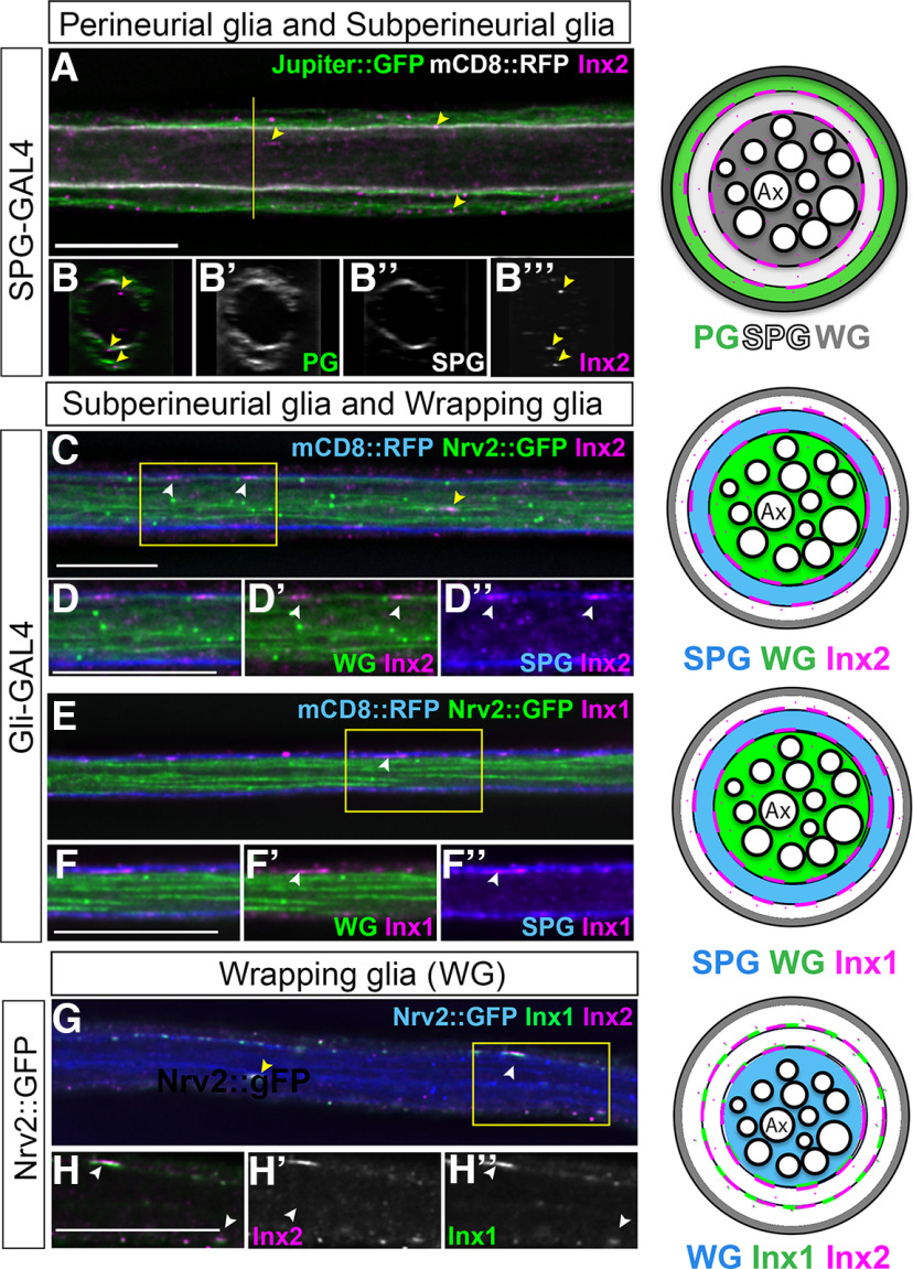
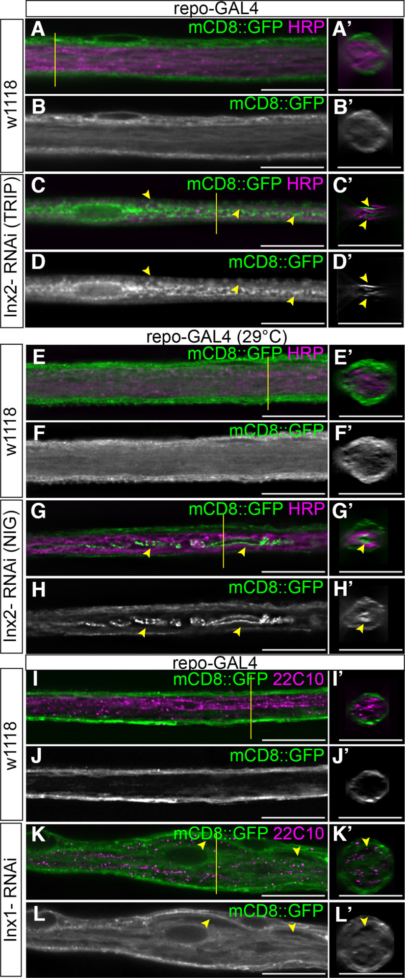
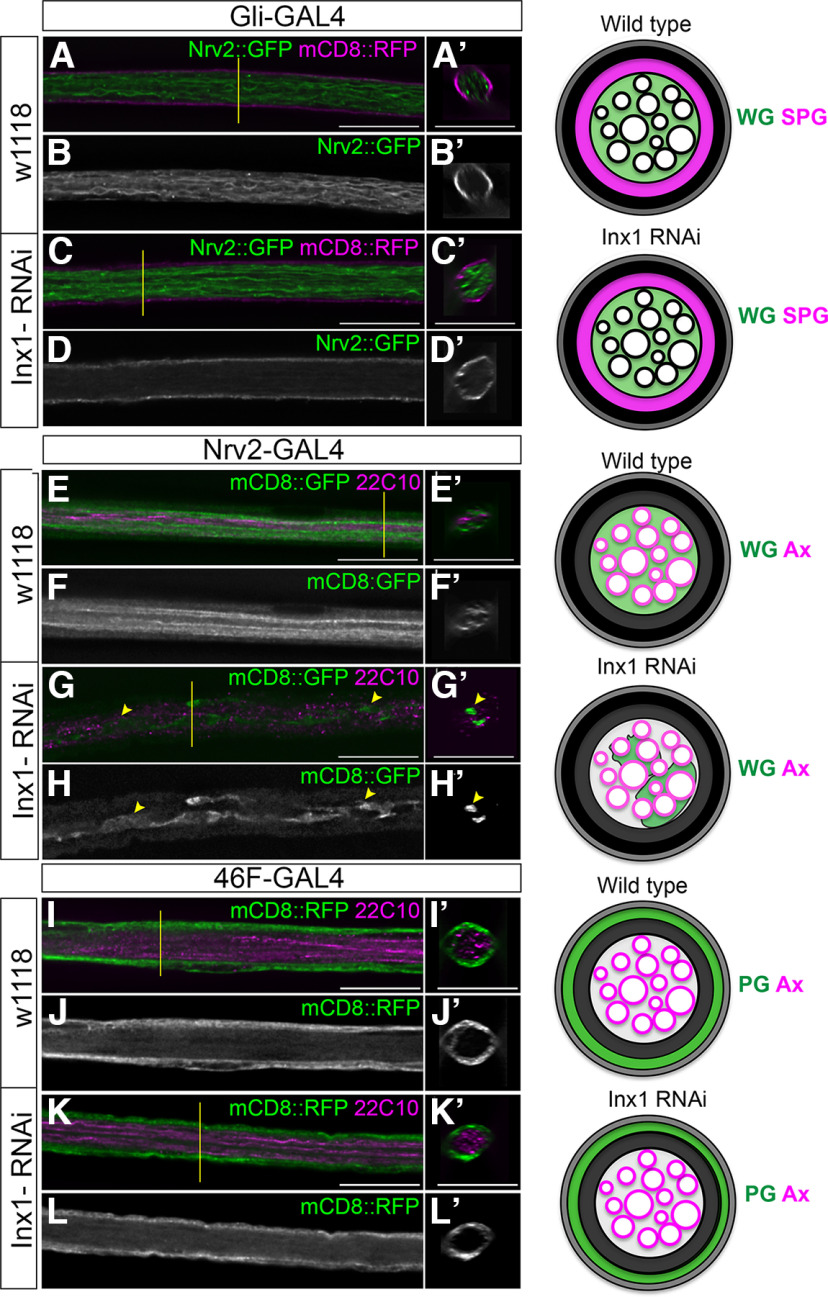
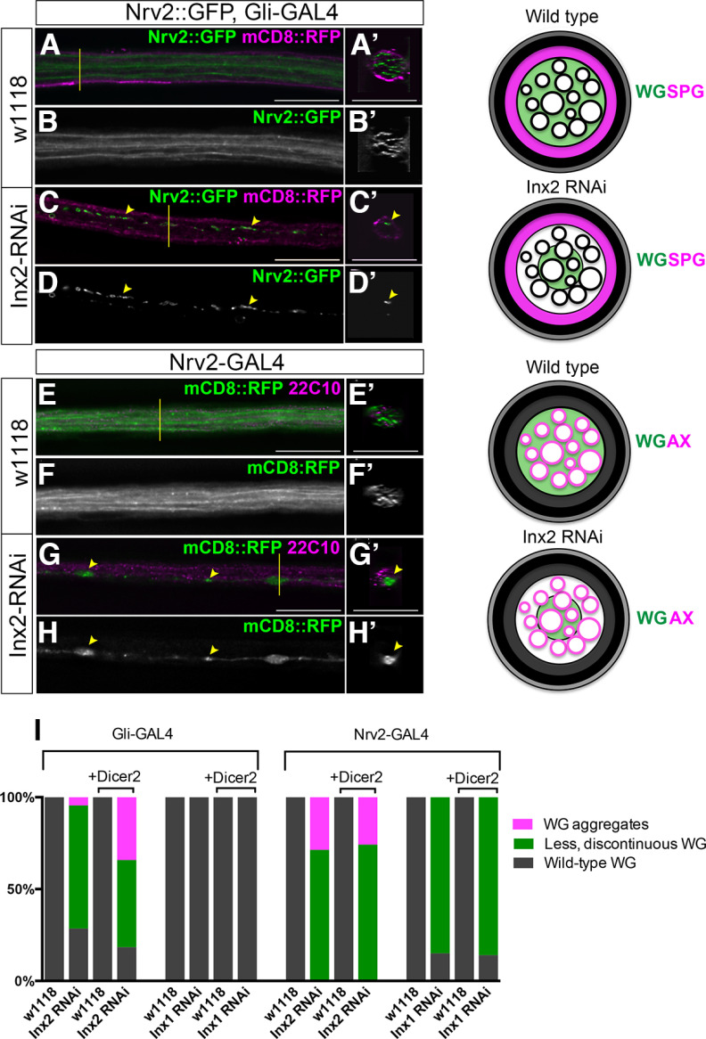
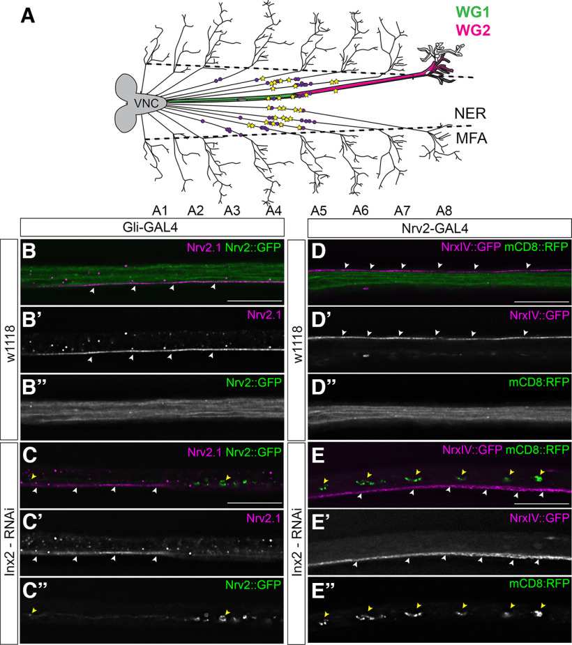
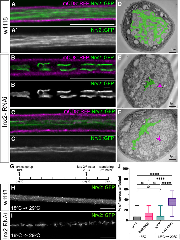
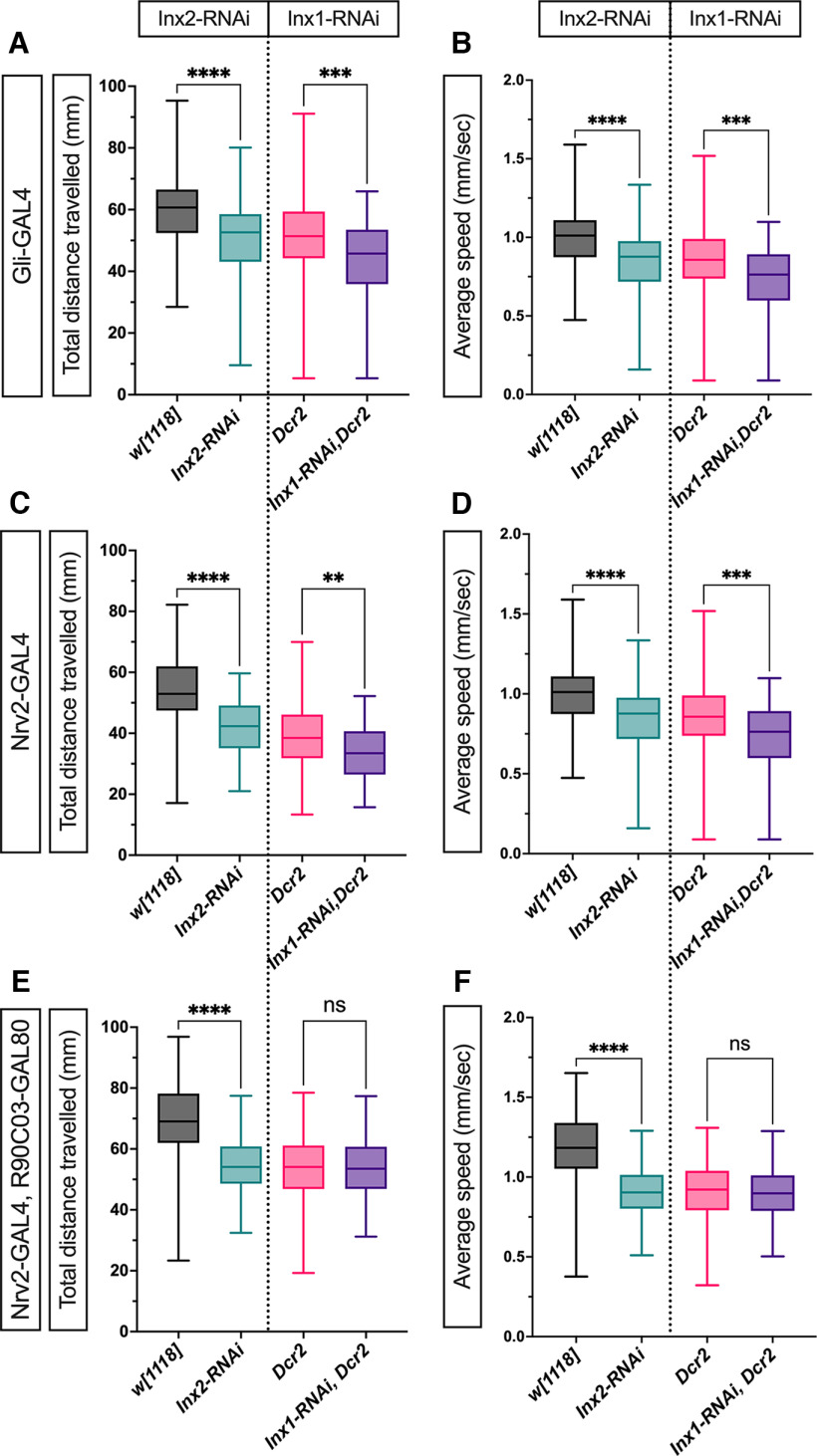
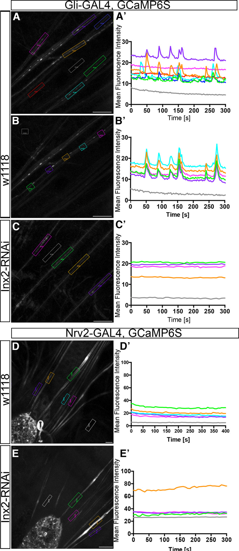
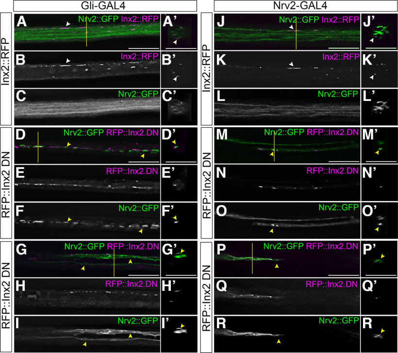
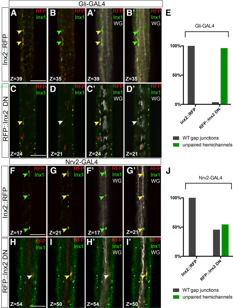
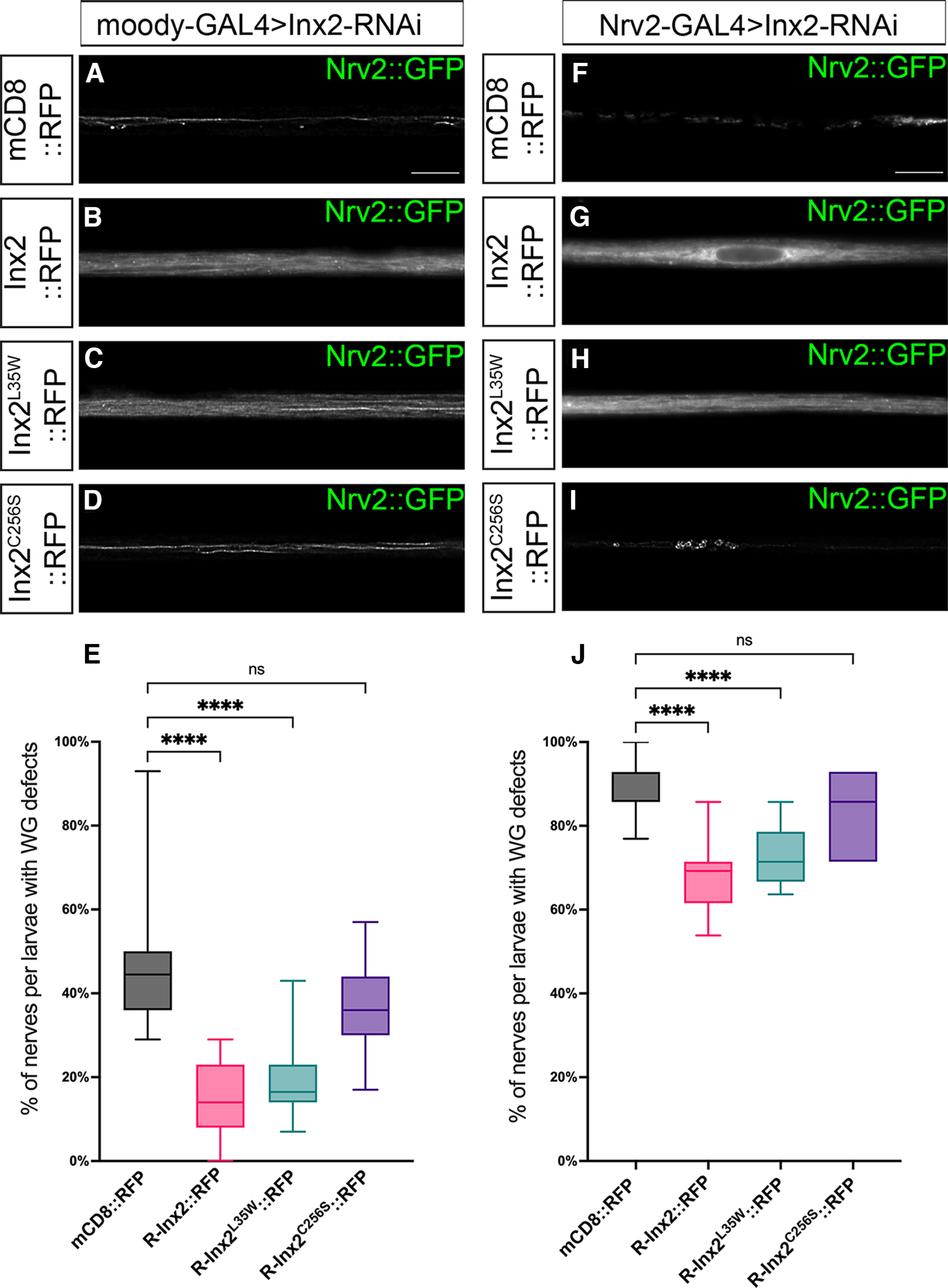
Glia are essential to protecting and enabling nervous system function and a key glial function is the formation of the glial sheath around peripheral axons. Each peripheral nerve in the Drosophila larva is ensheathed by three glial layers, which structurally support and insulate the peripheral axons. How peripheral glia communicate with each other and between layers is not well established and we investigated the role of Innexins in mediating glial function in the Drosophila periphery. Of the eight Drosophila Innexins, we found two (Inx1 and Inx2) are important for peripheral glia development. In particular loss of Inx1 and Inx2 resulted in defects in the wrapping glia leading to disruption of the glia wrap. Of interest loss of Inx2 in the subperineurial glia also resulted in defects in the neighboring wrapping glia. Inx plaques were observed between the subperineurial glia and the wrapping glia suggesting that gap junctions link these two glial cell types. We found Inx2 is key to Ca2+ pulses in the peripheral subperineurial glia but not in the wrapping glia, and we found no evidence of gap junction communication between subperineurial and wrapping glia. Rather we have clear evidence that Inx2 plays an adhesive and channel-independent role between the subperineurial and wrapping glia to ensure the integrity of the glial wrap.SIGNIFICANCE STATEMENT Gap junctions are critical for glia communication and formation of myelin in myelinating glia. However, the role of gap junctions in non-myelinating glia is not well studied, yet non-myelinating glia are critical for peripheral nerve function. We found the Innexin gap junction proteins are present between different classes of peripheral glia in Drosophila. Here Innexins form junctions to facilitate adhesion between the different glia but do so in a channel-independent manner. Loss of adhesion leads to disruption of the glial wrap around axons and leads to fragmentation of the wrapping glia membranes. Our work points to an important role for gap junction proteins in mediating insulation by non-myelinating glia.

Keywords: gap junction; glia; innexin; insulation.


Figures

Figure 1.
Figure 1.
Innexin 1 and Innexin 2 are expressed in the larval peripheral nerve. A, B, Longitudinal section (A) or cross section (B–B''') of a control peripheral nerve with the perineurial glia (PG) membrane and subperineurial glia (SPG) membranes labeled with Jupiter::GFP (green) and mCD8::RFP (white), respectively. Inx2 immunolabeling (magenta) identified puncta (yellow arrowheads) present in the PG and SPG, and within the wrapping glia (WG) in the center of the nerve. C, D, E, F, Longitudinal sections of control peripheral nerves with Inx2 (C, D) and Inx1 (E, F) immunolabeling (magenta) in the subperineurial glia and wrapping glia. SPG membrane are labeled with mCD8::RFP (blue), and WG membranes labeled with Nrv2::GFP (green). The yellow boxes were digitally magnified (200×) and shown in D–D'' and F–F'', respectively. White arrowheads in C, D and E, F indicate the Inx2 and Inx1 puncta, respectively, in the SPG. Inx2 puncta in the WG are indicated by the yellow arrowhead (C). Both Inx2 and Inx1 expression puncta are observed in the SPG-WG boundary (C, D'–D''' and E, F–F''', white arrowheads). G, H, Inx1(green) and Inx2 (magenta) form plaques along the SPG-WG boundary (G, H–H''', white arrowheads) and within the WG membrane (G, yellow arrowhead). Scale bars: 15 μm.
Figure 2.
Figure 2.
Knock-down of Inx2 in all glia leads to fragmentation of the inner glial membrane, whereas knock-down of Inx1 leads to glial swellings. Longitudinal sections of 3rd instar larval nerves with repo-GAL4 driving mCD8::GFP (green) to label glial membranes and axons labeled with anti-HRP or anti-22C10 (magenta). The yellow lines indicate the region from which the cross sections were taken. A, B, repo-GAL4. E, F, Control repo-GAL4 at 29°C. I, J, Control repo-GAL4. In controls the axons (magenta) are present in the center of the nerve and completely surrounded by the glial membrane (green). In the side projections (A'), the PG and SPG membranes surround the entire nerve and the WG membranes fill the core of the nerve. C, D, repo>Inx2-RNAi (TRIP). G, H, repo>Inx2-RNAi (NIG) at 29°C. Peripheral nerves are thinner compared with control nerves and the glial membranes (green) in the center of the nerve were disrupted with membrane fragments (C, G, arrowheads). In the side projections, the outer glial membranes still surround the nerves (C', G') but the glial membrane is collapsed or concentrated in the center of the nerve without ensheathing the axons (C', G', arrowheads). K, L, repo>Inx1-RNAi. Swellings were observed between the different glial membranes (green; K, L, yellow arrows) and the glial membranes surrounded the nerve and filled the center of the nerve (K', L', yellow arrows). Scale bars: 15 μm.
Figure 3.
Figure 3.
Knock-down of Inx1 in the wrapping glia leads to defects but knock-down in the other glial layers does not affect morphology. Nerves for 3rd instar larvae in longitudinal and cross sections. The yellow lines indicate the region from which the cross sections were taken. A schematic representation of the labeled glial layers in control and Inx1 knock-down nerves are shown to the right with each glial layer and axons (Ax) indicated. A–D, Subperineurial glia. A, B, Control Gli-GAL4. C, D, Gli>Inx1-RNAi. SPG membranes labeled with mCD8::RFP (magenta) and WG membranes labeled with Nrv2::GFP (green). The SPG and WG membranes in the Gli>Inx1-RNAi nerve were similar to the control nerve. WG extends processes along (A, C) and throughout the core of the nerve (A', C'). The thin SPG membrane flanks the nerve (A, C) surrounding the WG (A', C'). E–H, Wrapping glia. A, B, Control: Nrv2-GAL4. C, D, Nrv2>Inx1-RNAi. WG membranes were labeled with mCD8::RFP (green) and axons immunolabeled with 22C10 (magenta). Strands of WG wrap (green) around axons (magenta) in the control nerve (E) and cross sections (E'). WG strands were reduced and discontinuous in Nrv2>Inx1-RNAi nerves (G, yellow arrowhead) and the WG membrane does not fully wrap around axons in some regions of the nerve (G', yellow arrowhead). I–L, Perineurial glia. I, J, Control, 46F-GAL4. K, L, 46F>Inx1-RNAi. Nerves with PG membranes labeled with mCD8::RFP (green) and axons labeled with 22C10 (magenta). PG membranes surround the entire nerve in both control and 46F>Inx1-RNAi. Scale bars: 15 μm.
Figure 4.
Figure 4.
Knock-down of Inx2 in subperineurial glia or wrapping glia leads to fragmentation of the wrapping glia. Nerves for 3rd instar larvae in longitudinal and cross sections. The yellow lines indicate the region from which the cross sections were taken. A schematic representation of the labeled glial layers in control and Inx1 knock-down nerves are shown to the right with each glial layer and axons (Ax) indicated. Scale bars: 15 μm. A–D, Subperineurial glia (SPG). A, B, Control, Gli-GAL4 (A, B) and Gli>Inx2-RNAi (C, D). Nerves with SPG and wrapping glia membranes labeled with mCD8::RFP (magenta) and Nrv2::GFP (green), respectively. The WG (green) in control nerves extend processes along the entire length of the nerve and the thin SPG membrane (magenta) surrounds the WG (A'). WG membranes wrap around the peripheral axons (A, A'). WG membrane aggregates (green, C) are present along the length of the nerve in Gli>Inx2-RNAi. Remnants of the WG membrane (green) are found in the center of the nerve (C', arrowhead) and do not wrap around axons (magenta) in Gli>Inx2-RNAi. E–H, Wrapping glia (WG). Control (E) and Nrv2>Inx2-RNAi (G) peripheral nerves with WG membranes labeled with mCD8::RFP (green) and axons immunolabeled with 22C10 (magenta). The yellow lines indicate the region from which the cross sections were taken. Several strands of WG wrap (green) around axons (magenta) in the control peripheral nerve (E, E') and cross sections indicate that WG membrane surrounds axons (magenta). Loss of WG strands in Nrv2>Inx2-RNAi, with only a single WG strand (green) present in the peripheral nerve (G, G'). WG membrane in the Nrv2>Inx2-RNAi nerve is only present at the center with an uneven morphology and does not wrap around axons (magenta). I, Comparison of the WG phenotypes observed when Inx2 and Inx1 were knocked down in the SPG and the WG. The WG phenotypes were divided into three categories: wild-type WG (gray), less and discontinuous WG strands (green), WG aggregates (magenta). The specific percentage of nerves that fall under the three categories of WG phenotypes are detailed in Table 2.
Figure 5.
Figure 5.
Knock-down of Inx2 in the SPG or WG affects WG-WG boundaries but not septate junctions. A, Location of the wrapping glia phenotypes in the peripheral nervous system of 3rd instar larvae. Schematic representation of a peripheral nerves in a larva in which Inx2 is knocked down in either the SPG or WG layers. The two WG present in the nerve extension region (NER) of the peripheral nerve (WG1, WG2) are indicated. The location of the WG fragments in SPG and WG specific knock-down of Inx2 (yellow stars and purple circles, respectively) was mapped to show where majority of these phenotypes occur. WG fragments were only observed within the NER (marked with the dashed line) and predominantly in regions where WG1 and WG2 contact each other. B–E, Knock-down of Inx2 in the SPG and WG does not disrupt the SPG or septate junctions. Scale bars: 15 μm. B, C, Control (Gli-GAL4; A) and Gli>Inx2-RNAi (B) peripheral nerves with WG membranes labeled with Nrv2::GFP (green) and the SJ domain labeled with a Nrv2.1 antibody (magenta). D, E, Control (Nrv2-GAL4; C) and Nrv2>Inx2-RNAi (D) peripheral nerves with WG membranes labeled with mCD8::RFP (green) and the SJ domain labeled with NrxIV::GFP (magenta). WG (green) in the control nerves (A'', C'') extend processes along the entire length of the nerve and the SJ domain (magenta) is continuous along the length of the nerve (A', C', white arrowheads). With Inx2 knock-down WG membrane aggregates (B'', D'', yellow arrowheads) are present along the length of the nerve in Gli>Inx2-RNAi (B) and Nrv2>Inx2-RNAi (D). SJ morphology is not affected (B', D', white arrowheads).
Figure 6.
Figure 6.
Knock-down of Inx2 in the subperineurial glia generates wrapping glia phenotypes. A–F, 3rd instar peripheral nerves with SPG and WG membranes labeled with mCD8::RFP (magenta) and Nrv2::GFP (green), respectively, with ultrastructural images with the WG false-coloured green (D–F). A, D, Control (Gli-GAL4). A, WG strands (green) are present and extend along the length of the control peripheral nerve. D, In cross section, WG (green) extend processes and wrap around axons or bundles of axons. B, C, E, F, Gli>Inx2-RNAi. Loss of Inx2 in the SPG results in a range of WG phenotypes, with nerves showing wrapping glia membrane aggregates (B) or reduced wrapping glia strands (C). Representative TEM sections that correspond to the WG phenotypes with reduced strands (F) or reduction to single strands (E). Septate junctions are indicated in the TEM sections (D–F, magenta arrowheads). Scale bars: 15 μm (A–C) and 1 μm (D–F). G–J, 3rd instar peripheral nerves of larvae with WG membranes labeled with Nrv2::GFP with Gli-GAL4 driving expression in the SPG. G, Each cross was raised at 18°C continuously or shifted to 29°C at the end of the 2nd instar stage for 48 h before assessment of peripheral nerves at the wandering 3rd instar stage. H, Control (Gli-GAL4). WG strands were not disrupted in control (w1118) crosses shifted from 18°C to 29°C. I, Gli>Inx2-RNAi. Loss of Inx2 in the SPG disrupted WG processes including membrane fragmentation after the temperature shift. J, Quantification of the WG disruption in larvae raised continuously at 18°C versus larvae shifted from 18°C to 29°C. Gli-GAL4 crossed to w1118 (control) or Inx2-RNAi. The degree of WG defects was quantified and statistical significance was determined by a one-way ANOVA with Tukey's multiple comparisons test. Boxes indicate the 25th to 75th percentiles with the median indicated. The whiskers indicate the minimum to maximum values. ****p < 0.0001, ns = not significant. Scale bars: 15 μm.
Figure 7.
Figure 7.
Inx knock-down in the subperineurial and wrapping glia affects larval locomotion. Total distance traveled and average speed were determined for 3rd instar larvae. Boxes indicate the 25th to 75th percentiles with the median indicated. The whiskers indicate the minimum to maximum values. A, B, Subperineurial glia (Gli-GAL4). The total distance traveled (A) or average speed (B) in SPG control (Gli>; n = 86) compared with Gli>Inx2-RNAi (n = 211) and Gli>Dicer2 (n = 226) compared with Gli>Inx1-RNAi, Dicer2 (n = 69). C, D, Wrapping glia (Nrv2-GAL4). The total distance traveled (C) or average speed (D) in WG control (Nrv2>; n = 157) compared with Nrv2>Inx2-RNAi (n = 121) and Nrv2>Dicer2; n = 133) compared with Nrv2>Inx1-RNAi, Dicer2 (n = 50). E, F, Peripheral wrapping glia (Nrv2-GAL4, R90C03-GAL80). The total distance traveled (E) or average speed (F) in control (R90C03-GAL80, Nrv2>; n = 124) compared with R90C03-GAL80, Nrv2>Inx2-RNAi (n = 189) and R90C03-GAL80, Nrv2>Dicer2 (n = 205) compared with R90C03-GAL80, Nrv2>Inx1-RNAi, Dicer2 (n = 96). Statistical significance was determined by a one-way ANOVA with Tukey's multiple comparisons test for each driver crossed with: w1118; Inx2-RNAi; Dicer2; Inx1-RNAi, Dicer2. A, ****p < 0.0001, ***p = 0.0001. B, ****p > 0.0001, ***p = 0.0001. C, ****p < 0.0001, **p = 0.0037. D, ****p < 0.0001, **p < 0.0036. E, ****p < 0.0001, NS p = 0.7891. F, ****p < 0.0001, NS p = 0.9989.
Figure 8.
Figure 8.
Calcium pulses are confined to the subperineurial glia and require Inx2. 3rd instar larval nerves in live preparations with GCaMP6S expressed in subperineurial (A–C) or wrapping glia (D, E). The change in mean fluorescence intensity over time (seconds) is plotted in the graphs (A'–E') indicated by the regions of interest (ROIs). ROI 18 in each image (gray, A–E) was placed away from the peripheral nerve and represents the basal GCaMP6S signal. A–C, Subperineurial glia. GCaMP6S driven by Gli-GAL4 in control (Gli>GCaMP6S; A–B') or with Inx2-RNAi (Gli>GCaMP6S, Inx2-RNAi; C, C'). A, A', Calcium pulses were present in some but not all control nerves (ROIs: 1, 12–14, 16; green, purple, orange, light blue, and blue, respectively). B, B', Control nerve where the ROIs are placed along the same nerve. The change in mean fluorescence intensity occur as a pulse along the peripheral nerve. C, Knock-down of Inx2 in the SPG (Gli>Inx2-RNAi, GCaMP6S) blocked the pulses with no observed changes in mean fluorescence intensity. D, E, Wrapping glia. GCaMP6S driven by Nrv2-GAL4 in control (Nrv2>GCamp6S; D) or with Inx2-RNAi (Nrv2>GCaMP6C, Inx2-RNAi; E). Calcium pulses were not observed in the peripheral nerves (ROIs: 1–5; green, purple, orange, light blue, and pink, respectively) of control (D) or Inx2-RNAi expressing WG (E). The ROI 18 (gray; D, E) were placed in a region where peripheral nerves were absent to measure basal level of the GCaMP6S signal. Scale bars: 25 μm.
Figure 9.
Figure 9.
Dominant negative Inx2 triggers the same WG phenotypes as Inx2 RNAi. A–C, Gli>Inx2::RFP control nerves expressing control Inx2::RFP in the SPG (magenta) and WG membranes labeled with Nrv2::GFP (green). The WG membrane in the control nerve (A, green) extends its processes along the entire length of the nerve and Inx2::RFP plaques (magenta, white arrowheads) are present in the neighboring SPG (A, magenta). D–I, Gli>RFP::Inx2 DN nerves with the dominant negative Inx2 mutant (RFP::Inx2 DN, magenta) driven in the SPG and the WG membranes labeled with Nrv2::GFP (green). WG membrane aggregates (green, yellow arrowheads) are present along the length of some nerves in Gli>RFP::Inx2 DN larvae (D, F) whereas others have discontinuous WG membranes (G, I). RFP::Inx2 DN (magenta) is localized to the SPG-WG boundary (D–E', G–H'). J–L, Nrv2>Inx2::RFP control nerves expressing Inx2::RFP (magenta) and mCD8::GFP (green) in the WG. The WG membrane (green) extends processes along the entire length of the nerve and Inx2::RFP plaques (magenta, white arrowheads) are present in the WG, along the SPG-WG boundary. M–R, Nrv2>RFP::Inx2 DN peripheral nerves with the dominant negative Inx2 mutant (RFP::Inx2 DN, magenta) driven in the WG along with mCD8::GFP (green). Single, discontinuous WG strands (green, yellow arrowheads) were observed (M–O) with others nerves containing disrupted WG strands (P–R). Aggregates of WG membrane were found in the center of the nerve (M, M'). The yellow lines indicate the region from which the cross sections were taken. Scale bars: 15 μm.
Figure 10.
Figure 10.
Dominant negative Inx2 blocks coupling with Inx1 in the neighboring glia. 3rd instar nerves with Inx2::RFP or RFP::Inx2 DN (red) expressed in the SPG or WG with Inx1 immunolabeled (green). Individual Z-stacks are indicated a two focal planes to capture the association of Inx2 with Inx1 (first two panels) and in the context of the WG membrane (Nrv2::GFP, gray; second two panels). Scale bars: 7.5 μm. A, B, Gli>Inx2::RFP control nerves where Inx2::RFP (red) is expressed in the SPG with Inx1 immunolabelled (green). Inx2::RFP integrates with Inx1 in the SPG (yellow plaques; A, A', yellow arrowheads) that correspond to Inx1 immunolabeling (green plaques) in the underlying WG (B, B', green arrowheads). WG processes (Nrv2::GFP, gray) extend along the entire length of the nerve (A', B'). C, D, Gli>RFP::Inx2 DN nerves with dominant negative Inx2 mutant (RFP::Inx2 DN) driven in the SPG with Inx1 immunolabelling (green). RFP::Inx2 DN integrates with Inx1 in the SPG (yellow plaques; C, C', yellow arrowhead) but the absence of Inx1 in the underlying WG (D, D', white arrowhead) suggests a lack of coupling between the SPG and WG (C', D'). E, The percentage of wild-type gap junctions (gray) and unpaired hemichannels (green) observed in control (Gli>Inx2::RFP, n = 3 nerves) compared with nerves expressing dominant negative Inx2 in the SPG (Gli>RFP::Inx2 DN, n = 9 nerves). F, G, Nrv2>Inx2::RFP control nerves with Inx2::RFP (red) expressed in the WG (Nrv2::GFP, gray) and Inx1 immunolabelled (green). Inx2::RFP integrates with Inx1 in the WG (yellow plaque; G, G', yellow arrowheads) and corresponds to Inx1 the overlying SPG (green plaque; F, F', green arrowheads. WG processes extend along the entire length of the nerve (G'). H, I, Nrv2>RFP::Inx2 DN nerves with the dominant negative Inx2 mutant (RFP::Inx2 DN, red) driven in the WG and Inx1 immunolabelled (green). WG membranes were labeled with Nrv2::GFP (gray; H', I'). The dominant negative RFP::Inx2 DN integrates with Inx1 in the WG (yellow plaques; I, I', yellow arrowhead) but the absence of Inx1 (green) in the corresponding area in the SPG (H, H', white arrowhead) suggests a lack of coupling between the hemichannels in the SPG and WG (H', I'). Scale bars: 7.5 μm. J, The percentage of wild-type gap junctions (gray) and unpaired hemichannels (green) observed in control (Nrv2>Inx2::RFP, n = 2 nerves) compared with nerves expressing dominant negative Inx2 in the WG (Nrv2>RFP::Inx2 DN, n = 7 nerves).
Figure 11.
Figure 11.
Rescue of the Inx2-RNAi phenotype by both wild-type and a channel-deficient Inx2 mutant. 3rd instar nerves with the WG membrane marked by Nrv2::GFP. Scale bars: 15 μm. A–E, Subperineurial glia. Moody-GAL4 driven Inx2-RNAi (moody>Inx2-RNAi) with (A) mCD8::RFP, (B) Inx2::RFP, (C) Inx2::RFP[L35W], (D) Inx2::RFP[C256S]. Inx2-RNAi led to the disruption of the WG and these phenotypes were rescued by Inx2::RFP and Inx2::RFP[L35W] but not Inx2::RFP[C256WS]. E, The degree of rescue was quantified and statistical significance was determined by a one-way ANOVA with Tukey's multiple comparisons test. Boxes indicate the 25th to 75th percentiles with the median indicated. The whiskers indicate the minimum to maximum values. ****p < 0.0001, ns = not significant. F–J, Wrapping glia. Nrv2-GAL4 driven Inx2-RNAi (Nrv2>Inx2-RNAi) with (F) mCD8::RFP, (G) Inx2::RFP, (H) Inx2::RFP[L35W], (I) Inx2::RFP[C256S]. Inx2-RNAi lead to the disruption of the WG and these phenotypes were rescued by Inx2::RFP and Inx2::RFP[L35W] but not Inx2::RFP[C256WS]. J, The degree of rescue was quantified and statistical significance was determined by a one-way ANOVA with Tukey's multiple comparisons test. Boxes indicate the 25th to 75th percentiles with the median indicated. The whiskers indicate the minimum to maximum values. ****p < 0.0001, ns = not significant.

Similar articles

Cited by

References

    1. Baker MW, Yazdani N, Macagno ER (2013) Gap junction-dependent homolog avoidance in the developing CNS. J Neurosci 33:16673–16683. - PMC - PubMed
    1. Balice-Gordon RJ, Bone LJ, Scherer SS (1998) Functional gap junctions in the schwann cell myelin sheath. J Cell Biol 142:1095–1104. 10.1083/jcb.142.4.1095 - DOI - PMC - PubMed
    1. Bauer R, Lehmann C, Martini J, Eckardt F, Hoch M (2004) Gap junction channel protein innexin 2 is essential for epithelial morphogenesis in the Drosophila embryo. Mol Biol Cell 15:2992–3004. 10.1091/mbc.e04-01-0056 - DOI - PMC - PubMed
    1. Bauer R, Loer B, Ostrowski K, Martini J, Weimbs A, Lechner H, Hoch M (2005) Intercellular communication: the Drosophila innexin multiprotein family of gap junction proteins. Chem Biol 12:515–526. - PubMed
    1. Berridge MJ (2006) Calcium microdomains: organization and function. Cell Calcium 40:405–412. - PubMed

Publication types

LinkOut - more resources