

Prognostic CT features in patients with untreated thymic epithelial tumors
- PMID: 36801902
- PMCID: PMC9939415
- DOI: 10.1038/s41598-023-30041-z
Prognostic CT features in patients with untreated thymic epithelial tumors
Abstract
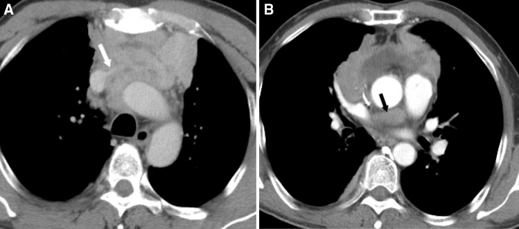
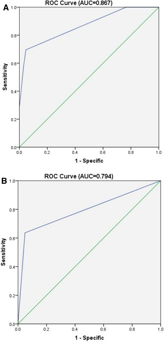
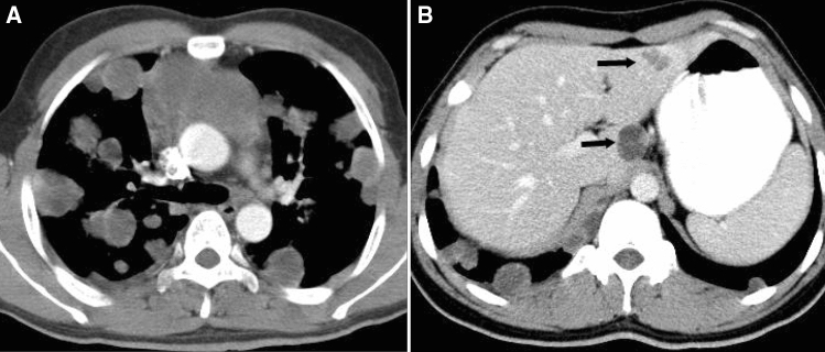
To determine the prognostic CT features in patients with untreated thymic epithelial tumors (TETs). Clinical data and CT imaging features of 194 patients with pathologically confirmed TETs were retrospectively reviewed. The subjects included 113 male and 81 female patients between 15 and 78 years of age, with a mean age of 53.8 years. Clinical outcomes were categorized according to whether relapse, metastasis or death occurred within 3 years after the first diagnosis. Associations between clinical outcomes and CT imaging features were determined using univariate and multivariate logistic regression analyses, while the survival status was analyzed by Cox regression. In this study, we analyzed 110 thymic carcinomas, 52 high-risk thymomas and 32 low-risk thymomas. Percentages of poor outcome and patient death in thymic carcinomas were much higher than those in patients with high-risk and low-risk thymomas. In the thymic carcinomas groups, 46 patients (41.8%) experienced tumor progression, local relapse or metastasis and were categorized as having poor outcomes; vessel invasion and pericardial mass were confirmed to be independent predictors by logistic regression analysis (p < 0.01). In the high-risk thymoma group, 11 patients (21.2%) were categorized as having poor outcomes, and the CT feature pericardial mass was confirmed to be an independent predictor (p < 0.01). In survival analysis, Cox regression showed that CT features of lung invasion, great vessel invasion, lung metastasis and distant organ metastasis were independent predictors for worse survival in the thymic carcinoma group (p < 0.01), while lung invasion and pericardial mass were independent predictors for worse survival in high-risk thymoma group. No CT features were related to poor outcome and worse survival in the low-risk thymoma group. Patients with thymic carcinoma had poorer prognosis and worse survival than those with high-risk or low-risk thymoma. CT can serve as an important tool for predicting the prognosis and survival of patients with TETs. In this cohort, CT features of vessel invasion and pericardial mass were related to poorer outcomes in those with thymic carcinoma and pericardial mass in those with high-risk thymoma. Features including lung invasion, great vessel invasion, lung metastasis and distant organ metastasis indicate worse survival in thymic carcinoma, whereas lung invasion and pericardial mass indicate worse survival in high-risk thymoma.
© 2023. The Author(s).
Conflict of interest statement
The authors declare no competing interests.
Figures

Similar articles
-
Does CT of thymic epithelial tumors enable us to differentiate histologic subtypes and predict prognosis?AJR Am J Roentgenol. 2004 Aug;183(2):283-9. doi: 10.2214/ajr.183.2.1830283. AJR Am J Roentgenol. 2004. PMID: 15269013
-
Thymic epithelial tumors: comparison of CT and MR imaging findings of low-risk thymomas, high-risk thymomas, and thymic carcinomas.Eur J Radiol. 2006 Oct;60(1):70-9. doi: 10.1016/j.ejrad.2006.05.003. Eur J Radiol. 2006. PMID: 16766154
-
Clinical characteristics, prognostic factors, and long-term outcomes associated with epithelial malignancies of the thymus: A 20-year single-institution experience.Cancer Rep (Hoboken). 2023 Mar;6(3):e1750. doi: 10.1002/cnr2.1750. Epub 2022 Nov 12. Cancer Rep (Hoboken). 2023. PMID: 36369906 Free PMC article. Review.
-
Correlation of clinical and computed tomography features of thymic epithelial tumours with World Health Organization classification and Masaoka-Koga staging.Eur J Cardiothorac Surg. 2022 Mar 24;61(4):742-748. doi: 10.1093/ejcts/ezab349. Eur J Cardiothorac Surg. 2022. PMID: 34329409
-
[Thymus cancers: A clinical observation].Pathologe. 2016 Feb;37(1):91-105; quiz 106. doi: 10.1007/s00292-016-0140-5. Pathologe. 2016. PMID: 26821326 Review. German.
Cited by
-
Current Treatment Approaches for Thymic Epithelial Tumors.Life (Basel). 2023 May 12;13(5):1170. doi: 10.3390/life13051170. Life (Basel). 2023. PMID: 37240815 Free PMC article. Review.
-
Advanced three-dimensional X-ray imaging unravels structural development of the human thymus compartments.Commun Med (Lond). 2024 Oct 22;4(1):204. doi: 10.1038/s43856-024-00623-7. Commun Med (Lond). 2024. PMID: 39438572 Free PMC article.
-
Machine learning for predicting the prognosis of patients with thymoma and thymic carcinoma.J Thorac Dis. 2025 Feb 28;17(2):824-835. doi: 10.21037/jtd-24-1263. Epub 2025 Feb 20. J Thorac Dis. 2025. PMID: 40083535 Free PMC article.
-
Development and validation of a scoring model for predicting surgical benefit in patients with Masaoka stage III/IV thymic carcinomas.Gland Surg. 2024 Dec 31;13(12):2395-2408. doi: 10.21037/gs-2024-527. Epub 2024 Dec 27. Gland Surg. 2024. PMID: 39822369 Free PMC article.
-
Hippo Pathway Dysregulation in Thymic Epithelial Tumors (TETs): Associations with Clinicopathological Features and Patients' Prognosis.Int J Mol Sci. 2025 Jun 20;26(13):5938. doi: 10.3390/ijms26135938. Int J Mol Sci. 2025. PMID: 40649713 Free PMC article.
References
-
- Filosso PL, et al. Comparison of outcomes between neuroendocrine thymic tumours and other subtypes of thymic carcinomas: A joint analysis of the European Society of Thoracic Surgeons and the International Thymic Malignancy Interest Group. Eur. J. Cardio-thorac. Surg. 2016;50:766–771. doi: 10.1093/ejcts/ezw107. - DOI - PMC - PubMed
Publication types
MeSH terms
Supplementary concepts
LinkOut - more resources
Full Text Sources
Medical
