. 2023 May;30(5):1221-1234.
doi: 10.1038/s41418-023-01133-0. Epub 2023 Feb 18.

CRISPR screens identify novel regulators of cFLIP dependency and ligand-independent, TRAIL-R1-mediated cell death

Affiliations

CRISPR screens identify novel regulators of cFLIP dependency and ligand-independent, TRAIL-R1-mediated cell death

Neil Kuehnle et al. Cell Death Differ. 2023 May.

Abstract

Kaposi's sarcoma-associated herpesvirus (KSHV) causes primary effusion lymphoma (PEL). PEL cell lines require expression of the cellular FLICE inhibitory protein (cFLIP) for survival, although KSHV encodes a viral homolog of this protein (vFLIP). Cellular and viral FLIP proteins have several functions, including, most importantly, the inhibition of pro-apoptotic caspase 8 and modulation of NF-κB signaling. To investigate the essential role of cFLIP and its potential redundancy with vFLIP in PEL cells, we first performed rescue experiments with human or viral FLIP proteins known to affect FLIP target pathways differently. The long and short isoforms of cFLIP and molluscum contagiosum virus MC159L, which are all strong caspase 8 inhibitors, efficiently rescued the loss of endogenous cFLIP activity in PEL cells. KSHV vFLIP was unable to fully rescue the loss of endogenous cFLIP and is therefore functionally distinct. Next, we employed genome-wide CRISPR/Cas9 synthetic rescue screens to identify loss of function perturbations that can compensate for cFLIP knockout. Results from these screens and our validation experiments implicate the canonical cFLIP target caspase 8 and TRAIL receptor 1 (TRAIL-R1 or TNFRSF10A) in promoting constitutive death signaling in PEL cells. However, this process was independent of TRAIL receptor 2 or TRAIL, the latter of which is not detectable in PEL cell cultures. The requirement for cFLIP is also overcome by inactivation of the ER/Golgi resident chondroitin sulfate proteoglycan synthesis and UFMylation pathways, Jagunal homolog 1 (JAGN1) or CXCR4. UFMylation and JAGN1, but not chondroitin sulfate proteoglycan synthesis or CXCR4, contribute to TRAIL-R1 expression. In sum, our work shows that cFLIP is required in PEL cells to inhibit ligand-independent TRAIL-R1 cell death signaling downstream of a complex set of ER/Golgi-associated processes that have not previously been implicated in cFLIP or TRAIL-R1 function.


Conflict of interest statement

The authors declare no competing interests.

Figures

Fig. 1
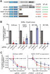
Fig. 1. cFLIP is broadly essential in PEL cell lines.
A Top: cell lysates from the indicated PEL cell lines or KSHV-negative BJAB cells were analyzed by western blotting for the indicated proteins. The cFLIP splice variants cFLIP-L and cFLIP-S are marked. Bottom: heatmap of depletion for sgRNAs targeting the indicated genes, using data from Manzano et al. (2018). Lower FDR-adjusted p values of depletion indicate significant sgRNA depletion in the screens, suggesting essential or fitness roles. B Cas9-expressing PEL cell lines were transduced with the indicated sgRNAs at MOI 3 and selected with puromycin. Graphs show the endpoints (days 8–10) of cumulative cell growth curves relative to an sgRNA targeting the safe harbor locus AAVS1 (see Material and methods). sgRNAs targeting PSMD1 and IRF4 are controls for other essential genes. Error bars represent SD (n = 4–6 independent repeats). For western blot control, see Supplementary Fig. S1C. C BCBL-1/Cas9 expressing ZsGreen, sgRNA-resistant cFLIP-L, or sgRNA-resistant cFLIP-S were challenged with the indicated sgRNAs. Error bars represent SD (n = 3 independent repeats). For western blot control, see Supplementary Fig. S1E. In panels B and C, statistical significance for loss of cell viability compared to sgAAVS1 was analyzed using one-sided, one-sample t-testing (* denotes FDR-adjusted p ≤ 0.05), FDR-adjusted p values are listed in Supplementary Table S6. Rescue in panel C was significant as determined using a one-sided, independent two-sample t-test, with FDR-adjusted p values listed in Supplementary Table S6.
Fig. 2
Fig. 2. cFLIP and KSHV vFLIP are functionally distinct in PEL cell lines.
A Schematic representation of human CASP8, the cFLIP-L/S splice variants, KSHV vFLIP, and the MCV FLIP proteins MC159L and MC160L. Also shown are the characterized activities of each protein toward CASP8/the extrinsic apoptosis pathway and the NF-κB pathway. B BCBL-1/Cas9 expressing ZsGreen, KSHV vFLIP, MCV MC159L, or MCV MC160L were challenged with the indicated sgRNAs. Analyses of cumulative growth curve experiments on day 6 after sgRNA transduction are shown. Error bars represent SD (n = 3 independent repeats). For western blot controls, see Supplementary Fig. S2. Statistical significance for loss of cell viability compared to sgAAVS1 was analyzed using one-sided, one-sample t-testing (* denotes FDR-adjusted p ≤ 0.05). Rescue by expression of KSHV vFLIP and MCL159L was significant as determined using a one-sided, independent two-sample t-test. FDR-adjusted p values are listed in Supplementary Table S6. C Extended growth curves for the ZsGreen and KSHV vFLIP data in panel B show that vFLIP rescue is significant, but not efficient. For details on statistical analysis see Supplementary Table S6 and the Material and methods section.
Fig. 3
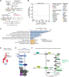
Fig. 3. Genome-wide synthetic rescue screens uncover components of the death program repressed by cFLIP in PEL cells.
A Experimental design for CRISPR-based synthetic rescue screening. B Results of the BCBL-1 resistance screens. Shown are the gene-level ranks output by MAGeCK-RRA based on library sgRNA enrichment after the toxic cFLIP shRNA challenge (y-axis) or the toxic cFLIP sgRNA challenge (x-axis) relative to matched negative control perturbations. For clarity, only the top 500 hits in each screen are displayed. UFL1 was highlighted as a UFMylation pathway gene but does not pass our analysis cutoff. C DAVID pathway analysis of the 23 high confidence hits obtained by intersecting the top 150 hits each from the BCBL-1 sgRNA and shRNA screens. Numbers at right show number of hits from each pathway. For complete DAVID output, see Supplementary Table S3. D Diagram of canonical death signaling via TRAIL through TRAIL receptors. Hits are colored as in panel B. E Diagram of the UFMylation and ER-associated translation pathways, screening hits are colored as in panel B. UFM1 is cytoplasmic, but the UFM1 conjugation machinery is anchored on the cytoplasmic face of the ER membrane by adaptors ODR4 and DDRGK1 (UFBP1). RPL26 is the main characterized substrate for UFMylation and impacts translational pausing during ER-associated degradation. The SRP complex is involved in protein translocation and initiates translational pausing. F Diagram of the Chondroitin sulfate (CS) proteoglycan synthesis pathway, screen hits are colored as in panel B.
Fig. 4
Fig. 4. Cellular FLIP protects PEL cells from CASP8-induced cell death.
A BCBL-1/Cas9 cells were transduced with the indicated sgRNAs, selected with hygromycin, and knockout of CASP8 was confirmed by western blot. B The cell lines from panel A were challenged with puromycin-resistant lentiviral sgRNA vectors at equal MOI and cumulative growth curve analyses were performed as previously. Error bars represent SD from 3 independent repeats. For western blot controls, see Supplementary Fig. S4D. Statistical significance for loss of cell viability compared to sgAAVS1 was analyzed using one-sided, one-sample t-testing (* denotes FDR-adjusted p ≤ 0.05). Rescue by CASP8 KO was significant as determined using a one-sided, independent two-sample t-test. FDR-adjusted p values are listed in Supplementary Table S6. C Cells were challenged with control or cFLIP sgRNAs as in panel B, but not selected with puromycin, and stained with Annexin V 48 h after transduction (n = 2 independent repeats, error bars indicate range).
Fig. 5
Fig. 5. Cellular FLIP protects PEL cells from a ligand-independent, intracellular TRAIL-R1-mediated cell death program.
A BCBL-1/Cas9 cells were transduced with the indicated sgRNAs, selected with hygromycin, and knockout of TRAIL-R1 was confirmed by western blot. B Cell lines depicted in panel A were challenged with puromycin-resistant lentiviral sgRNA vectors at equal MOI and cumulative growth curve analyses were performed as previously. Error bars represent SD from 3 independent repeats. For western blot control, see Supplementary Fig. S5A. Statistical significance for loss of cell viability compared to sgAAVS1 was analyzed using one-sided, one-sample t-testing (* denotes p ≤ 0.05). Rescue by TRAIL-R1 KO, but not TRAIL-R2 or TRAIL KO, was significant as determined using one-sided, independent two-sample t-tests. FDR-adjusted p values are listed in Supplementary Table S6. C BC-3 or BCBL-1 cells were plated at 2 × 105 cells/mL, and supernatants or lysates were harvested 3 days later and used for anti-TRAIL ELISA assay. Known concentrations of recombinant human TRAIL confirm sensitivity down to the ~10 pg/mL range. D Knockout cell lines were fixed in 4% FA and stained for TRAIL-R1 with or without permeabilization by saponin. Shown at left are unstained control cells. Quantification over 3 independent repeats can be found in Supplementary Fig. S6C. E Representative images to establish colocalization of TRAIL-R1 with itself, using a positive control antibody directed against a different epitope, the ERGIC marker ERGIC53, the ER marker ERp72, and the Golgi marker Giantin. TRAIL-R1 is shown in green with other markers in magenta to depict overlapping signal as white. See Supplementary Fig. S5E for results from a second ERGIC53 antibody and TRAIL-R1 KO cells. Scale bar = 10 μm. Mse and Rb indicate antibodies raised in mice or rabbits, respectively. F 3D colocalization of TRAIL-R1 with the indicated proteins was quantified using Pearson’s correlation coefficients (PCCs, n = 3 independent repeats, except n = 2 for mouse anti-ERGIC53). Representative images are shown in panel E and Supplementary Fig. S5D, E. We quantified an average of ~1800 cells per replicate. Statistical significance of the difference from TRAIL-R1 self-colocalization detected with two different TRAIL-R1-specific antibodies was determined by two-sided, independent two-sample t-test (*≤0.05). All markers other than ERp72 and Giantin (indicated by ns) were significantly less colocalized with TRAIL-R1 than TRAIL-R1 self-colocalization.
Fig. 6
Fig. 6. Knockdown of genes that participate in UFMylation, the chondroitin sulfate synthesis pathway, or of JAGN1 and CXCR4 overcome cFLIP dependency in PEL cells.
A Single-gene knockout pools were generated as previously for two genes each in the UFMylation and chondroitin sulfate biosynthesis pathway along with CXCR4 and JAGN1 and these cell lines were used to perform cumulative growth curves following transduction with a second sgRNA as previously. Statistical significance of rescue from cFLIP sg1 induced cell death was determined using one-sided, independent two-sample t-tests (* denotes FDR-adjusted p ≤ 0.05). FDR-adjusted p values and statistical significance of loss of viability are listed in Supplementary Table S6. Western controls are in Supplementary Fig. S6B. B Single-gene KO pools were analyzed for TRAIL-R1 and pro-caspase 8 expression by western blotting.

Similar articles

Cited by

References

    1. Cesarman E, Chang Y, Moore PS, Said JW, Knowles DM. Kaposi’s sarcoma-associated herpesvirus-like DNA sequences in AIDS-related body-cavity-based lymphomas. N Engl J Med. 1995;332:1186–91. doi: 10.1056/NEJM199505043321802. - DOI - PubMed
    1. Nador RG, Cesarman E, Chadburn A, Dawson DB, Ansari MQ, Sald J, et al. Primary effusion lymphoma: a distinct clinicopathologic entity associated with the Kaposi’s sarcoma-associated herpes virus. Blood. 1996;88:645–56. doi: 10.1182/blood.V88.2.645.bloodjournal882645. - DOI - PubMed
    1. Lurain K, Polizzotto MN, Aleman K, Bhutani M, Wyvill KM, Goncalves PH, et al. Viral, immunologic, and clinical features of primary effusion lymphoma. Blood. 2019;133:1753–61. doi: 10.1182/blood-2019-01-893339. - DOI - PMC - PubMed
    1. Wies E, Mori Y, Hahn A, Kremmer E, Sturzl M, Fleckenstein B, et al. The viral interferon-regulatory factor-3 is required for the survival of KSHV-infected primary effusion lymphoma cells. Blood. 2008;111:320–7. doi: 10.1182/blood-2007-05-092288. - DOI - PubMed
    1. Godfrey A, Anderson J, Papanastasiou A, Takeuchi Y, Boshoff C. Inhibiting primary effusion lymphoma by lentiviral vectors encoding short hairpin RNA. Blood. 2005;105:2510–8. doi: 10.1182/blood-2004-08-3052. - DOI - PubMed

Publication types

MeSH terms