. 2023 Feb 18;28(1):3.
doi: 10.1007/s10911-023-09529-x.

Valine Treatment Enhances Antimicrobial Component Production in Mammary Epithelial Cells and the Milk of Lactating Goats Without Influencing the Tight Junction Barrier

Affiliations

Valine Treatment Enhances Antimicrobial Component Production in Mammary Epithelial Cells and the Milk of Lactating Goats Without Influencing the Tight Junction Barrier

Yusaku Tsugami et al. J Mammary Gland Biol Neoplasia. .

Erratum in

Abstract

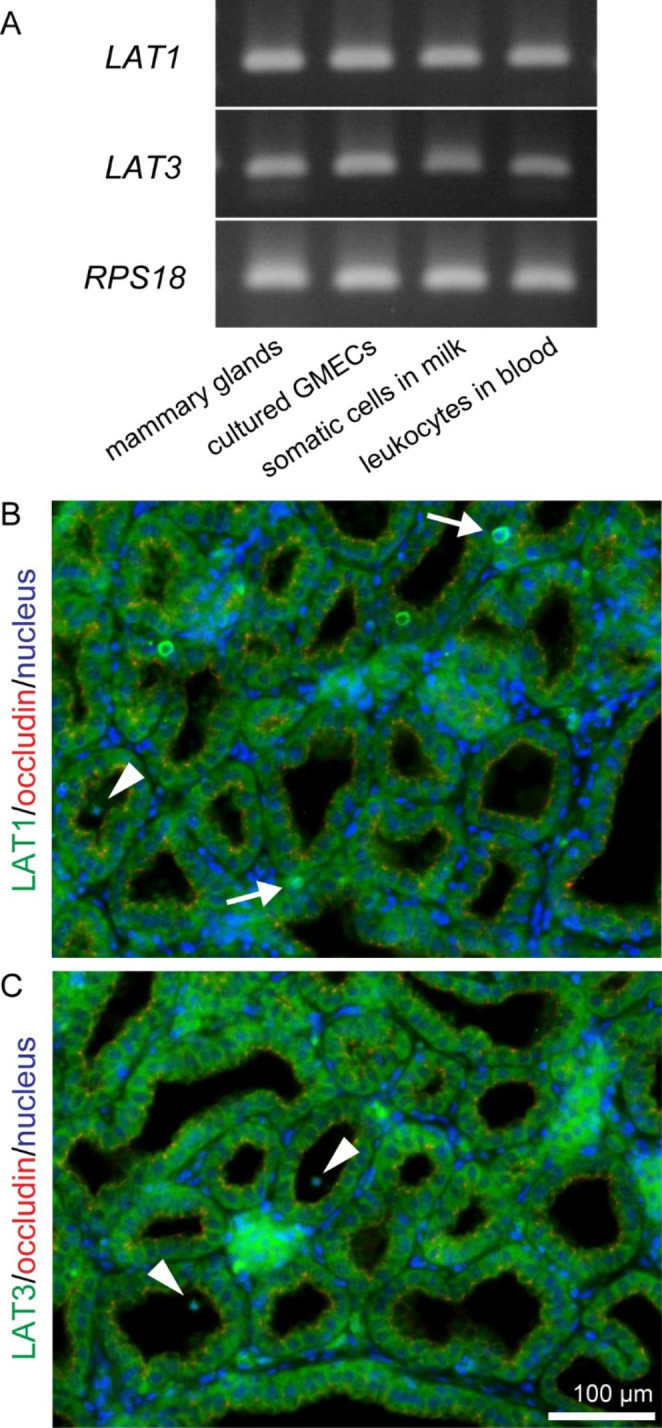
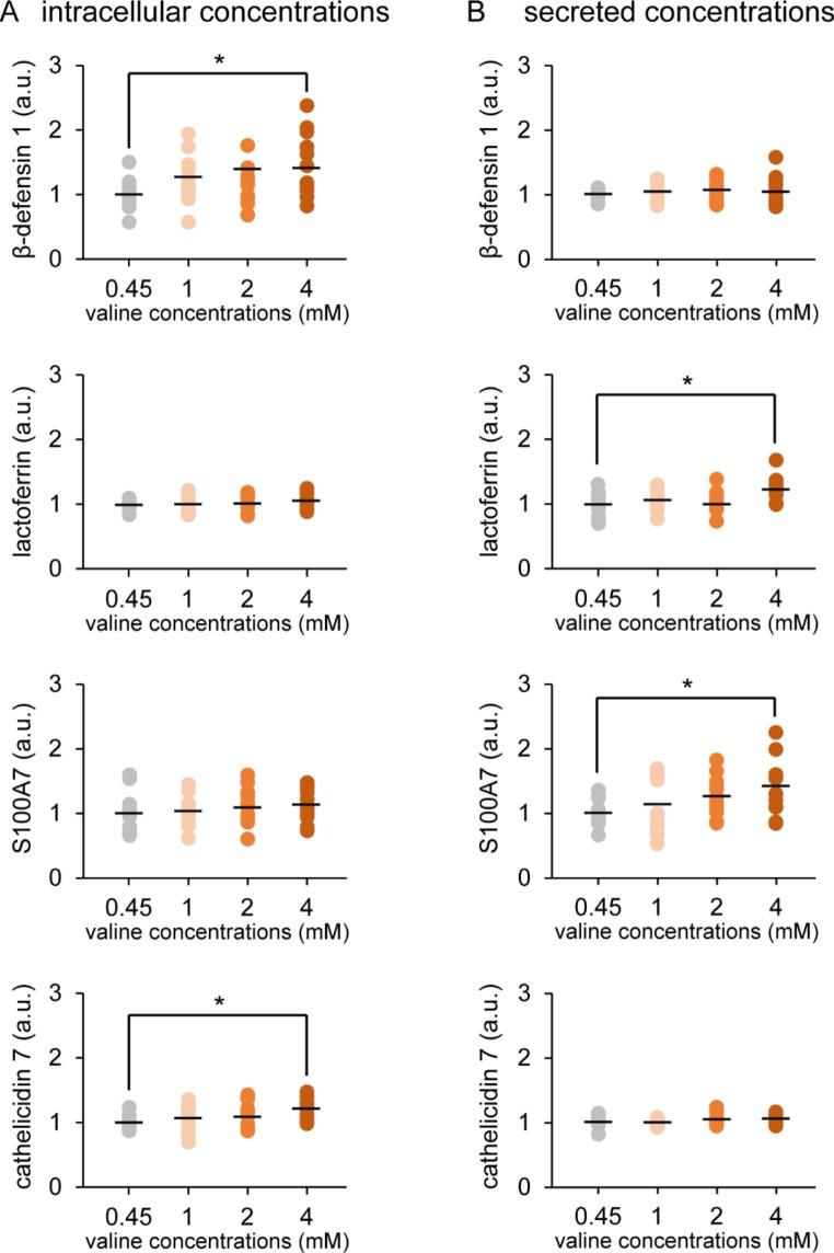
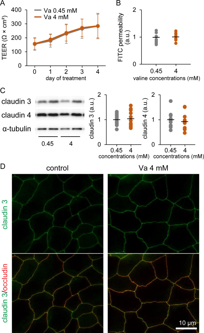
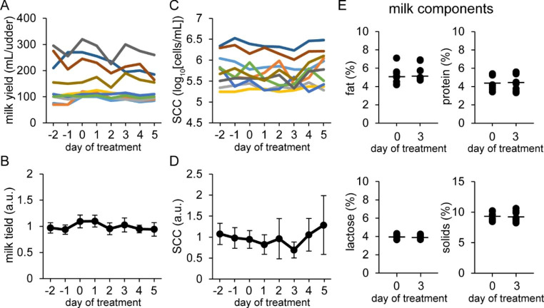
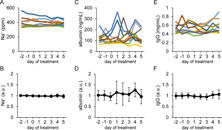
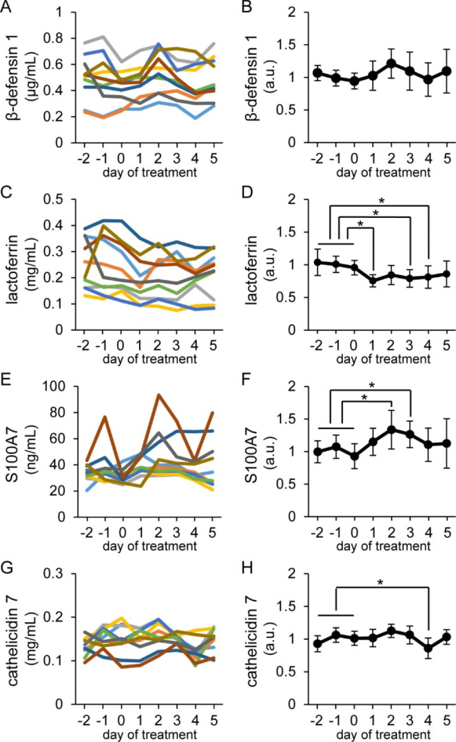
The production of antimicrobial components and the formation of less-permeable tight junctions (TJs) are important in the defense system of lactating mammary glands and for safe dairy production. Valine is a branched-chain amino acid that is actively consumed in the mammary glands and promotes the production of major milk components like β-casein; additionally, branched-chain amino acids stimulate antimicrobial component production in the intestines. Therefore, we hypothesized that valine strengthens the mammary gland defense system without influencing milk production. We investigated the effects of valine in vitro using cultured mammary epithelial cells (MECs) and in vivo using the mammary glands of lactating Tokara goats. Valine treatment at 4 mM increased the secretion of S100A7 and lactoferrin as well as the intracellular concentration of β-defensin 1 and cathelicidin 7 in cultured MECs. In addition, an intravenous injection of valine increased S100A7 levels in the milk of Tokara goats without influencing milk yield and milk components (i.e., fat, protein, lactose, and solids). In contrast, valine treatment did not affect TJ barrier function either in vitro or in vivo. These findings indicate that valine enhances antimicrobial component production without influencing milk production and TJ barrier function in lactating mammary glands; thus, valine contributes to safe dairy production.

Keywords: Antimicrobial component; Branched-chain amino acid; Mammary gland; Tight junction; Valine.


Conflict of interest statement

The authors declare that they have no conflicts of interest.

Figures

Fig. 1
Fig. 1
mRNA expression and localization of LAT1 and LAT3 in goat mammary glands. (A) Bands show the results of RT-PCR for LAT1, LAT3, and RPS18 in mammary glands, cultured goat mammary epithelial cells (GMECs), somatic cells in milk, and leukocytes in the blood of Tokara goats. Immunofluorescence images show the localization of LAT1 (green) (B) and LAT3 (green) (C) in mammary gland tissue. Occludin (red) was used as a marker of most apical regions in lateral membranes. Single cells in the mammary stromal regions (arrows) and the mammary alveolus (arrowheads) showed positive reactions against LAT1 or LAT3 antibodies. Scale bar, 100 μm
Fig. 2
Fig. 2
Influence of valine on antimicrobial component production in cultured goat mammary epithelial cells (GMECs). Graphs show the intracellular concentrations (A) and secreted concentrations into the medium (B) of β-defensin 1, lactoferrin, S100A7, and cathelicidin 7. Antimicrobial component concentrations were detected using ELISA in GMECs treated with valine at the given concentrations. Control GMECs were affected by valine at 0.45 mM. Asterisks show significant differences (p < 0.05) between groups according to Tukey’s test (n = 12)
Fig. 3
Fig. 3
Influence of valine on tight junction (TJ) barrier function in cultured goat mammary epithelial cells (GMECs). Graphs show the transepithelial electrical resistance (TEER) (A) and fluorescein isothiocyanate (FITC) permeability (B) in GMECs treated with 4 mM valine for 4 days (n = 10–12). (C) Western blotting of claudin 3 and claudin 4 in GMECs treated with valine at 4 mM for 3 days. Graphs show the results of densitometry analysis, with α-tubulin used as an internal control (n = 12). (D) Localization of claudin 3 (green) in GMECs treated valine at 4 mM for 4 days. Occludin (red) was used as the TJ marker. Scale bar, 10 μm
Fig. 4
Fig. 4
Influence of intravenous injections of valine on milk yield, somatic cell count (SCC) in milk and milk components, and the tight junction barrier in mammary glands Tokara goats were intravenously injected with 6 mL of valine at 80 mg/mL on days 0–2. Upper graphs show changes in milk yield (A) and SCC (C) in individual udders, whereas lower graphs (B and D) show changes in the relative values of these variables compared with the average of pretreatment (days − 2 to 0), i.e., the control. Colored lines show the changes in milk yield and SCC derived from the same udder. (E) Milk components (fat, protein, lactose, and solids) on days 0 and 3. Data are means ± SD (n = 10)
Fig. 5
Fig. 5
Influence of intravenous injections of valine on the blood-derived components in milk Tokara goats were intravenously injected with 6 mL of valine at 80 mg/mL on days 0–2. Upper graphs show changes in Na+ level (A), albumin level (C), and IgG level (E) in milk of individual udders, whereas lower graphs (B, D, and F) show changes in the relative values of these variables compared with the average of pretreatment (days − 2 to 0), i.e., the control. Colored lines show the changes in blood-derived components in milk derived from the same udder. Data are means ± SD (n = 10)
Fig. 6
Fig. 6
Influence of intravenous injections of valine on antimicrobial component concentrations in milk Tokara goats were intravenously injected with 6 mL of valine at 80 mg/mL on days 0–2. Left graphs show changes in β-defensin 1 (A), lactoferrin (C), S100A7 (E), and cathelicidin 7 (G) levels in individual udders, whereas right graphs (B, D, F, and H) show changes in the relative values of these variables compared with the average of pretreatment (days − 2 to 0), i.e., the control. Colored lines show the changes in antimicrobial components in milk derived from the same udder. Data are means ± SD (n = 10). Asterisks indicate significant differences (p < 0.05 versus the control) according to Dunnett’s test

Similar articles

Cited by

References

    1. Jamali H, Barkema HW, Jacques M, Lavallée-Bourget E, et al. Invited review: incidence, risk factors, and effects of clinical mastitis recurrence in dairy cows. J Dairy Sci. 2018;101:4729–46. doi: 10.3168/jds.2017-13730. - DOI - PubMed
    1. Murphy JM. The genesis of bovine udder infection and mastitis; the occurrence of streptococcal infection in a cow population during a seven-year period and its relationship to age. Am J Vet Res. 1947;8:29–42. - PubMed
    1. Ruegg PL. A 100-Year review: Mastitis detection, management, and prevention. J Dairy Sci. 2017;100:10381–97. doi: 10.3168/jds.2017-13023. - DOI - PubMed
    1. Isobe N. Control mechanisms for producing antimicrobial factors in ruminant mammary gland. Anim Sci J. 2017;88:937–43. doi: 10.1111/asj.12808. - DOI - PubMed
    1. Baumgartner HK, Rudolph MC, Ramanathan P, Burns V, et al. Developmental expression of claudins in the mammary gland. J Mammary Gland Biol Neoplasia. 2017;22:141–57. doi: 10.1007/s10911-017-9379-6. - DOI - PMC - PubMed

Publication types