. 2023 Feb 21;11(2):e0408222.
doi: 10.1128/spectrum.04082-22. Online ahead of print.

Plasma Gelsolin Enhances Phagocytosis of Candida auris by Human Neutrophils through Scavenger Receptor Class B

Affiliations

Plasma Gelsolin Enhances Phagocytosis of Candida auris by Human Neutrophils through Scavenger Receptor Class B

Łukasz Suprewicz et al. Microbiol Spectr. .

Abstract

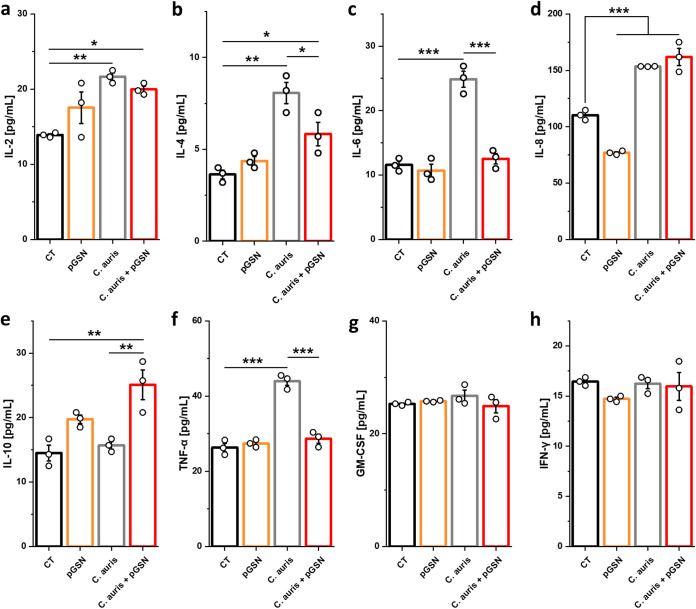
In addition to its role as an actin-depolymerizing factor in the blood, plasma gelsolin (pGSN) binds bacterial molecules and stimulates the phagocytosis of bacteria by macrophages. Here, using an in vitro system, we assessed whether pGSN could also stimulate phagocytosis of the fungal pathogen Candida auris by human neutrophils. The extraordinary ability of C. auris to evade immune responses makes it particularly challenging to eradicate in immunocompromised patients. We demonstrate that pGSN significantly enhances C. auris uptake and intracellular killing. Stimulation of phagocytosis was accompanied by decreased neutrophil extracellular trap (NET) formation and reduced secretion of proinflammatory cytokines. Gene expression studies revealed pGSN-dependent upregulation of scavenger receptor class B (SR-B). Inhibition of SR-B using sulfosuccinimidyl oleate (SSO) and block lipid transport-1 (BLT-1) decreased the ability of pGSN to enhance phagocytosis, indicating that pGSN potentiates the immune response through an SR-B-dependent pathway. These results suggest that the response of the host's immune system during C. auris infection may be enhanced by the administration of recombinant pGSN. IMPORTANCE The incidence of life-threatening multidrug-resistant Candida auris infections is rapidly growing, causing substantial economic costs due to outbreaks in hospital wards. Primary and secondary immunodeficiencies in susceptible individuals, such as those with leukemia, solid organ transplants, diabetes, and ongoing chemotherapy, often correlate with decreased plasma gelsolin concentration (hypogelsolinemia) and impairment of innate immune responses due to severe leukopenia. Immunocompromised patients are predisposed to superficial and invasive fungal infections. Morbidity caused by C. auris among immunocompromised patients can be as great as 60%. In the era of ever-growing fungal resistance in an aging society, it is critical to seek novel immunotherapies that may help combat these infections. The results reported here suggest the possibility of using pGSN as an immunomodulator of the immune response by neutrophils during C. auris infection.

Keywords: Candida auris; inflammation; neutrophils; phagocytosis; plasma gelsolin.


Conflict of interest statement

The authors declare no conflict of interest.

Figures

FIG 1
FIG 1
Plasma gelsolin enhances uptake and intracellular killing of C. auris cells by human neutrophils. (a) Live cell imaging data of the uptake of zymosan pHrodo particles after 2 h by human neutrophils that were serum starved for 1 h and either preincubated with pGSN for 1 h or had pGSN simultaneously added with the zymosan particles (n = 4). Results were normalized to the untreated control (0), set as 1. (b) Representative images of the uptake of zymosan particles (green) by pGSN-pretreated neutrophils; bar, ~100 μm. (c to f) Percentage uptake and phagocytic index for pGSN-preincubated human neutrophils ingesting C. auris cells (n = 5) at MOIs of 1 (c and e) and 5 (d and f). Neutrophils taking up at least one fungal cell were manually tracked to allow a quantitative analysis of percentage uptake during the 2-h coincubation period (c and d). The number of fungal cells ingested (phagocytic index) by the neutrophils was manually counted after 2 h of coincubation (e and f). The survival of C. auris cells after infection of pGSN-pretreated (1 h) human neutrophils (1 h serum starved) at MOIs of 1 (g) and 5 (h) for 2 h was evaluated by plating dilutions of samples on Sabouraud agar (n = 4). (i) Images of neutrophils that were serum starved, pretreated or not with pGSN, cocultured with Calcofluor white (blue)-stained C. auris cells for 2 h, fixed, and immunolabeled for actin with Texas red phalloidin (red). White arrows indicate C. auris cells. Bar, ~15 μm. Data are presented as the mean ± standard error of the mean (SEM). **, P < 0.01; ***, P < 0.001. Significance was determined by Student’s t test (a) or one-way analysis of variance (ANOVA) with Tukey’s test (c to h). Human albumin was used as a negative control.
FIG 2
FIG 2
Plasma gelsolin prevents NETotic death of human neutrophils upon C. auris infection. (a) Live cell imaging data of extracellular DNA release (eDNA) by human neutrophils that were either preincubated with pGSN for 1 h or had pGSN simultaneously added to them with the C. auris cells at an MOI of 5 for 2 h (n = 4). (b) Representative images of pGSN-pretreated neutrophils releasing eDNA (orange) upon C. auris infection. Bar, ~250 μm. (c) Images of NETs released by human neutrophils that were pretreated as previously described; myeloperoxidase (MPO) is shown in green, while blue indicates DNA stained with DAPI. Bar, ~50 μm. (d) Percentage of NETs (n = 4) and (e) release of the reactive oxygen species (n = 3) from human neutrophils (pGSN pretreated, as previously) upon C. auris infection at an MOI of 5. Data are presented as the mean ± SEM. Results were normalized to the untreated control (0 or CT), set as 1 (a and e). *, P < 0.05; **, P < 0.01; ***, P < 0.001. Significance was determined by Student’s t test (a) or one-way ANOVA with Tukey’s test (d and e). PMA (phorbol-12-myristate-13-acetate) was used as a positive control.
FIG 3
FIG 3
Plasma gelsolin inhibits the expression of proteins involved in NET formation. (a) Immunoblot analysis for the indicated proteins using lysates from human neutrophils that were serum starved for 1 h, then preincubated with pGSN at 250 μg/mL for 1 h, washed, and finally infected with live C. auris yeast cells (MOI = 5) for 2 h. Data are expressed as the mean ± SEM and represent three independent experiments. Results were normalized to the expression of the β-actin and are presented relative to those of the negative control (CT), set as 1. *, P < 0.05; **, P < 0.01; ***, P < 0.001. Significance was determined by Student’s t test (b). PMA (phorbol-12-myristate-13-acetate) and human albumin were used as the positive and negative controls, respectively.
FIG 4
FIG 4
Plasma gelsolin triggers an anti-inflammatory phenotype in human neutrophils during C. auris infection. Production of IL-2 (a), IL-4 (b), IL-6 (c), IL-8 (d), IL-10 (e), TNF-α (f), GM-CSF (g), and IFN-γ (h), as determined by magnetic bead-based enzyme-linked immunosorbent assay (ELISA), in the culture supernatants of human neutrophils that were serum starved for 1 h, preincubated with pGSN at 250 μg/mL for 1h, washed, and infected with live C. auris yeast cells (MOI = 5) for 2 h (n = 3). Data are expressed as the mean ± SEM. *, P < 0.05; **, P < 0.01; ***, P < 0.001. Significance was determined by one-way ANOVA with Tukey’s test.
FIG 5
FIG 5
Plasma gelsolin enhances phagocytosis of C. auris through stimulation of the scavenger receptor class B type I (SR-BI). (a) Heat map of changes in gene expression of selected phagocytosis-related genes upon incubation with pGSN, C. auris cells, and preincubation with pGSN followed by addition of the yeast (n = 3). Results are presented as the log2 fold change (log2FC). (b to g) Effect of scavenger receptor class B type I (SR-BI) inhibitors SSO at 200 μM and BLT-1 at 1 μM compared to the vehicle control on fungal uptake (n = 5) (b and c), the phagocytic index (n = 5) (d and e), and yeast survival (n = 4) (f and g). Human neutrophils were preincubated with the indicated SR-BI inhibitors during serum starvation for 1 h; then, pGSN was added for 1 h, the cells were carefully washed, and C. auris was introduced to the neutrophils at an MOI of 1 for 2 h. (c, e, and g) Trend of the effect of SR-BI inhibitors compared to vehicle controls on fungal uptake, the phagocytic index, and fungal survival. Data are expressed as the mean ± SEM. *, P < 0.05; **, P < 0.01; ***, P < 0.001. Significance was determined by one-way ANOVA with Tukey’s test. The black asterisk refers to the comparison with the untreated condition within each group (vehicle or SSO- or BLT-1-treated cells), while the blue asterisk refers to the significance compared to the corresponding concentration in the vehicle control. Human albumin was used as a negative control.

Similar articles

Cited by

  • Innate immune response to Candida auris.
    Holt AM, Nett JE. Holt AM, et al. Curr Opin Microbiol. 2024 Aug;80:102510. doi: 10.1016/j.mib.2024.102510. Epub 2024 Jul 3. Curr Opin Microbiol. 2024. PMID: 38964276 Free PMC article. Review.

References

    1. Piktel E, Levental I, Durnaś B, Janmey PA, Bucki R. 2018. Plasma gelsolin: indicator of inflammation and its potential as a diagnostic tool and therapeutic target. Int J Mol Sci 19:2516. doi:10.3390/ijms19092516. - DOI - PMC - PubMed
    1. Bucki R, Byfield FJ, Kulakowska A, McCormick ME, Drozdowski W, Namiot Z, Hartung T, Janmey PA. 2008. Extracellular gelsolin binds lipoteichoic acid and modulates cellular response to proinflammatory bacterial wall components. J Immunol 181:4936–4944. doi:10.4049/jimmunol.181.7.4936. - DOI - PubMed
    1. Cheng Y, Hu X, Liu C, Chen M, Wang J, Wang M, Gao F, Han J, Zhang C, Sun D, Min R. 2017. Gelsolin inhibits the inflammatory process induced by LPS. Cell Physiol Biochem 41:205–212. doi:10.1159/000456043. - DOI - PubMed
    1. Peng X, Zhang X, Wang L, Zhu Q, Luo J, Wang W, Wang X. 2011. Gelsolin in cerebrospinal fluid as a potential biomarker of epilepsy. Neurochem Res 36:2250–2258. doi:10.1007/s11064-011-0549-4. - DOI - PubMed
    1. Bucki R, Kulakowska A, Byfield FJ, Zendzian-Piotrowska M, Baranowski M, Marzec M, Winer JP, Ciccarelli NJ, Górski J, Drozdowski W, Bittman R, Janmey PA. 2010. Plasma gelsolin modulates cellular response to sphingosine 1-phosphate. Am J Physiol Cell Physiol 299:C1516–C1523. doi:10.1152/ajpcell.00051.2010. - DOI - PMC - PubMed