. 2023 Feb 28;120(9):e2102569120.
doi: 10.1073/pnas.2102569120. Epub 2023 Feb 21.

Deficiency of the minor spliceosome component U4atac snRNA secondarily results in ciliary defects in human and zebrafish

Affiliations

Deficiency of the minor spliceosome component U4atac snRNA secondarily results in ciliary defects in human and zebrafish

Deepak Khatri et al. Proc Natl Acad Sci U S A. .

Abstract

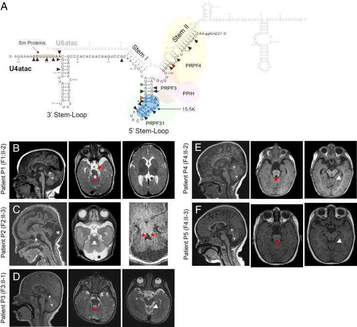
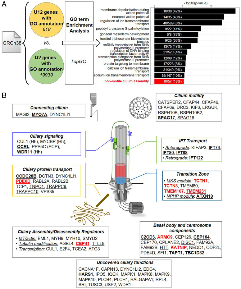
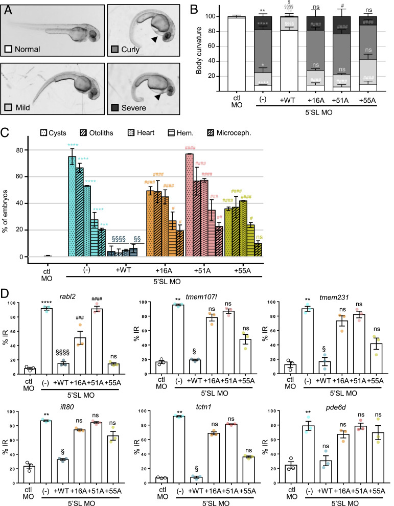
In the human genome, about 750 genes contain one intron excised by the minor spliceosome. This spliceosome comprises its own set of snRNAs, among which U4atac. Its noncoding gene, RNU4ATAC, has been found mutated in Taybi-Linder (TALS/microcephalic osteodysplastic primordial dwarfism type 1), Roifman (RFMN), and Lowry-Wood (LWS) syndromes. These rare developmental disorders, whose physiopathological mechanisms remain unsolved, associate ante- and post-natal growth retardation, microcephaly, skeletal dysplasia, intellectual disability, retinal dystrophy, and immunodeficiency. Here, we report bi-allelic RNU4ATAC mutations in five patients presenting with traits suggestive of the Joubert syndrome (JBTS), a well-characterized ciliopathy. These patients also present with traits typical of TALS/RFMN/LWS, thus widening the clinical spectrum of RNU4ATAC-associated disorders and indicating ciliary dysfunction as a mechanism downstream of minor splicing defects. Intriguingly, all five patients carry the n.16G>A mutation, in the Stem II domain, either at the homozygous or compound heterozygous state. A gene ontology term enrichment analysis on minor intron-containing genes reveals that the cilium assembly process is over-represented, with no less than 86 cilium-related genes containing at least one minor intron, among which there are 23 ciliopathy-related genes. The link between RNU4ATAC mutations and ciliopathy traits is supported by alterations of primary cilium function in TALS and JBTS-like patient fibroblasts, as well as by u4atac zebrafish model, which exhibits ciliopathy-related phenotypes and ciliary defects. These phenotypes could be rescued by WT but not by pathogenic variants-carrying human U4atac. Altogether, our data indicate that alteration of cilium biogenesis is part of the physiopathological mechanisms of TALS/RFMN/LWS, secondarily to defects of minor intron splicing.

Keywords: U4atac; genetic disease; minor introns; primary cilium; splicing.


Conflict of interest statement

The authors declare no competing interest.

Figures

Fig. 1.
Fig. 1.
RNU4ATAC is mutated in five cases of patients with JBTS-like traits. (A) Schema of the structure of the U4atac/U6atac bi-molecule with the main interacting proteins, showing with black arrowheads the nucleotides mutated in patients with RNU4ATAC-associated syndromes (9). The red arrowhead points to 16G [mutated in all five patients with JBTS-like disorder (n.16G>A)], the orange arrowheads to 33C and 51G [mutated in JBTS-like patients P3 (n.51G>A) and P4/P5 (n.33C>G)], the green arrowhead to 55G, the second most frequently mutated nucleotide after 51G in TALS patients. (B–F) Cerebral examination of all five RNU4ATAC-associated JBTS-like patients, performed at 7 mo of age for patient P1 (male, F1:II-2) (B), at 4 d of age in a premature baby born at 31.4 GW for patient P2 (female, F2:II-3) (C), at 1 y of age for patients P3 (female, F3:II-1) (D) and P4 (male, F4:II-2) (E), and at 2 y of age for patient P5 (female, F4:II-3) (F). Full descriptions of T1- or T2-weighted mid-sagittal (Left) and axial (Middle and Right) MRI are available in case reports, in SI Appendix. Main features: double arrows indicate vermian hypoplasia and/or dysplasia, asterisks arachnoid cyst, and red stars the « MTS ».
Fig. 2.
Fig. 2.
The non-motile cilium assembly pathway is enriched among U12 genes. (A) GO term enrichment among U12-type intron-containing genes. Comparison of 618 U12 and 19,939 U2 genes with GO annotations, using the TopGO R package, allowed to identify 243 enriched biological processes with a P value ≤0.05, of which the 13 most significant are shown. Numbers in bars indicate the ratio of U12 genes belonging to each GO term (in brackets, the percentage). (B) Schema representing the localization and/or function of the 86 cilium-related U12 genes. Underlined are the "Gold_Standard" genes (17), in bold the genes associated with a ciliopathy, and in red those associated with JBTS. Hh, Hedgehog pathway; PCP, Planar Cell Polarity pathway.
Fig. 3.
Fig. 3.
JBTS-like and TALS RNU4ATAC mutations lead to U12-type IR in ciliary genes and result in ciliary function alteration in patient’s fibroblasts. (A) Table summarising the results of the analysis of U12-type IR in the transcriptomic data of patient P1 vs. control (ctl1) fibroblasts, in the same format as those we previously published (12). Indicated are the number and percentage of U12 genes presenting differences of percent-spliced-in (PSI) values (dPSI) greater than 10%, between 10 and 1%, or below 1% between patient and control cells. (B) Sashimi plots of U12-type intron splicing of TMEM107 and TCTN1 genes, based on the present RNA-seq data of P1 and ctl1 fibroblasts, and on the previous pooled data of TALS fibroblasts (12). On lines, the number of reads split across splice junctions; in brackets, the range of coverage of each base of the depicted region. (C) RT-PCR using primers depicted in the schemas. ACTB is the actin beta housekeeping gene. (D and E) Graphs of qRT-PCR analyses showing the mean ± SEM of at least three independent experiments. Only significant comparisons are shown; *P < 0.05, **P < 0.01, ***P < 0.001 using Kruskal–Wallis test with Dunn’s multiple comparisons. (F) Confocal images of an age- and sex-matched control, TALS6 and P1 patients’ fibroblasts derived from skin biopsy. Shifted channels of adenylate cyclase (AC3, green) and Arl13b (cilium marker, magenta) staining are shown. (Scale bar, 10 µm.) (G) Quantification of the percentage of AC3-positive cilia seen in (F). Graphs show the mean ± SEM of at least 5 fields in five independent experiments (total number of analyzed cells >200). Only significant comparisons are shown; ****P < 0.0001, using Kruskal–Wallis test with Dunn’s multiple comparisons. (H) qRT-PCR analysis of the expression of the Hh target gene GLI1 upon SAG treatment (+SAG). Graphs show the mean ± SEM of four to five independent experiments. Only significant comparisons are shown; **P < 0.01, *= 0.0499, using Kruskal–Wallis test with Dunn’s multiple comparisons.
Fig. 4.
Fig. 4.
Deficiency of u4atac leads to cilium alterations in zebrafish. (A–F) Global morphology of control (ctl) (Upper) and u4atac 5’SL (Lower) morpholino (MO)-injected embryos at 48 hpf. u4atac morphants are characterized by body curvature and cardiac edema (A’, arrowhead); cysts (B’, asterisks) in glomeruli; anomalies of otoliths in OV (C’); alterations of cardiac morphology (D’); blood hemorrhages in brain (E’, arrowhead); and microcephaly (F’). Dorsal views in B and B’ and ventral views in D and D’, with anterior to the top. (G) Percentage of embryos displaying each of the phenotypes shown in A'F', following injection of control MO, u4atac 5’SL MO alone or with human WT U4atac snRNA. Graph shows the mean ± SEM of three independent experiments (total number of embryos >300). P < 0.01, t-tests comparing 5’SL MO to ctl MO, and 5’SL MO + WT to 5’SL MO. (H) Live confocal imaging of motile cilia in CC of MO-injected Tg(bactin:arl13bGFP) transgenic embryos, at 48 hpf. Slow acquisition speed of motile cilia produces imaging artifacts, delineating the range of motion of each cilium (yellow angles). (H’) Quantification of the angle of motile cilium range of motion in control MO (n = 22), u4atac 5’SL MO alone (n = 22), or with human WT U4atac snRNA (n = 20) injected embryos, as described in (H). (I–K) Immunostaining of cilia (acetylated α-tubulin) in the proximal straight tubule (I), the OV (J), and at the nasal pit (K) of control MO, u4atac 5’SL MO alone, or with human WT U4atac snRNA injected embryos, at 24 (J) or 48 (I and K) hpf. In brackets, the number of embryos exhibiting the phenotype shown in the image over the total number of analyzed embryos. Dotted lines in I delineate the tubule lumen. (J’) Quantification of the cilium number in OV of control MO (n = 15), u4atac 5’SL MO alone (n = 15), or with human WT U4atac snRNA (n = 16) injected embryos, as described in (J). For quantification (H’ and J’), box-and-whisker plots show in the box the median and the 25th to 75th percentiles and in whiskers the minimum to the maximum values, which were obtained in two independent experiments. For H’, values are the mean range of motion of all cilia measured in one single embryo. Only significant comparisons are shown; ****P < 0.0001, ***P < 0.005, **P < 0.01 by Kruskal–Wallis test with Dunn’s multiple comparisons test. Images show the maximum intensity projection (H, I, K) or a single optical slice selected in the middle of z-stack (J). (Scale bars, 10 µm.) a, atrium; v, ventricle; hem, hemorrhages.
Fig. 5.
Fig. 5.
Coinjected JBTS-like and TALS U4atac mutants lead to ciliopathy-related phenotypes in zebrafish. (A) Global morphology of embryos coinjected with 5’SL MO and JBTS-like (16A) or TALS (51A, 55A)-related U4atac variants. A varying severity of body curvature could be observed (mild, curly, severe), as well as the presence of cardiac edema (black arrowhead). (B and C) Percentage of embryos exhibiting the various severities of body curvature (B) shown in (A), and each of the phenotypes (C) shown in Fig. 4 AF, following coinjection of 5’SL MO with human WT or mutated U4atac snRNA. Graphs show the mean ± SEM of three batches of 60 embryos issued from independent experiments (total number of embryos: 180). Statistical analysis with ctl MO (*), 5’SL MO (§), or 5’SL MO+WT hU4atac (#). ****P < 0.0001, ***P < 0.001, **P < 0.01, *P < 0.05, ns nonsignificant, by two-way ANOVA test with Sidak’s (*, §) or Dunnett’s (#) multiple comparisons test. (D) qRT-PCR analysis of U12-type IR in six cilium-related genes in embryos coinjected with 5’SL MO and human WT or mutated U4atac snRNA. Graphs show the mean ± SEM of three independent experiments. Statistical analysis with ctl MO (*), 5’SL MO (§), or 5’SL MO+WT hU4atac (#). ****P < 0.0001, ***P < 0.001, **P < 0.01, *P < 0.05, ns nonsignificant, by Kruskal–Wallis test with Dunn’s multiple comparisons test.

References

    1. Edery P., et al. , Association of TALS developmental disorder with defect in minor splicing component U4atac snRNA. Science 332, 240–243 (2011). - PubMed
    1. He H., et al. , Mutations in U4atac snRNA, a component of the minor spliceosome, in the developmental disorder MOPD I. Science 332, 238–240 (2011). - PMC - PubMed
    1. Merico D., et al. , Compound heterozygous mutations in the noncoding RNU4ATAC cause Roifman syndrome by disrupting minor intron splicing. Nat. Commun. 6, 8718 (2015). - PMC - PubMed
    1. Farach L. S., et al. , The expanding phenotype of RNU4ATAC pathogenic variants to Lowry wood syndrome. Am. J. Med. Genet. A 176, 465–469 (2018). - PMC - PubMed
    1. Hagiwara H., et al. , Immunodeficiency in a patient with microcephalic osteodysplastic primordial dwarfism type I as compared to Roifman syndrome. Brain Dev. 43, 337–342 (2020). - PubMed

Publication types

Substances

Supplementary concepts

LinkOut - more resources