. 2023 Feb 20;11(1):29.
doi: 10.1186/s40478-023-01519-8.

Bi-allelic variations in CRB2, encoding the crumbs cell polarity complex component 2, lead to non-communicating hydrocephalus due to atresia of the aqueduct of sylvius and central canal of the medulla

Affiliations

Bi-allelic variations in CRB2, encoding the crumbs cell polarity complex component 2, lead to non-communicating hydrocephalus due to atresia of the aqueduct of sylvius and central canal of the medulla

Aude Tessier et al. Acta Neuropathol Commun. .

Abstract

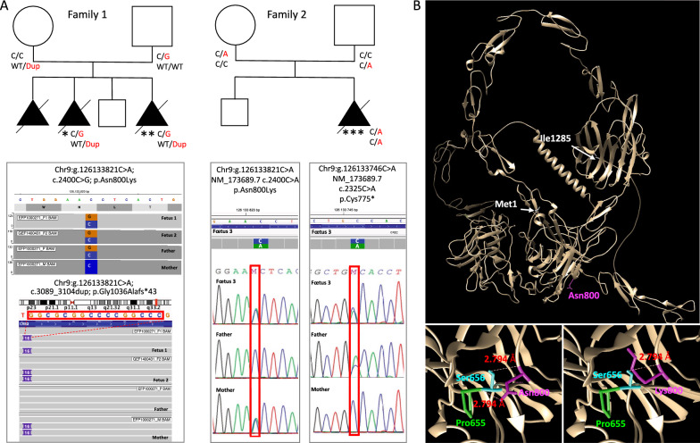
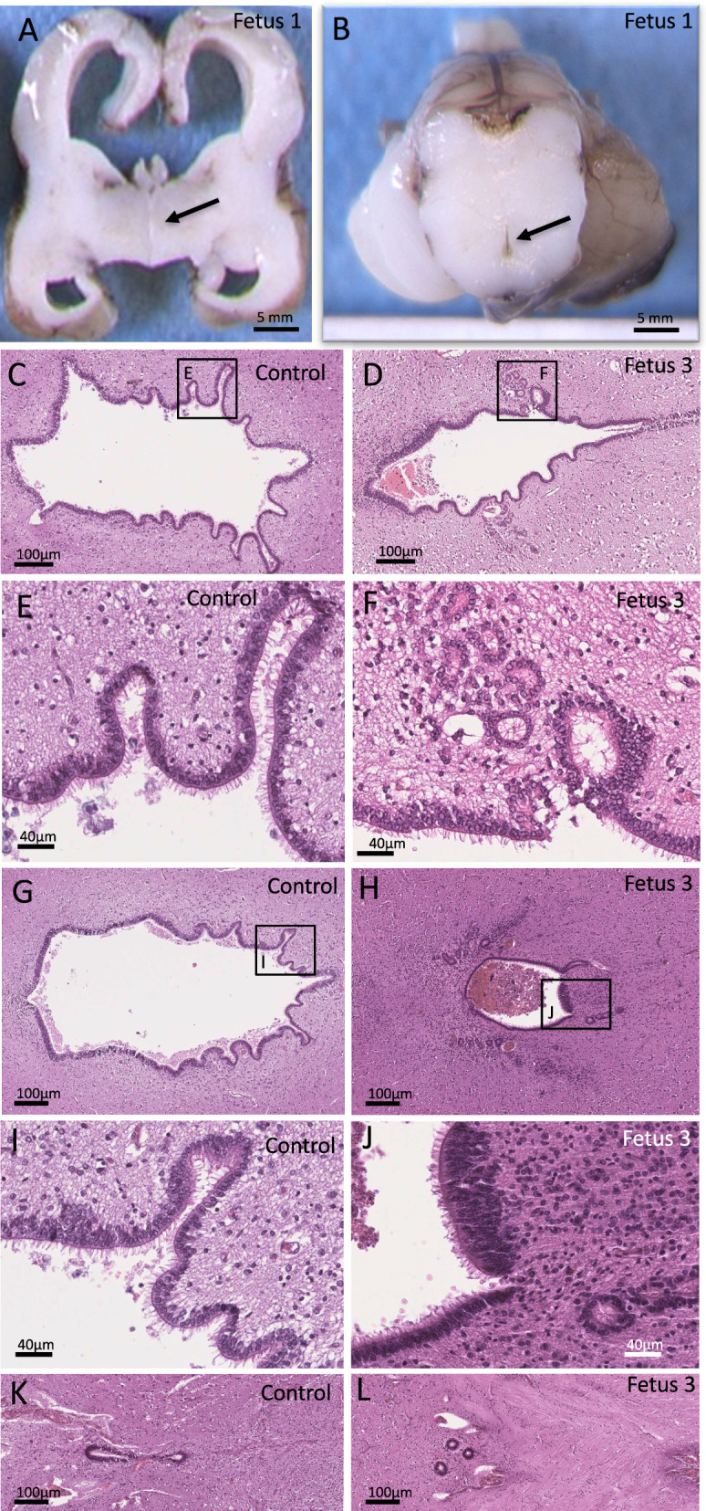
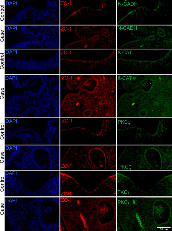
Congenital hydrocephalus is a common condition caused by the accumulation of cerebrospinal fluid in the ventricular system. Four major genes are currently known to be causally involved in hydrocephalus, either isolated or as a common clinical feature: L1CAM, AP1S2, MPDZ and CCDC88C. Here, we report 3 cases from 2 families with congenital hydrocephalus due to bi-allelic variations in CRB2, a gene previously reported to cause nephrotic syndrome, variably associated with hydrocephalus. While 2 cases presented with renal cysts, one case presented with isolated hydrocephalus. Neurohistopathological analysis allowed us to demonstrate that, contrary to what was previously proposed, the pathological mechanisms underlying hydrocephalus secondary to CRB2 variations are not due to stenosis but to atresia of both Sylvius Aqueduct and central medullar canal. While CRB2 has been largely shown crucial for apico-basal polarity, immunolabelling experiments in our fetal cases showed normal localization and level of PAR complex components (PKCι and PKCζ) as well as of tight (ZO-1) and adherens (β-catenin and N-Cadherin) junction molecules indicating a priori normal apicobasal polarity and cell-cell adhesion of the ventricular epithelium suggesting another pathological mechanism. Interestingly, atresia but not stenosis of Sylvius aqueduct was also described in cases with variations in MPDZ and CCDC88C encoding proteins previously linked functionally to the Crumbs (CRB) polarity complex, and all 3 being more recently involved in apical constriction, a process crucial for the formation of the central medullar canal. Overall, our findings argue for a common mechanism of CRB2, MPDZ and CCDC88C variations that might lead to abnormal apical constriction of the ventricular cells of the neural tube that will form the ependymal cells lining the definitive central canal of the medulla. Our study thus highlights that hydrocephalus related to CRB2, MPDZ and CCDC88C constitutes a separate pathogenic group of congenital non-communicating hydrocephalus with atresia of both Sylvius aqueduct and central canal of the medulla.

Keywords: Apical constriction; Aqueduct of sylvius atresia; CCDC88C; CRB2; Cell polarity; Cell–cell junction; Central canal of the medulla; Congenital hydrocephalus; MPDZ; Ventriculomegaly.


Conflict of interest statement

The authors declare that they have no conflict of interest.

Figures

Fig. 1
Fig. 1
CRB2 variations in the 2 reported families. A Pedigree of both families with 3 female fetuses carrying bi-allelic compound heterozygous CRB2 variations. CRB2 variants are reported on the basis of Human Genome Assembly GRCh37 (hg19), and the CRB2 reference sequence used was GenBank: NM_173689.7. Fetus 1 (*) and 2 (**) from family 1 harbor two compound heterozygous variations c.2400C > G p.(Asn800Lys), inherited from their father and c.3089_3104dup p.(Gly1036Alafs*43) inherited from their mother. Fetus 3 (***) from family 2 harbor two compound heterozygous variations c.2325C > A p.(Cys775*) inherited from the father and c.2400C > A p.(Asn800Lys) inherited from the mother. B 3D modeling of human CRB2 (Uniprot Q5IJ48) based on the AphaFold structure prediction and using UCSF Chimera showing that Asn800 is involved in two hydrogen binding interactions with Ser656 and Pro655 and that modifying Asn800 to Lys leads to the loss of the hydrogen bond with Pro655 suggesting altered stability of the mutated protein
Fig. 2
Fig. 2
Main neuropathological findings in fetuses with CRB2 pathogenic variants. Macroscopic coronal section of the brain of fetus 1 showing severe bilateral ventricular dilatation, thinning of the cerebral parenchyma and undiscernible third ventricle (arrow) (A). Macroscopic transversal section of the midbrain showing atretic aqueduct of Sylvius while the pons and cerebellum appear normal (B). Hematoxylin and eosin staining of transversal sections along the mesencephalon and medulla of a control fetus (C, E, G, I, K) and fetus 3 (D, F, H, J, L) showing small dysmorphic aqueduct with multiple indentations and rosettes (D, H) with otherwise normally ciliated ependymal cells in fetus 3 (F, J) as compared to the age-matched control (C, E, G, I). Transversal section of the medulla showing atresia of the central medullar canal, reduced to 3 rosettes (L) as compared to an age-matched control in which the central canal of the medulla is normally visualized (K)
Fig. 3
Fig. 3
Confocal analysis performed after double immunostaining on sections of the midbrain from fetus 3 and an age-matched control with ZO-1 antibody (red) combined with either N-cadherin (green) or β-catenin (green) or PKCζ (green) or PKCι (green) showing that mutated and control fetuses show similar staining for all markers
Fig. 4
Fig. 4
Schematic representation of CRB2 protein showing its known functional domains and the position of all variations previously reported in the literature (above) and the three variations we report in this study (below)

References

    1. Rekate HL. The definition and classification of hydrocephalus: a personal recommendation to stimulate debate. Cerebrosp Fluid Res. 2008;5:2. doi: 10.1186/1743-8454-5-2. - DOI - PMC - PubMed
    1. Tully HM, Dobyns WB. Infantile hydrocephalus: a review of epidemiology, classification and causes. Eur J Med Genet. 2014;57:359–368. doi: 10.1016/j.ejmg.2014.06.002. - DOI - PMC - PubMed
    1. McAllister JP. Pathophysiology of congenital and neonatal hydrocephalus. Semin Fetal Neonatal Med. 2012;17:285–294. doi: 10.1016/j.siny.2012.06.004. - DOI - PubMed
    1. Marivin A, Morozova V, Walawalkar I, Leyme A, Kretov DA, Cifuentes D, et al. GPCR-independent activation of G proteins promotes apical cell constriction in vivo. J Cell Biol. 2019;218:1743–1763. doi: 10.1083/jcb.201811174. - DOI - PMC - PubMed
    1. Marivin A, Garcia-Marcos M. DAPLE and MPDZ bind to each other and cooperate to promote apical cell constriction. Mol Biol Cell. 2019;30:1900–1910. doi: 10.1091/mbc.E19-02-0091. - DOI - PMC - PubMed

Publication types