Review
. 2023 Feb:205:124-135.
doi: 10.1016/j.biochi.2022.07.019. Epub 2022 Nov 8.

Structure and function of prodrug-activating peptidases

Affiliations
Review

Structure and function of prodrug-activating peptidases

José A Velilla et al. Biochimie. 2023 Feb.

Abstract

Bacteria protect themselves from the toxicity of antimicrobial metabolites they produce through several strategies. In one resistance mechanism, bacteria assemble a non-toxic precursor on an N-acyl-d-asparagine prodrug motif in the cytoplasm, then export it to the periplasm where a dedicated d-amino peptidase hydrolyzes the prodrug motif. These prodrug-activating peptidases contain an N-terminal periplasmic S12 hydrolase domain and C-terminal transmembrane domains (TMDs) of varying lengths: type I peptidases contain three transmembrane helices, and type II peptidases have an additional C-terminal ABC half-transporter. We review studies which have addressed the role of the TMD in function, the substrate specificity, and the biological assembly of ClbP, the type I peptidase that activates colibactin. We use modeling and sequence analyses to extend those insights to other prodrug-activating peptidases and ClbP-like proteins which are not part of prodrug resistance gene clusters. These ClbP-like proteins may play roles in the biosynthesis or degradation of other natural products, including antibiotics, may adopt different TMD folds, and have different substrate specificity compared to prodrug-activating homologs. Finally, we review the data supporting the long-standing hypothesis that ClbP interacts with transporters in the cell and that this association is important for the export of other natural products. Future investigations of this hypothesis as well as of the structure and function of type II peptidases will provide a complete account of the role of prodrug-activating peptidases in the activation and secretion of bacterial toxins.

Keywords: Bacterial toxin; Beta-lactamase; Membrane-embedded peptidase; Non-ribosomal peptide synthesis; Prodrug resistance mechanism; Sequence similarity network.


Figures

Fig. 1.
Fig. 1.. Peptidases remove N-acyl-d-asparagine motifs from prodrug precursors to activate bacterial toxins.
(A) Chemical structures of the prodrug toxins colibactin [44,45], xenocoumacin 1 [23], amicoumacin A [46], zwittermicin A [13], paenilamicin [15], and edeine A1 [16]. (B) Schematic of a prodrug resistance mechanism used in the production of toxic metabolites. In the first step of biosynthesis, an NRPS assembles an N-acyl-d-asparagine prodrug motif (green). The motif is then modified by other enzymes in the pathway to generate a non-toxic precursor. The precursor is exported to the periplasm, where cleavage of the prodrug motif by a dedicated peptidase converts it to the active toxin. Prodrug-activating peptidases can be classified as type I (purple), which consist of an N-terminal S12 hydrolase domain followed by a three-helix TMD, or type II, which contain an additional ABC half-transporter (blue). While the amicoumacin and colibactin BGCs (activated by the type I peptidases AmiB and ClbP respectively) contain MFS or MATE type transporters that export the precursors to the periplasm, the BGCs of zwittermicin, paenilamicin, and edeine do not contain additional transporter genes, suggesting that type II peptidases both export and activate prodrug precursors. (C) Sequence similarity network (SSN) for 730 ClbP homologs, with the 271 identified prodrug-activating peptidases colored by identified BGC. The SSN was built with an expectation value cutoff of 1e-90. Peptidases involved in amicoumacin, xenocoumacin, colibactin, edeine, paenilamicin, and zwittermicin biosynthesis, respectively, generally cluster together. Some homologs, like ones from colibactin BGCs, are scattered in several clusters.
Fig. 2.
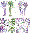
Fig. 2.. Structure of ClbP, the type I prodrug-activating peptidase that activates colibactin.
(A) ClbP forms a dimer which subtends a dome-shaped cavity lined with the two active sites at opposite ends. The two subunits are colored differently, and the gray box marks the dimer interface detailed in panel B. (B) ClbP dimer interface as viewed from outside the cell from a plane perpendicular to the membrane. The dimerization interface is composed of interactions between pairs of interlocking loops spanning α8 and β11, and the α11 helix. Red spheres mark the hydroxyl oxygens of the two catalytic S95 residues. The black oval denotes a two-fold crystallographic symmetry axis. (C) View of a single ClbP subunit. The interface between the periplasmic and TM domains is important for the function and stability of ClbP. The black box marks the interdomain interface detailed in panel D. (D) The interface between periplasmic and TM domains forms the substrate-binding site. Positions conserved throughout prodrug-activating peptidases are labeled. Part of a precolibactin substrate—modeled based on ClbP structures with bound prodrug motif analogs—is shown as sticks with the N-myristoyl-d-asparagine prodrug motif in green and the start of the toxin in black, with the black dots indicating the direction of extension. The two atoms forming the scissile peptide bond are marked as spheres.
Fig. 3.
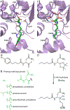
Fig. 3.. A network of conserved polar residues recognizes the N-acyl-d-asparagine prodrug motif.
(A) An N-acyl-d-asparaginate product analog (green sticks) bound to the ClbP active site (PDB ID: 7MDF). Residues conserved in prodrug-activating peptidases that interact with the prodrug motif are shown as sticks. (B) The chemical structure of the product analog consistent with the electron density observed in the active site (a substrate analog used in cocrystallization was hydrolyzed during the crystallization process). (C) Crystal structure of wildtype ClbP bound to a boronic acid prodrug mimic which potently inhibits the enzyme by forming a covalent adduct with the catalytic serine (PDB ID: 7MDC). (D) Chemical structure of the inhibitor and its enzyme adduct. (E) Acyl groups at the N-terminus of the prodrug motifs of different toxin precursors. These fatty acyl groups extend towards the membrane and may interact with hydrophobic residues in the groove between TM1 and TM3.
Fig. 4.
Fig. 4.. Prodrug-activating peptidases are predicted to adopt a common fold with the largest variations in the loops ClbP uses for dimerization.
(A) The highest ranking AlphaFold prediction for the type I peptidases ClbP, AmiB, and XcnG and for the peptidase modules of the type II peptidases EdeA, PamJ, and ZmaM are superimposed and colored according to their pLDDT confidence score in a spectrum where pLDDT values below 75 (reflecting greater uncertainty) are colored magenta and values larger than 90 (representing high confidence) are colored in cyan. Superposition of the AlphaFold model of ClbP onto a high-resolution crystal structure of ClbP (grey; PDB ID: 7MDF) shows that the model is an accurate prediction of the experimentally determined structure (RMSD = 1.8 Å). All models were generated at a time when the coordinates of full-length ClbP had not been released to the PDB. The two loops that form the dimer interface in ClbP are boxed. The arrow points to the TM2 kink, which is predicted in all models but at distinct angles. (B) The models are tiled to highlight the differences in both length and predicted structures (and lower confidence scores) of the two loop regions that form the dimer interface in ClbP (which are boxed in the ClbP panel). Type I peptidases are at the top, Type II peptidases, for which the models are most similar to each other, are at the bottom.
Fig. 5.
Fig. 5.. ClbP-like homologs not part of prodrug resistance BGCs may have novel architectures and substrates.
(A) Sequence logos representing the conservation of the S12 catalytic motifs among all the membrane-embedded β-lactamases in our SSN. (B-C) Sequence logos detailing the conservation of N-acyl-d-asparagine binding positions among prodrug-activating peptidases (B) and among other ClbP-like proteins (C). The d-asparagine-binding residues are very highly conserved in prodrug-activating peptidases, but much more variable in ClbP-like proteins, suggesting they process different substrates. Highlighted positions are shown as sticks in the ClbP structures in Figure 3A-B. (D) Color-coding of the nodes illustrates the variety of sidechains found at the N331 position across the SSN of membrane-embedded S12 hydrolase homologs.
Fig. 6
Fig. 6. Distribution of TMD topologies in membrane-embedded β-lactamases.
(A) SSN of membrane-embedded β-lactamases (PF00144) colored according to the predicted topology of their TMD. The number of TM helices was predicted using TMHMM, discounting any N-terminal predicted TM helix as they correspond to signal peptides in most sequences. Prodrug-activating peptidases have either type I (3 TMs; blue) or type II (9 TMs; cyan) topology, while ClbP-like proteins which are not part of an NRPS-containing BGC are more diverse, including predicted TMDs of length consistent with type I (“ClbP-like”; yellow) or type II peptidases, and TMDs of intermediate lengths. Homologs with intermediate TMDs include a subset with 4 TM helices and an inserted domain (orange), and a second subset with a small cytoplasmic domain and three N-terminal helices preceding a ClbP-like fold (red). AlphaFold predictions of homologs from the most common predicted topologies suggest there is at least one novel TMD fold for “4-helix” ClbP-like proteins. (B) Genomic neighborhoods (+/− 10 genes) for the ClbP homologs illustrated in panel A and representative members of additional SSN groups (nodes indicated with black outlines). Possible tailoring enzymes (epimerases, kinases, phosphatases, hydrolases, and acyltransferases) are seen in a handful of genomic neighborhoods, but most genes encoding ClbP-like proteins are proximal to transporters and regulators rather than extended biosynthetic gene clusters. By comparison, ZmaM and XcnG homologs are associated with larger mixed NRPS-PKS biosynthetic gene clusters (bottom), and genomic neighborhoods encoding ZmaM homologs tend to lack genes encoding other transporters.

References

    1. Patin NV, Duncan KR, Dorrestein PC, Jensen PR, Competitive strategies differentiate closely related species of marine actinobacteria, ISME J 10 (2016) 478–490. 10.1038/ismej.2015.128. - DOI - PMC - PubMed
    1. Stubbendieck RM, Straight PD, Multifaceted Interfaces of Bacterial Competition, J Bacteriol 198 (2016) 2145–2155. 10.1128/JB.00275-16. - DOI - PMC - PubMed
    1. Schmidt R, Ulanova D, Wick LY, Bode HB, Garbeva P, Microbe-driven chemical ecology: past, present and future, ISME J 13 (2019) 2656–2663. 10.1038/s41396-019-0469-x. - DOI - PMC - PubMed
    1. Yan Y, Liu N, Tang Y, Recent developments in self-resistance gene directed natural product discovery, Nat Prod Rep 37 (2020) 879–892. 10.1039/c9np00050j. - DOI - PMC - PubMed
    1. Cundliffe E, Demain AL, Avoidance of suicide in antibiotic-producing microbes, J Ind Microbiol Biotechnol 37 (2010) 643–672. 10.1007/s10295-010-0721-x. - DOI - PubMed

MeSH terms

LinkOut - more resources