. 2023 Feb 17;30(1):12.
doi: 10.1186/s12929-023-00906-6.

CLEC5A mediates Zika virus-induced testicular damage

Affiliations

CLEC5A mediates Zika virus-induced testicular damage

Hsin-Wei Wang et al. J Biomed Sci. .

Abstract

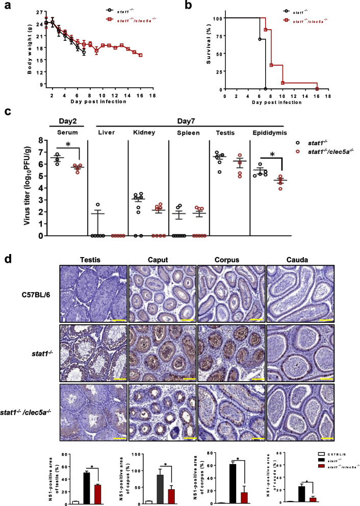
Background: Zika virus (ZIKV) infection is clinically known to induce testicular swelling, termed orchitis, and potentially impact male sterility, but the underlying mechanisms remain unclear. Previous reports suggested that C-type lectins play important roles in mediating virus-induced inflammatory reactions and pathogenesis. We thus investigated whether C-type lectins modulate ZIKV-induced testicular damage.

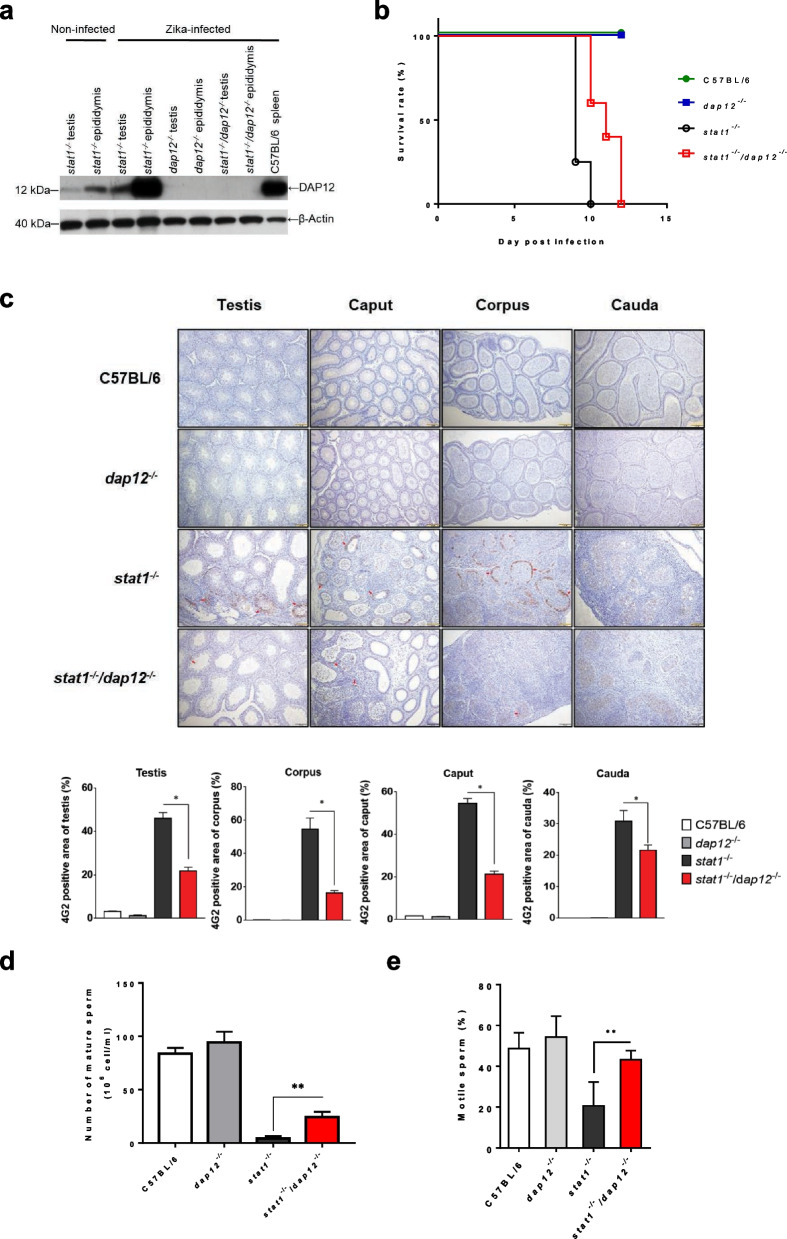
Methods: C-type lectin domain family 5 member A (CLEC5A) knockout mice were generated in a STAT1-deficient immunocompromised background (denoted clec5a-/-stat1-/-) to enable testing of the role played by CLEC5A after ZIKV infection in a mosquito-to-mouse disease model. Following ZIKV infection, mice were subjected to an array of analyses to evaluate testicular damage, including ZIKV infectivity and neutrophil infiltration estimation via quantitative RT-PCR or histology and immunohistochemistry, inflammatory cytokine and testosterone detection, and spermatozoon counting. Furthermore, DNAX-activating proteins for 12 kDa (DAP12) knockout mice (dap12-/-stat1-/-) were generated and used to evaluate ZIKV infectivity, inflammation, and spermatozoa function in order to investigate the potential mechanisms engaged by CLEC5A.

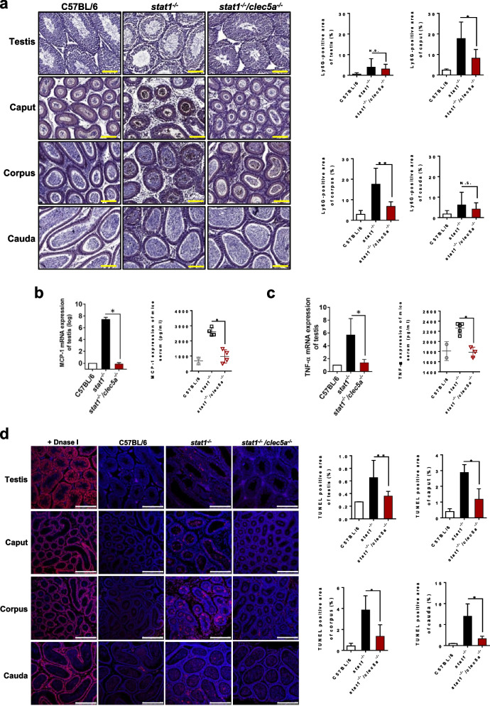
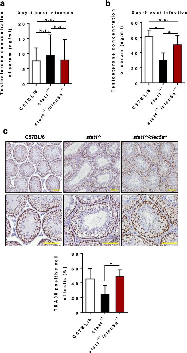
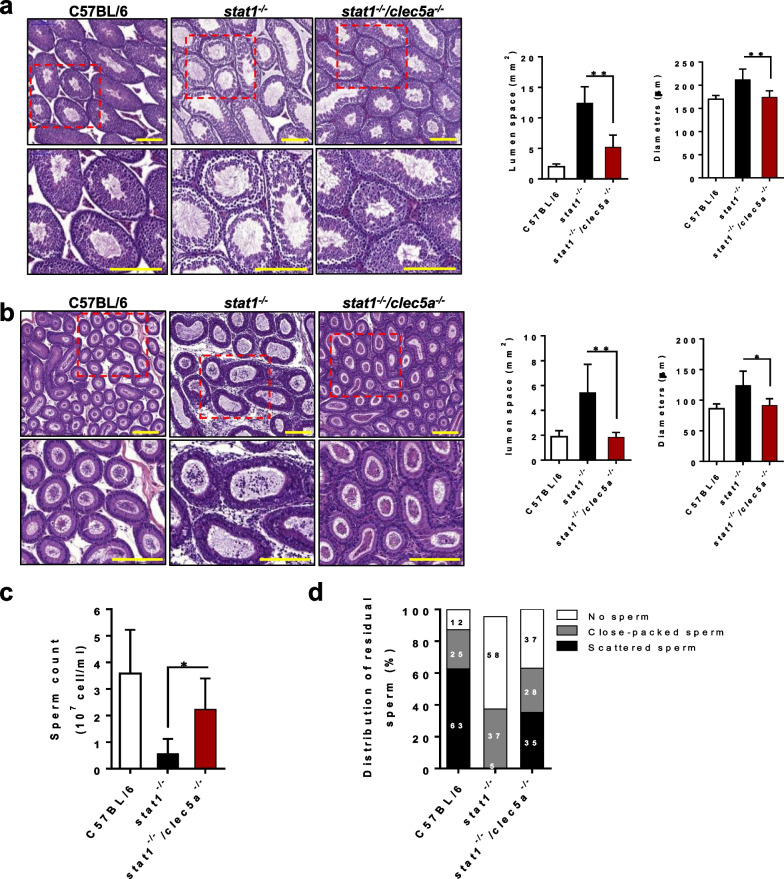
Results: Compared to experiments conducted in ZIKV-infected stat1-/- mice, infected clec5a-/-stat1-/- mice showed reductions in testicular ZIKV titer, local inflammation and apoptosis in testis and epididymis, neutrophil invasion, and sperm count and motility. CLEC5A, a myeloid pattern recognition receptor, therefore appears involved in the pathogenesis of ZIKV-induced orchitis and oligospermia. Furthermore, DAP12 expression was found to be decreased in the testis and epididymis tissues of clec5a-/-stat1-/- mice. As for CLEC5A deficient mice, ZIKV-infected DAP12-deficient mice also showed reductions in testicular ZIKV titer and local inflammation, as well as improved spermatozoa function, as compared to controls. CLEC5A-associated DAP12 signaling appears to in part regulate ZIKV-induced testicular damage.

Conclusions: Our analyses reveal a critical role for CLEC5A in ZIKV-induced proinflammatory responses, as CLEC5A enables leukocytes to infiltrate past the blood-testis barrier and induce testicular and epididymal tissue damage. CLEC5A is thus a potential therapeutic target for the prevention of injuries to male reproductive organs in ZIKV patients.

Keywords: CLEC5A; Inflammation; Mosquito; Mouse; Testicular damage; Zika virus.


Conflict of interest statement

The authors declare that they have no competing interests.

Figures

Fig. 1
Fig. 1
Clec5a knockout attenuates ZIKV infectivity in the testicular tissues of mice. Bodyweight (recorded daily) (a) and survival (monitored every 2 days) (b) in male stat1−/− and stat1−/−clec5a−/− mice infected with ZIKV via mosquitoes. c ZIKV titer in serum samples (n ≥ 3) collected at 2 days post-infection (dpi) and in various tissue samples (n ≥ 4) collected at 7 dpi. Plaque assay was used to quantify infectious ZIKV and the limit of detection was estimated by counting numbers of plaque formation in original lysates. No plaque formation was observed in the livers, kidneys and spleens of some ZIKV-infected mice. d Immunohistochemical staining of tissues from the testes and epididymis (caput, corpus, and cauda) collected from ZIKV-infected mice (n ≥ 4) at 7 dpi and from uninfected C57BL/6 mice (n = 3), which were used as controls. Quantitative data are presented as mean ± standard deviation (SD). *p < 0.05. Scale bars: 100 µm
Fig. 2
Fig. 2
Loss of CLEC5A reduces testicular inflammation and damage. a Neutrophil infiltration in the testes and epididymis (caput, corpus, and cauda) collected from ZIKV-infected stat1−/− mice (n ≥ 8) and stat1−/−clec5a−/− mice (n ≥ 8) at 7 dpi and from uninfected C57BL/6 control mice (n ≥ 6) was examined and quantified via immunohistochemical staining with anti-Ly6G antibody. Levels of b chemokine MCP-1 and c proinflammatory cytokine TNF-α in serum and testes from infected mutant mice or uninfected WT mice. The serum was collected at 2 dpi, and the testicular tissues were collected at 7 dpi. The concentrations of MCP-1 and TNF-α in the serum were determined via a standard ELISA assay, and the RNA levels of MCP-1 and TNF-α expressed in the testicular tissues were assessed by quantitative real-time PCR assay. d Apoptosis assay using tissue sections from the testes and epididymis (caput, corpus, and cauda) of ZIKV-infected stat1−/− mice (n ≥ 4) and stat1−/−clec5a−/− mice (n ≥ 4), with uninfected C57BL/6 mice (n = 3) used as a negative control. DNase I-treated samples served as a positive control. The graphs show the ratio of TUNEL-staining intensity in the ZIKV-infected and uninfected testicular tissue to that in the DNase I-treated samples. Neutrophil invasion and apoptosis in the testicular tissues were evaluated at 7 dpi. Quantitative data are presented as mean ± SD. *p < 0.05; **p < 0.01; N.S., not significant. Scale bars: (a) 100 μm; (d) 250 μm
Fig. 3
Fig. 3
CLEC5A deficiency improves ZIKV-damaged testicular function. Testosterone levels in serum samples from ZIKV-infected mice at 1 dpi (a) and 6 dpi (b). c Immunohistochemical analysis of testicular tissue stained with anti-TRA98. Uninfected C57BL/6 mice (n = 5) served as a negative control for the ZIKV-infected stat1−/− mice (n = 4) and stat1−/−clec5a−/− mice (n = 6) at 7 dpi. Quantitative data are presented as mean ± SD. *p < 0.05; N.S., not significant. Scale bars: 100 μm
Fig. 4
Fig. 4
Loss of CLEC5A attenuates oligospermia following ZIKV infection. Hematoxylin-and-eosin staining histological analysis of tissue sections of the seminiferous tubules (a) in the testis and (b) the mesonephric tubules in the epididymis. c Sperm count for epididymis tissues from mice. d Spermatid density and distribution in the epididymis of ZIKV-infected stat1−/− mice (n ≥ 4) and stat1−/−clec5a−/− mice (n ≥ 5) (7 dpi), with uninfected C57BL/6 mice (n = 6) as negative controls, quantified using Panoramic Viewer software. For a statistical analysis of the distribution of sperm, more than two randomly selected fields of view of the section of the epididymis tissue were assessed for each mouse, and more than three mice were analyzed for each group. Quantitative data are presented as mean ± SD. *p < 0.05; **p < 0.01. Scale bars: 200 μm
Fig. 5
Fig. 5
CLEC5A function is related to malfunctioning spermatozoa following ZIKV infection. a A cytomorphological study of mature spermatozoa harvested from the cauda of the epididymis was conducted using Papanicolaou stain. Aberrant morphology in the head or tail of spermatozoa (red arrows) was observed in ZIKV-infected stat1−/− mice and stat1−/−clec5a−/− mice compared to that in uninfected C57BL/6 mice. The proportions of normal and abnormal sperm were quantified for statistical analysis. b Sperm motility assay. At least three randomly selected fields of view were analyzed for each group (a). c Immunofluorescence staining of sperm allowed the visualization of ZIKV (green, anti-NS1-eGFP), cytoplasm (red, CellTracker), and nuclei (blue, DAPI). ZIKV-positive sperm were counted from more than 150 sperm cells in three random fields of view per mouse and are presented as percentages of all cells. Samples were taken from ZIKV-infected stat1−/−(n = 12) and stat1−/−clec5a−/− mice (n = 8) (7 dpi) and uninfected C57BL/6 mice (n = 3), which served as negative controls. Quantitative data are presented as mean ± SD. *p < 0.05; ***p < 0.001; N.D., not detected. Scale bars: (a) 50 μm; (c) 25 μm
Fig. 6
Fig. 6
Loss of DAP12 compromises ZIKV infectivity and spermatozoa function in ZIKV-damaged testicular tissues. a Western blot analysis of DAP12 protein expressed in various testicular tissues of male stat1−/−, dap12−/−, or stat1−/−dap12−/− mice infected with ZIKV at 7 dpi. Spleen tissues with known high DAP12 expression were collected from C57BL/6 mice as a positive control. b Survival (monitored daily) in male mutant or C57BL/6 mice infected with ZIKV via mosquito. c Immunohistochemical staining (with anti-4G2) of tissues from the testes and epididymis (caput, corpus, and cauda) collected from ZIKV-infected mice (n ≥ 3) at 7 dpi and from uninfected C57BL/6 mice (n = 3) as controls. Scale bars: 100 µm. Quantitative data show the percentage of ZIKV-infected area in different tissues. Sperm count for epididymis tissues (d) and sperm motility (e) in mutant or C57BL/6 mice with ZIKV infection. For be, stat1−/−, dap12−/−, or stat1−/−dap12−/− mutant mice were used. At least three randomly selected fields of view were analyzed for each group. Quantitative data are presented as mean ± SD. *p < 0.05. **p < 0.01

References

    1. Armah HB, et al. Systemic distribution of West Nile virus infection: postmortem immunohistochemical study of six cases. Brain Pathol. 2007;17:354–362. - PMC - PubMed
    1. Shamim M, Naqvi SZ. Dengue fever associated with acute scrotal oedema: two case reports. J Pak Med Assoc. 2011;61:601–603. - PubMed
    1. Zheng B, et al. Japanese Encephalitis Virus infection induces inflammation of swine testis through RIG-I-NF-kB signaling pathway. Vet Microbiol. 2019;238:108430. - PubMed
    1. Chan JF, et al. Zika Virus Infection in Dexamethasone-immunosuppressed Mice Demonstrating Disseminated Infection with Multi-organ Involvement Including Orchitis Effectively Treated by Recombinant Type I Interferons. EBioMedicine. 2016;14:112–122. - PMC - PubMed
    1. Reynolds MR, et al. Vital Signs: Update on Zika Virus-Associated Birth Defects and Evaluation of All US Infants with Congenital Zika Virus Exposure - US Zika Pregnancy Registry, 2016. MMWR Morb Mortal Wkly Rep. 2017;66:366–373. - PMC - PubMed